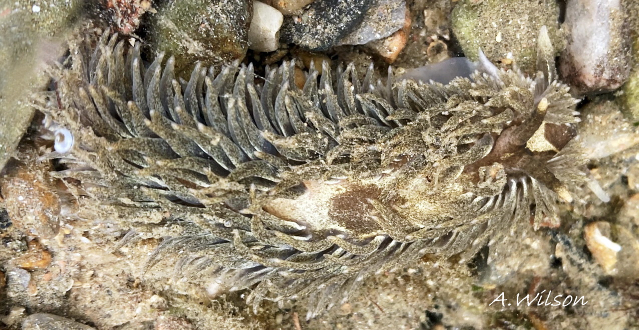
Specimen with cerata darkened by contents of digestive gland and some surface pigment. Despite the dark shade, large white cnidosacs are clearly visible in apices of cerata. This form of A. filomenae with dark chocolate pigment on basal 75% of rhinophores, on head with contrasting white Y, and a patch on the notum, occurs fairly frequently and is the form in Alder & Hancock’s image, . Many flattened cerata on the anterior half of the slug’s right side have thin edges towards the camera.
Plymouth, England. May 2020 © A. Wilson, CC BY_NC.
06 Aeolidia filomenae

