Click image to enlarge with full caption. Main text below slider.
01 Aeolidia filomenae
02 Aeolidia filomenae
03 Aeolidia filomenae
04 Aeolidia filomenae
05 Aeolidia filomenae
06 Aeolidia filomenae
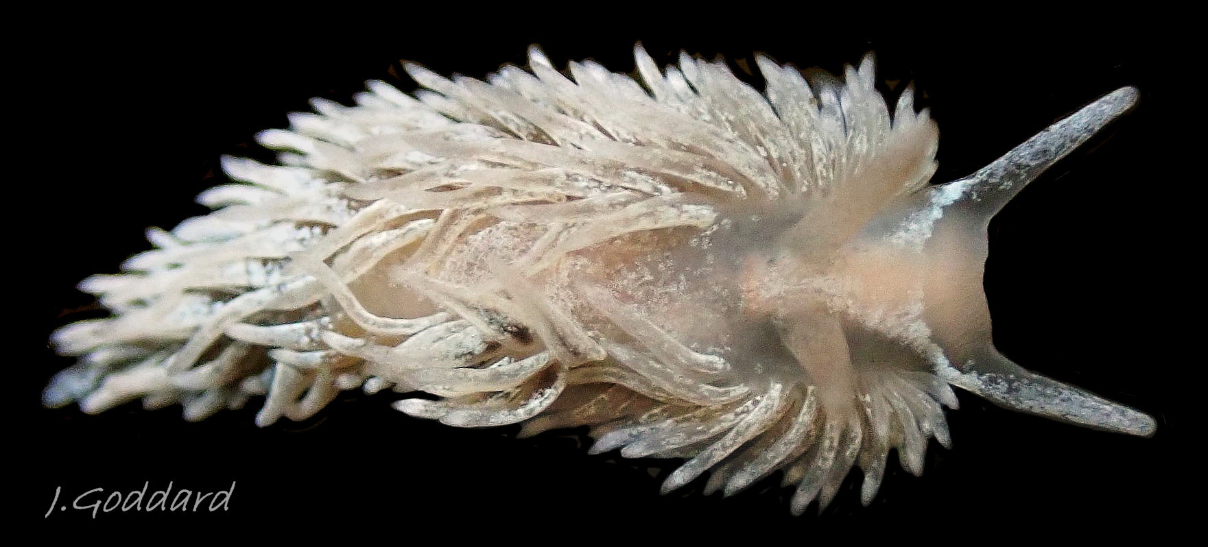
07 Aeolidia filomenae
08 Aeolidia filomenae
09 Aeolidia filomenae
10 Aeolidia filomenae
11 Aeolidia filomenae
12 Aeolidia filomenae
13 Aeolidia filomenae
14 Aeolidia filomenae
15 Aeolidia filomenae
16 Aeolidia filomenae
17 Aeolidia filomenae
18 Aeolidia filomenae COMPARE Aeolidia papillosa
19 Aeolidia filomenae COMPARE Aeolidia papillosa
20 Aeolidia filomenae COMPAREAeolidia papillosa
21 Aeolidia filomenae COMPARE Aeolidia papillosa
22 Aeolidia filomenae COMPARE Aeolidia papillosa
23 Aeolidia filomenae COMPARE Aeolidia papillosa
24 Aeolidia filomenae COMPARE Aeolidiella alderi
25 Aeolidia filomenae COMPARE Aeolidiella glauca
26 Aeolidia filomenae COMPARE Aeolidiella sanguinea
27 Aeolidia filomenae
28 Aeolidia filomenae
29 Aeolidia filomenae
30 Aeolidia filomenae
31 Aeolidia filomenae
32 Aeolidia filomenae COMPARE Aeolidia papillosa
33 Aeolidia filomenae COMPARE Aeolidia papillosa
Aeolidia filomenae Kienberger, Carmona, Pola, Padula, Gosliner & Cervera, 2016.
Synonyms: Limax papillosus Linnaeus, 1761 (partim); Eolis papillosa (Linnaeus, 1761) (partim); Aeolidia papillosa (Linnaeus, 1761) (partim).
Current taxonomy: World Register of Marine Species www.marinespecies.org/aphia.php?p=taxdetails&id=880371
GLOSSARY BELOW
Preface
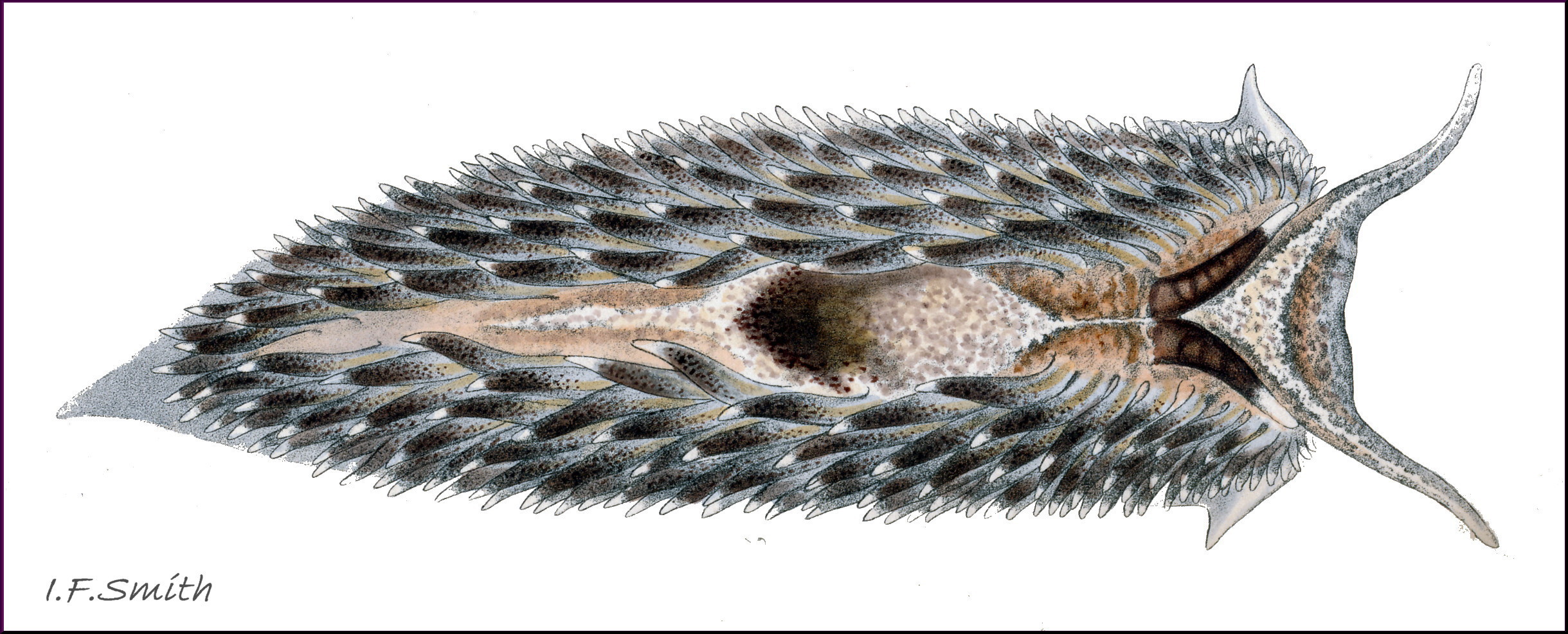
In 2016, Kienberger et al. segregated with molecular sequencing what had previously been generally accepted in Europe as A. papillosa into Aeolidia papillosa sensu stricto (Linnaeus, 1761) and Aeolidia filomenae Kienberger et al., 2016. Descriptions published before 2016 combine features of the two species and often, such as in Alder & Hancock (1845-1855) 07 Aeolidia filomenae and Thompson & Brown (1984), illustrated descriptions of A. papillosa with images of probable A. filomenae. This account draws on the descriptions in Kienberger et al. (2016).
Description
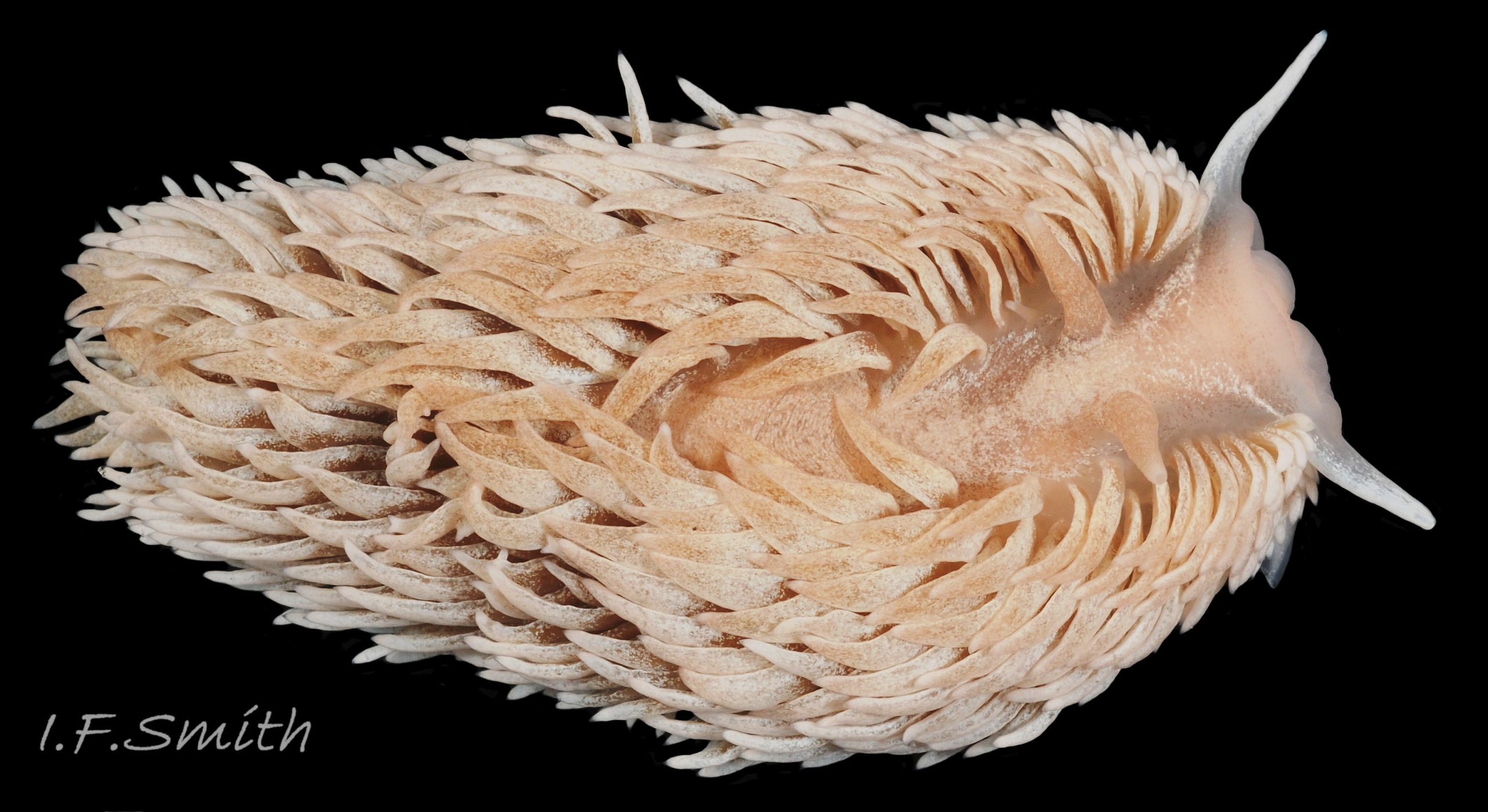
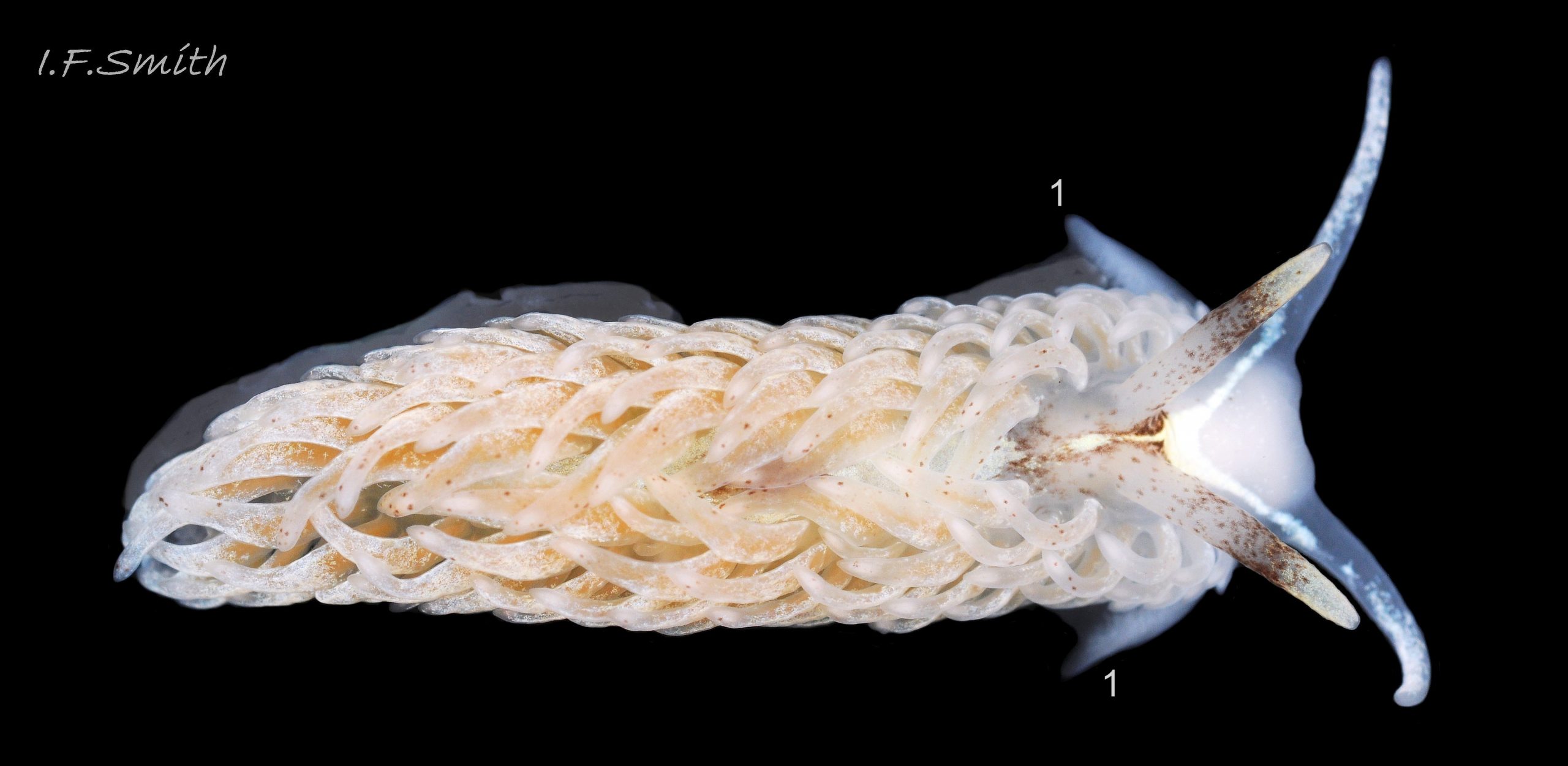
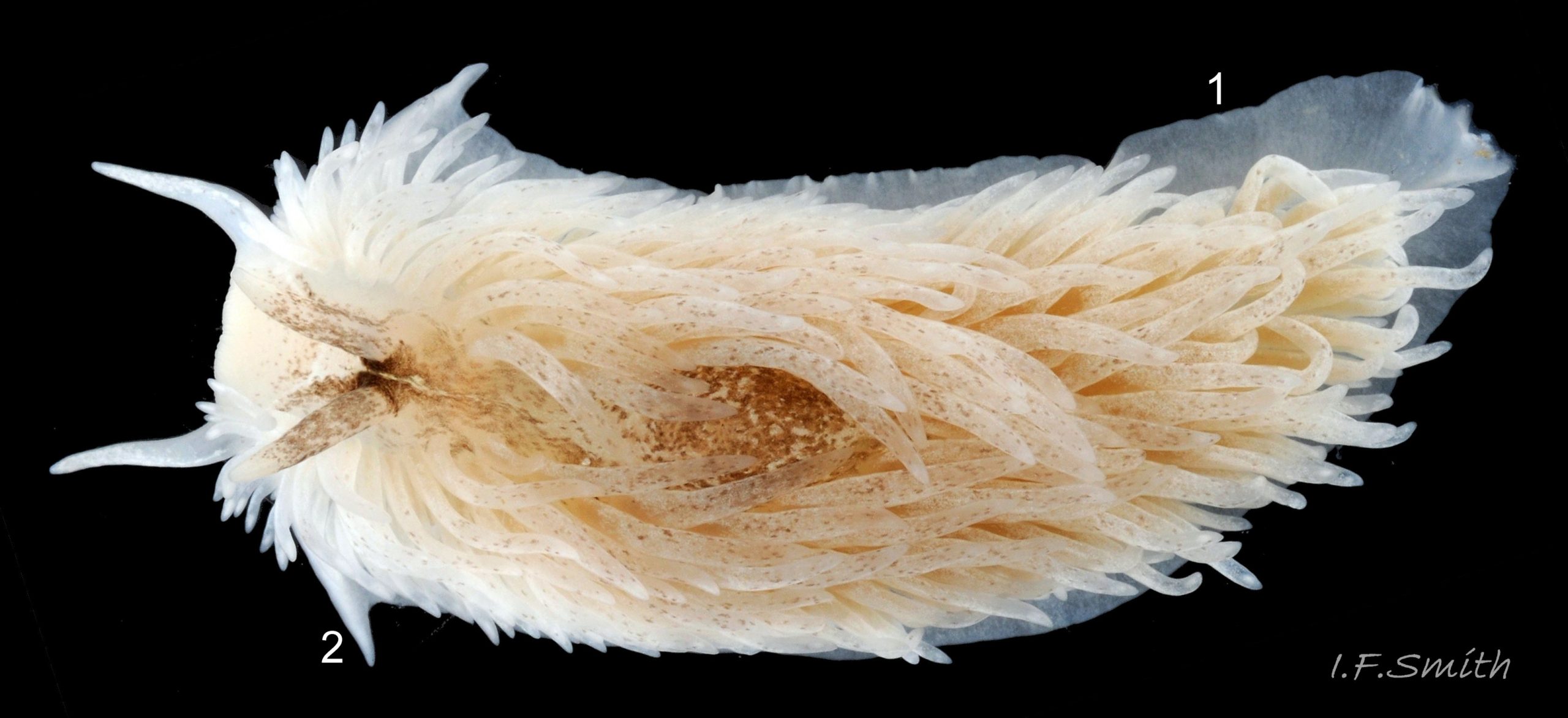
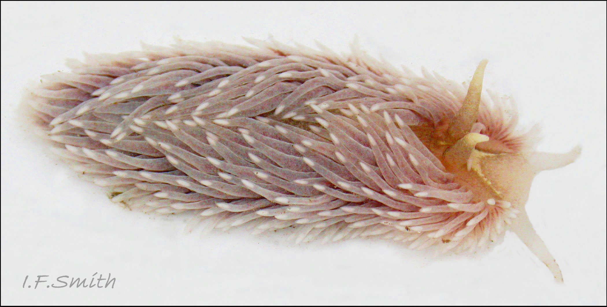
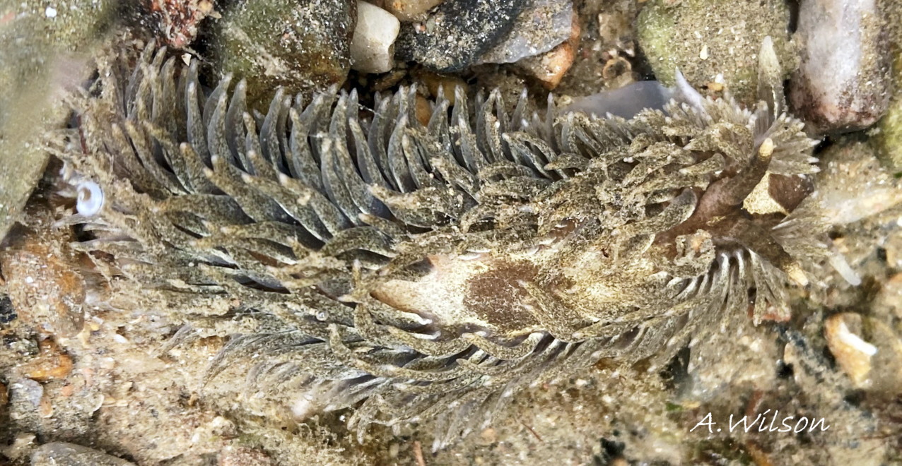
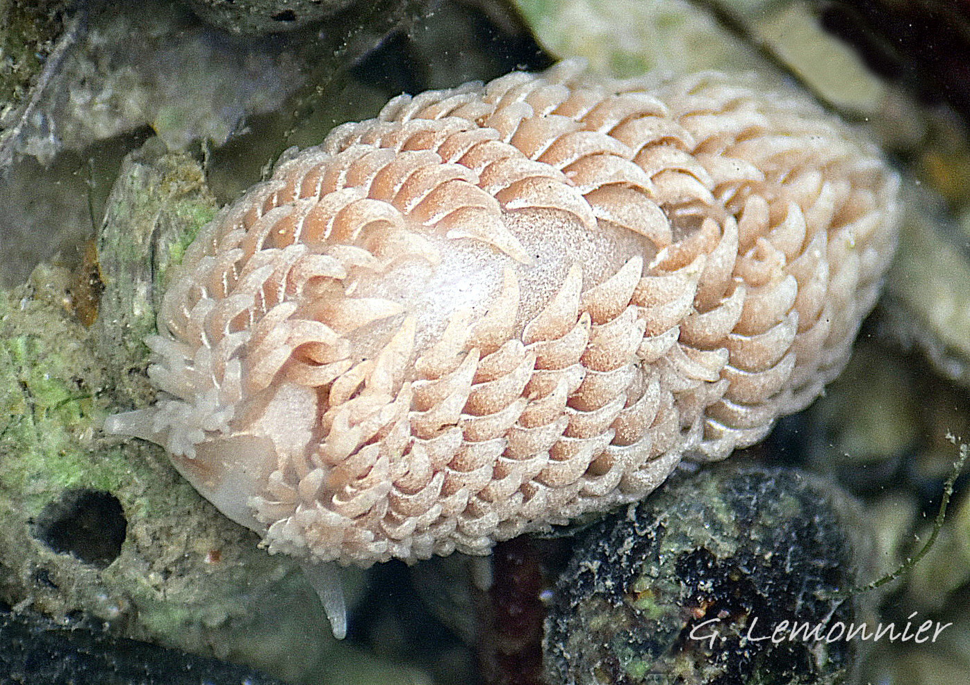
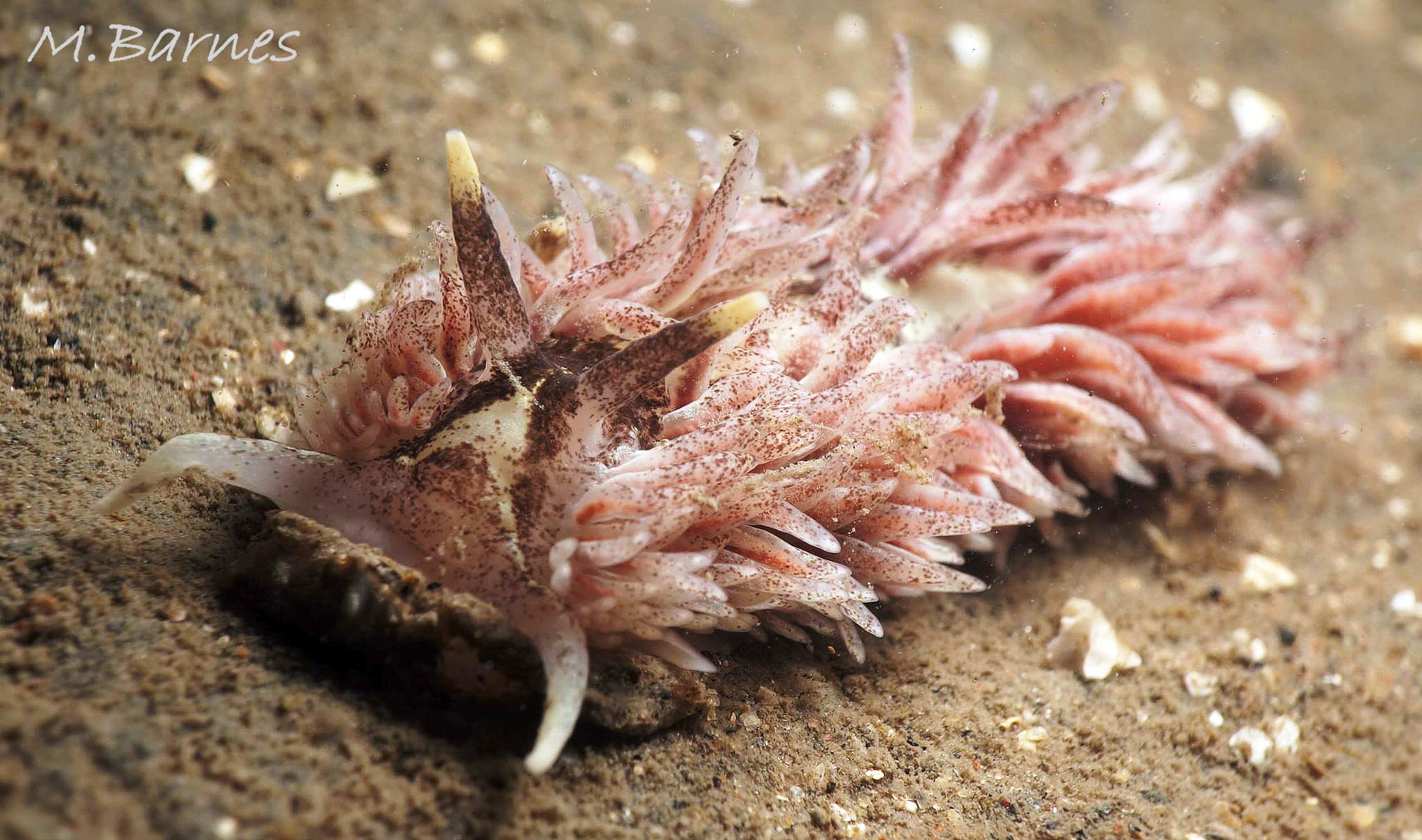
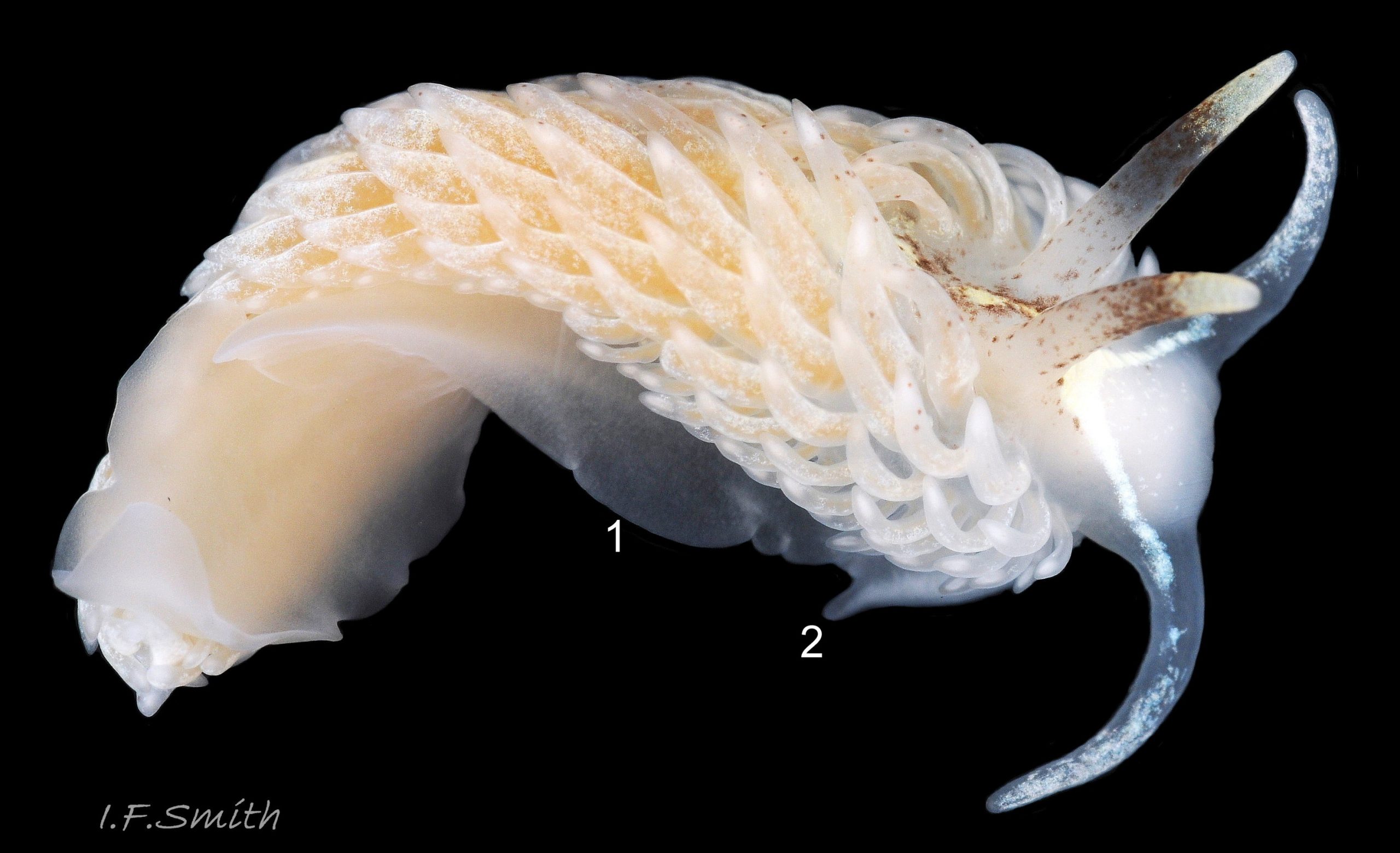
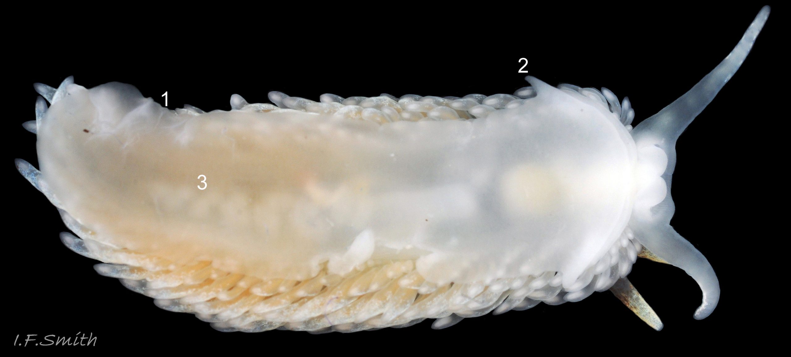
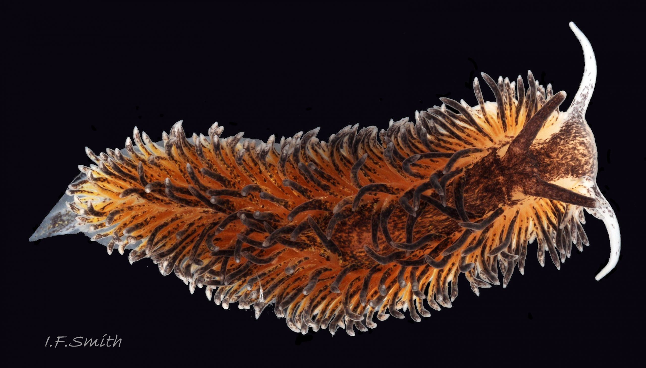
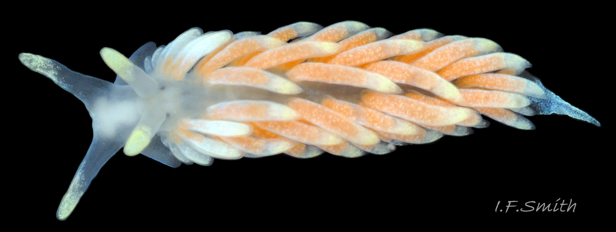
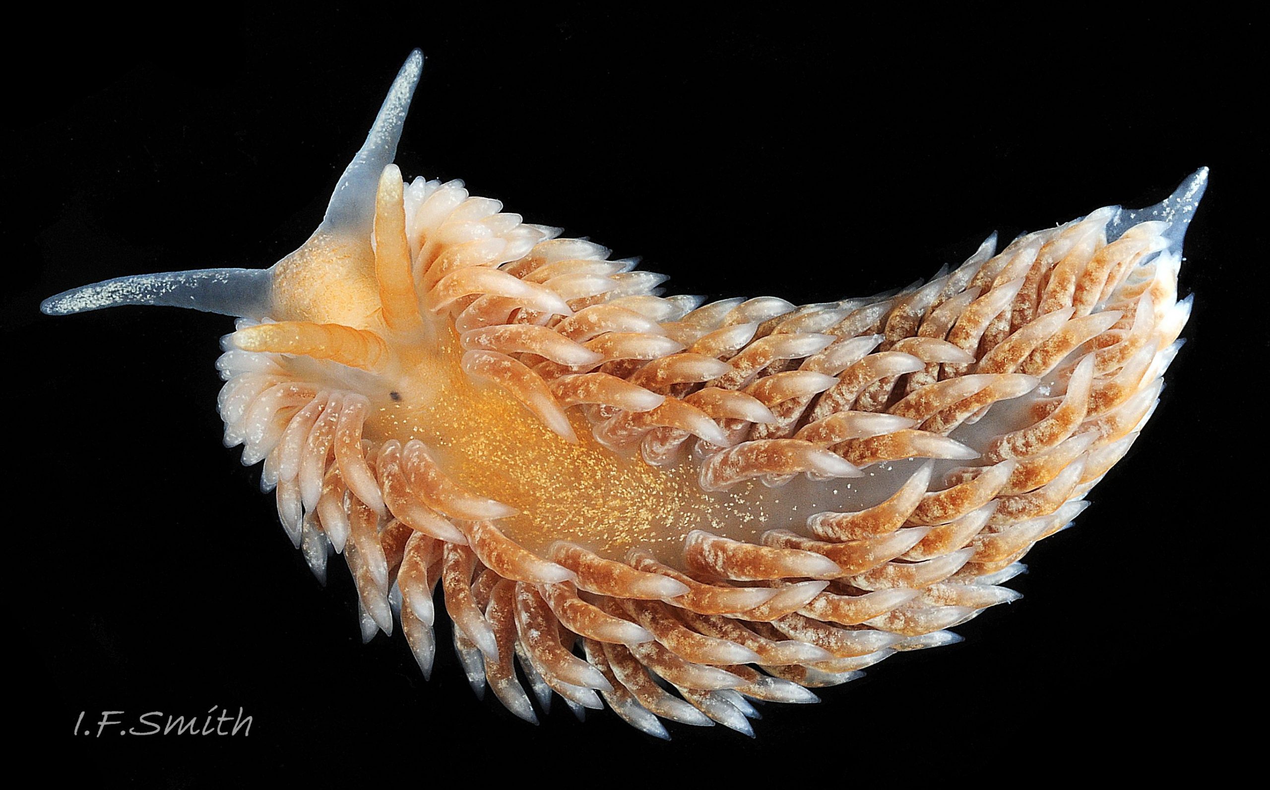
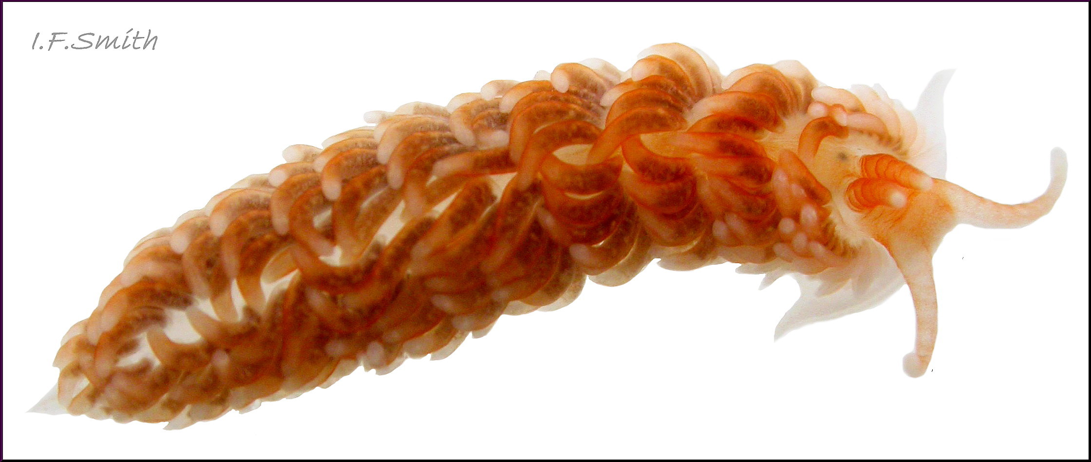
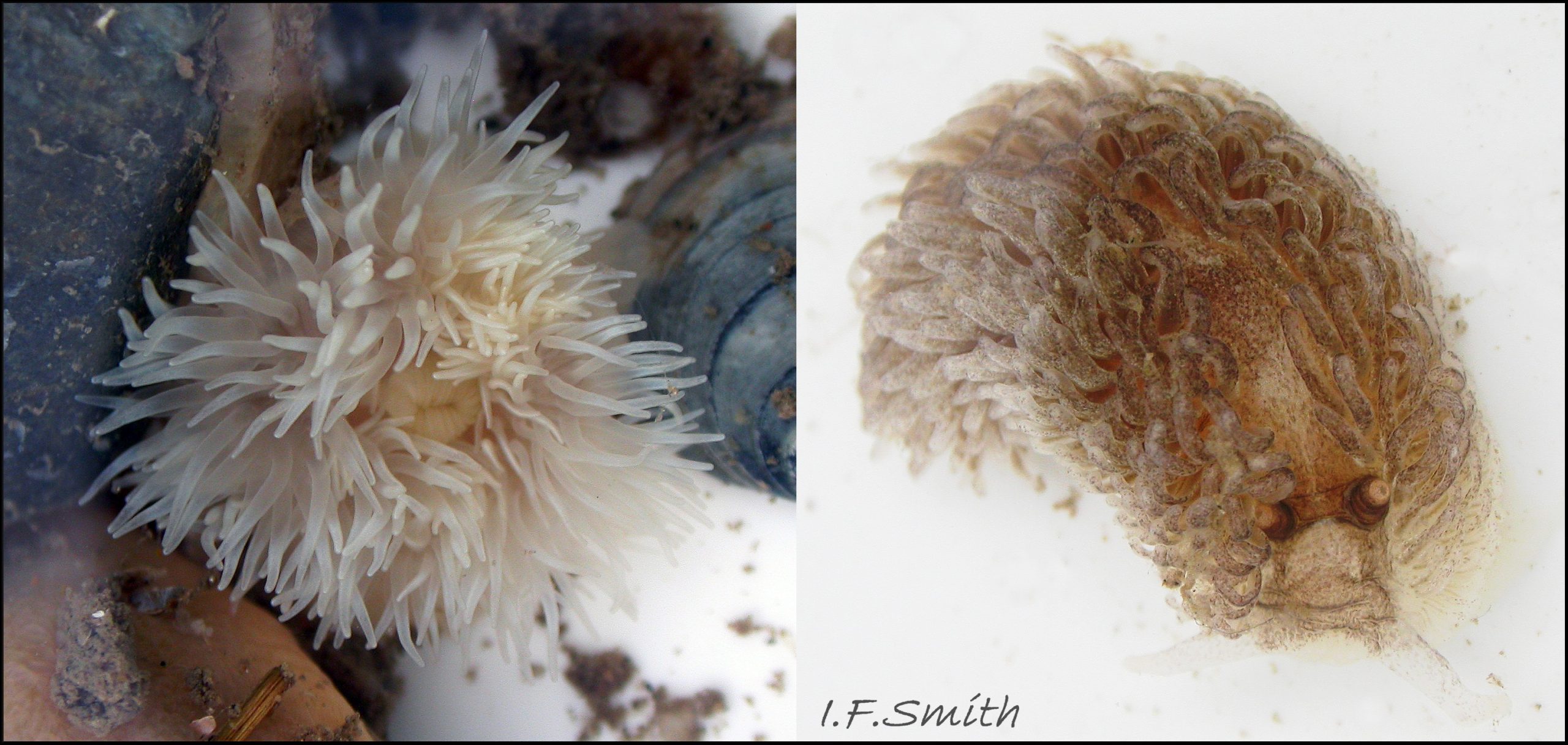
A. filomenae grows to at least 70 mm long 01 Aeolidia filomenae . Individuals can vary their width from broad 02 Aeolidia filomenae to relatively narrow 03 Aeolidia filomenae . The ground colour of the notum varies from white 02 Aeolidia filomenae through light beige and pink 01 Aeolidia filomenae to greenish with white or brown flecks 04 Aeolidia filomenae which sometimes concentrate into dense brown on the head, rhinophores and pericardial area 05 Aeolidia filomenae & 06 Aeolidia filomenae . There is often a white Y–shaped mark on the oral tentacles and head with its stem passing back between the rhinophores. The mark can be clearly defined intense opaque white 08 Aeolidia filomenae , less obtrusive beige or light brown, or be partly obscured by coloured flecks 01 Aeolidia filomenae .
Dense cerata usually conceal dorsal surfaces except on the head and a bare zone extending back from the rhinophores 01 Aeolidia filomenae .
The cleioproctic anus is located between the ninth and tenth row of the right side (Kienberger et al. 2016). The genital aperture is situated on the right side between the fourth and fifth anterior rows of cerata (Kienberger et al. 2016) which usually conceal it.
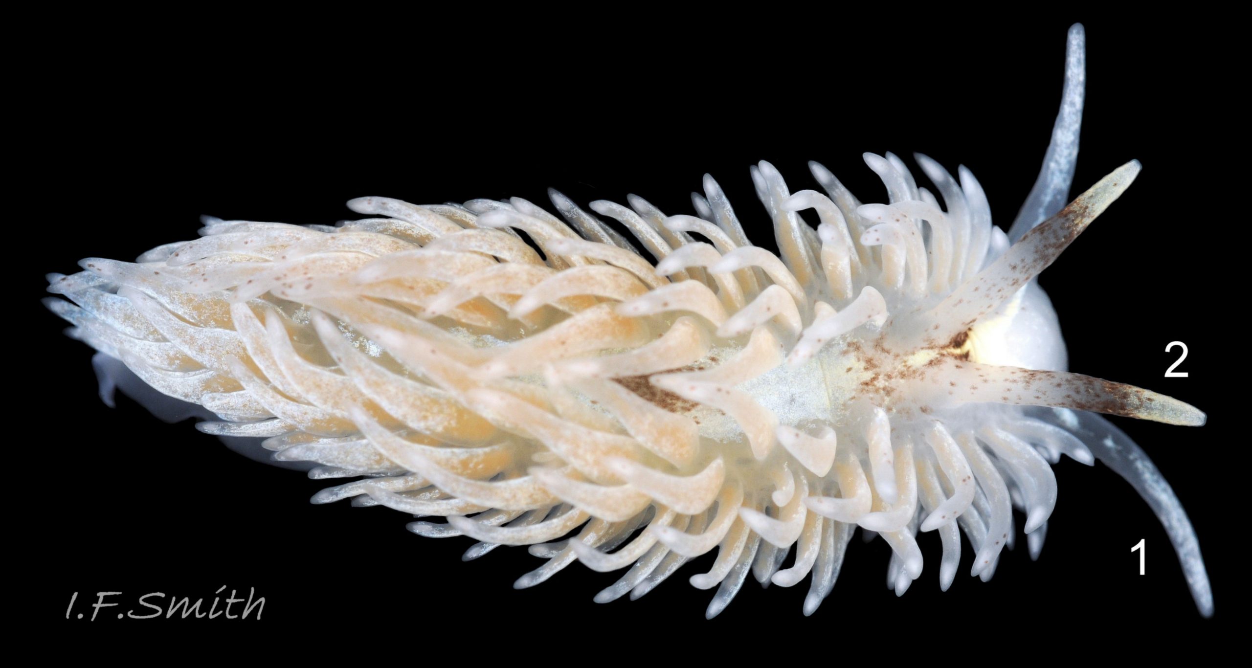
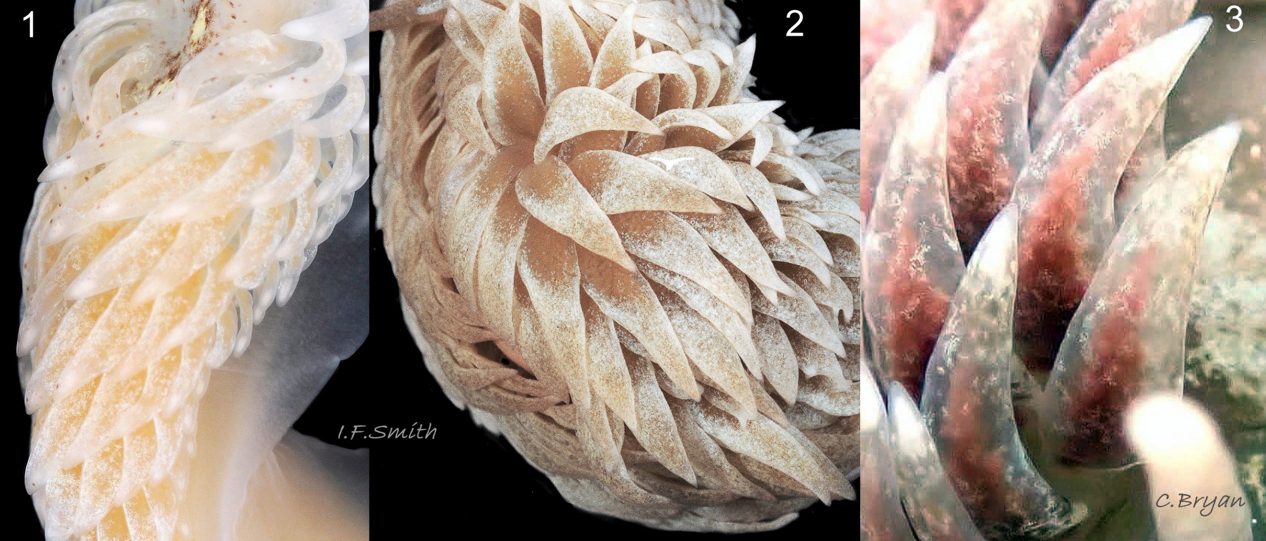
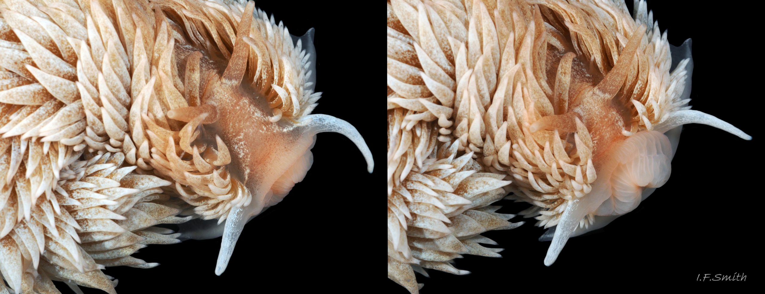
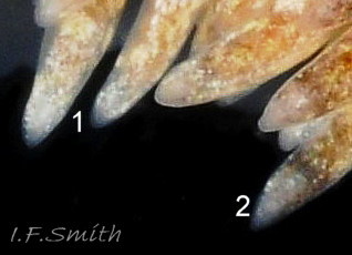
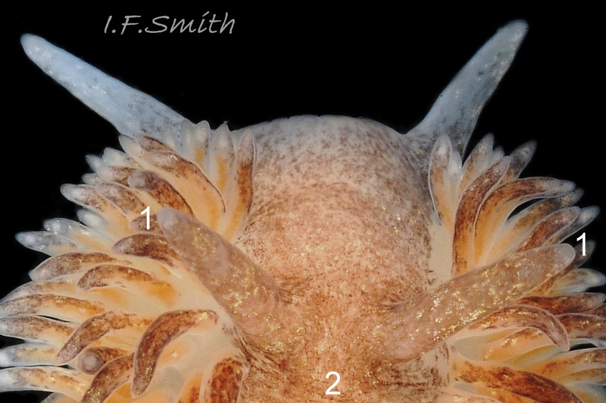
The cerata are arranged in up to 16, often neatly arranged, oblique rows on each side of the body ( 09 Aeolidia filomenae and Kienberger et al. 2016). Those at the posterior and anterior ends of the body are smaller than the others. They are flattened, broader at their base, and curved inwards 01 Aeolidia filomenae & 02 Aeolidia filomenae . They may inflate a little to give a thicker cross section 10 Aeolidia filomenae . They are translucent, usually with little surface pigment to obstruct view of the internal digestive gland which is often white-beige, but can be other colours 11 Aeolidia filomenae . A large opaque white cnidosac is often visible at the apex of each ceras 05 Aeolidia filomenae & 08 Aeolidia filomenae . The general colour of individuals depends largely on the colour of the digestive gland in the cerata 10 Aeolidia filomenae & 11 Aeolidia filomenae but can be modified by, often white, pigment freckles 01 Aeolidia filomenae , 06 Aeolidia filomenae , & 10 Aeolidia filomenae . The cerata are usually, but not always, paler than the body.
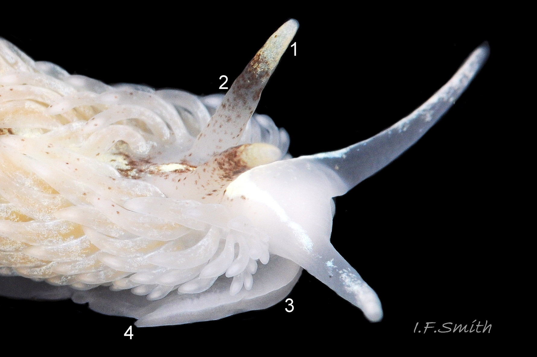
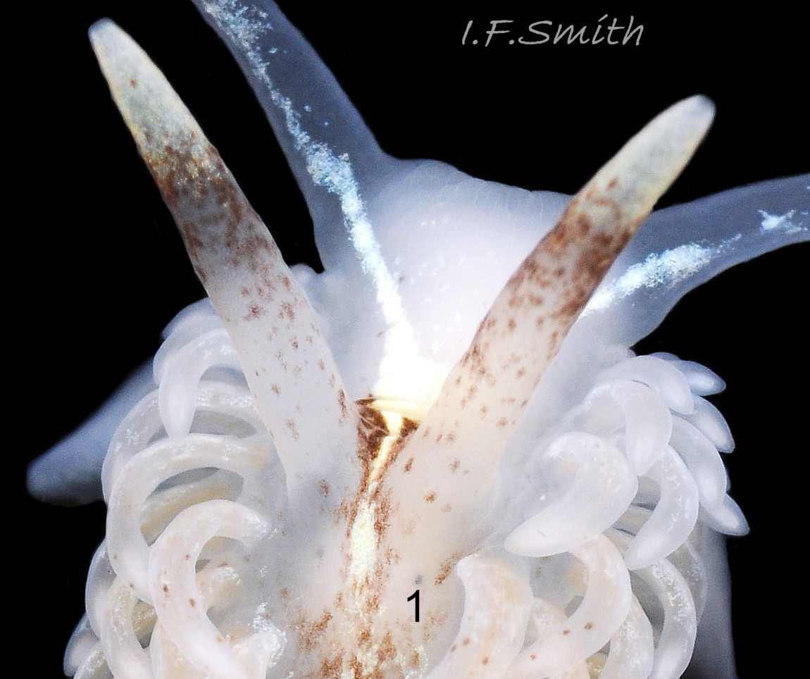
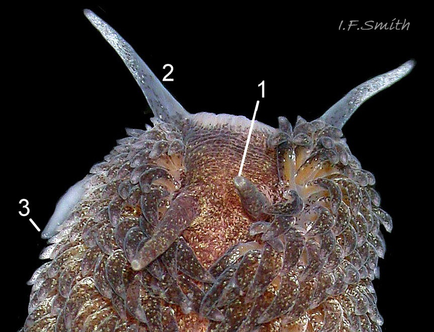
The blunt, conical, smooth rhinophores are translucent whitish with opaque white or yellowish-white freckles which often concentrate to colour the distal quarter 12 Aeolidia filomenae . Most also have brown spots of varying extent and intensity on the basal three-quarters 10 Aeolidia filomenae . The eyes are often visible at the base of the rhinophores in lighter
specimens 13 Aeolidia filomenae .
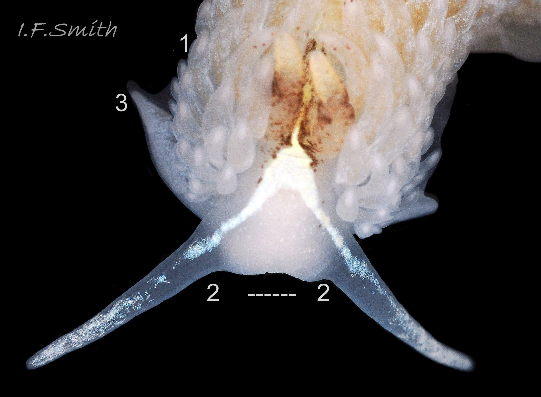
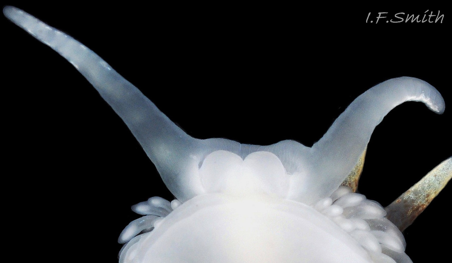
The head has translucent, whitish, oral tentacles, with opaque white pigment, which often forms the linear branches of a cephalic Y mark 08 Aeolidia filomenae . Some also have a few spots of brown pigment 10 Aeolidia filomenae . The oral tentacles are slightly longer than the rhinophores 02 Aeolidia filomenae and are placed apart on the anterior edge of the head at a distance, when it is spread, equal to about three times the thickness of a tentacle base 08 Aeolidia filomenae (IFS, pers. obs.). Ventrally the head is mostly occupied by the bilobed outer lips 14 Aeolidia filomenae which expand when projected forwards 15 Aeolidia filomenae .
The broad, thin, translucent foot 16 Aeolidia filomenae has small, triangular, propodial tentacles 08 Aeolidia filomenae on the bilaminate anterior 12 Aeolidia filomenae . Internal viscera, including spheroid ovotestes , may be visible through the sole 17 Aeolidia filomenae .
Key identification features
Features vary and may sometimes overlap with similar species. Identifications should be made on the basis of more than a single feature.
Aeolidia filomenae Kienberger, Carmona, Pola, Padula, Gosliner & Cervera, 2016.
Features as in Kienberger et al. (2016). [IFS pers. obs. in square brackets.]
1) Longest specimen described in Kienberger et al. (2016) was 45 mm; maximum possible length was not stated. 70 mm specimen in 01 Aeolidia filomenae .
2) Body white 02 Aeolidia filomenae , light beige, pink 01 Aeolidia filomenae or greenish with white or brown flecks [which may concentrate into dense brown on the head, rhinophores and pericardial area 05 Aeolidia filomenae & 06 Aeolidia filomenae ].
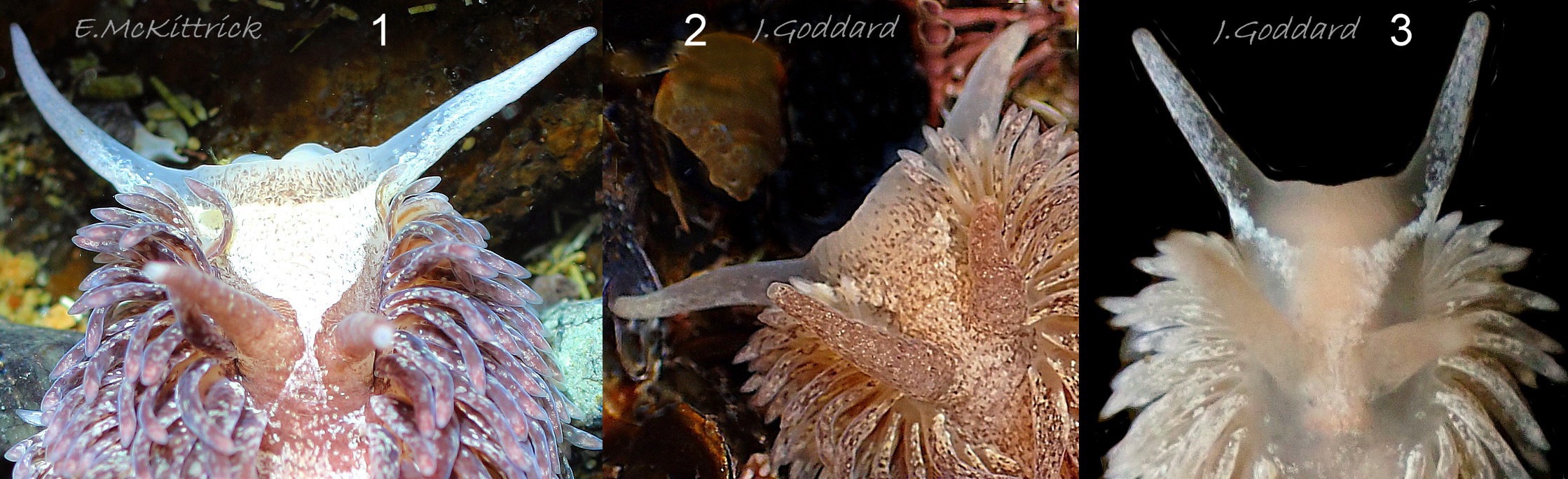
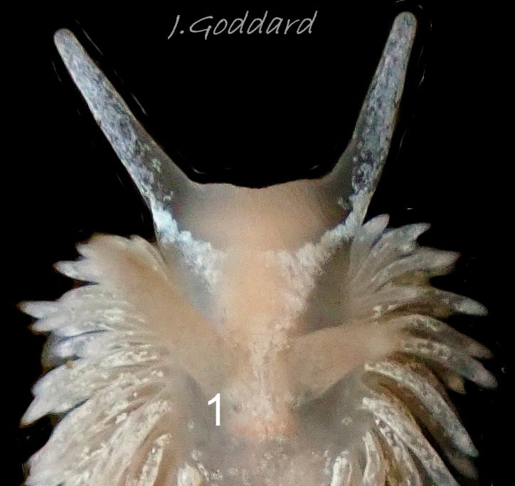
3) White ‘Y’ mark on oral tentacles and head, with stem passing back between the rhinophores, is often present. Varies from very evident and intense opaque white 08 Aeolidia filomenae to unobtrusive beige or light brown, and may be partly covered by white or beige flecks 01 Aeolidia filomenae . [See appendix.]
4) Cerata are typically flattened, broader at their base, and curved inwards 01 Aeolidia filomenae & 02 Aeolidia filomenae . This is the primary feature for identification (L. Carmona, pers. comm. 7 October 2022) [They may inflate a little to give a thicker cross section 10 Aeolidia filomenae .]
5) Cerata [usually have much less surface pigment than on A. papillosa and] are usually lighter than the rest of the body 08 Aeolidia filomenae . [But some have dark cerata 06 Aeolidia filomenae .]
6) Apices of cerata are white, [often revealing large white cnidosacs 08 Aeolidia filomenae ].
7) Blunt, conical, smooth rhinophores are translucent whitish with opaque white or yellowish-white freckles which often concentrate to colour the distal quarter 12 Aeolidia filomenae . [Most also have brown spots of varying extent and intensity on the basal three-quarters 10 Aeolidia filomenae .]
8) Internal eyes sometimes faintly visible at base of rhinophores in lighter specimens 13 Aeolidia filomenae .
9) [Distance between oral tentacles at base about three times thickness of tentacle base. 08 Aeolidia filomenae ,]
Pre 2016 authors such as Alder & Hancock (1845-1855) 07 Aeolidia filomenae and Thompson & Brown (1984), illustrated their descriptions of A. papillosa with images of probable A. filomenae.
Similar species
Aeolidia papillosa
Features as in Kienberger et al. (2016). [IFS pers. obs. in square brackets.]
1) Maximum length 120 mm.
2) Body colour extremely variable: from light white-beige, through mustard brownish, to reddish brown or dark brown.
3) “A white Y–shaped or triangular mark extending from the oral tentacles to the pericardial area between the rhinophores may be present”. [Pale marks on the head are frequent on A. papillosa in America 18 Aeolidia filomenae, where A. filomenae is absent, but seem to be rare in Europe. See appendix below.]
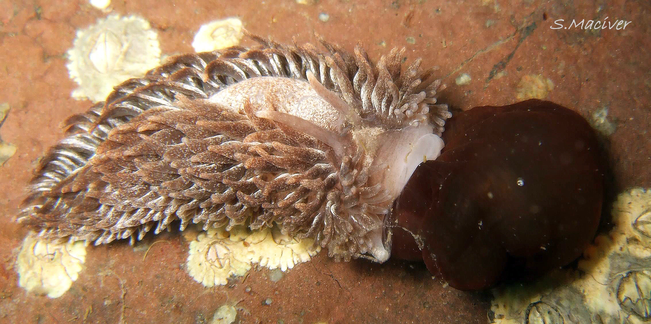
4) Elongate thin cerata, not flattened, usually with uniform diameter for most of length 19 Aeolidia filomenae.
5) Cerata usually darker than body 19 Aeolidia filomenae.
6) [Dull whitish cnidosac visible if not obscured by white apical pigment 20 Aeolidia filomenae.]
7) Rhinophores dark, [often have a small, terminal, translucent, pale spot on the truncated tip 21 Aeolidia filomenae & 22 Aeolidia filomenae].
8) Internal eyes at base of rhinophores, visible rarely if ever in Europe flic.kr/p/2nToCc3 . [Sometimes visible in America 23 Aeolidia filomenae].
9) [Distance between oral tentacles at base about three times thickness of tentacle base, 21 Aeolidia filomenae ].
Aeolidiella alderi (Cocks, 1852)
24 Aeolidia filomenae A. glauca (Alder & Hancock, 1845) 25 Aeolidia filomenae and A. sanguinea (Norman, 1877) 26 Aeolidia filomenae .
1) Extreme maximum length 46 mm (A. sanguinea, others shorter).
2) Body translucent white or pale shade of other colour. Any opaque marks are scattered, small and not dark.
3) No white ‘Y’ or triangular mark on head.
4) Cerata not flattened, with uniform diameter for most of length
5) Cerata have more saturated colour than body.
6) Apices of cerata are white or pale.
7) Rhinophores translucent, no dark pigment marks.
8) Internal eyes visible at base of rhinophores
9) Distance between oral tentacles at base about same as thickness of single tentacle base (IFS pers. obs.).
Habits and ecology
Information in this section is mainly from Thompson & Brown (1984) who did not differentiate A. papillosa from A. filomenae, so details may apply to either one or both.
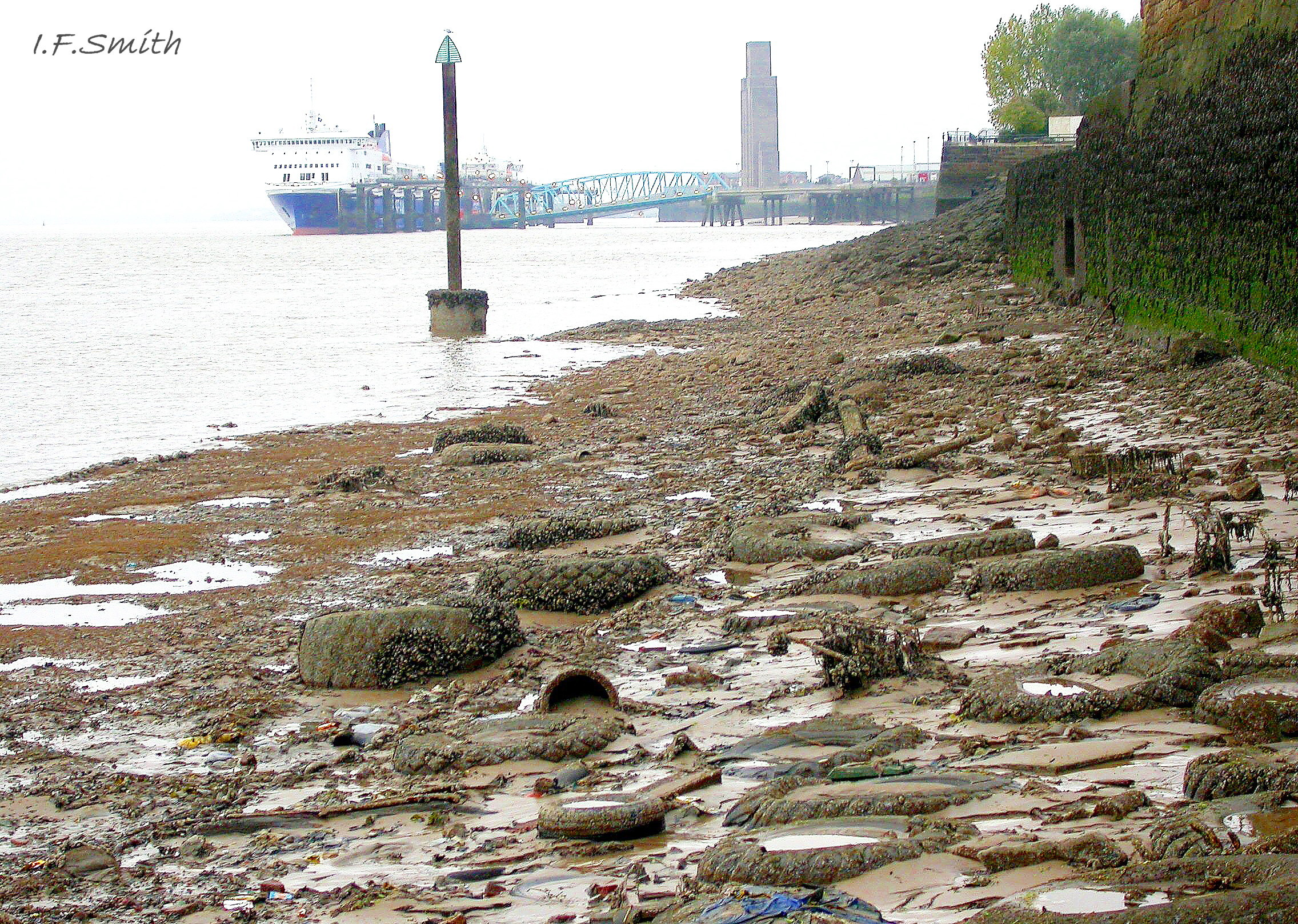
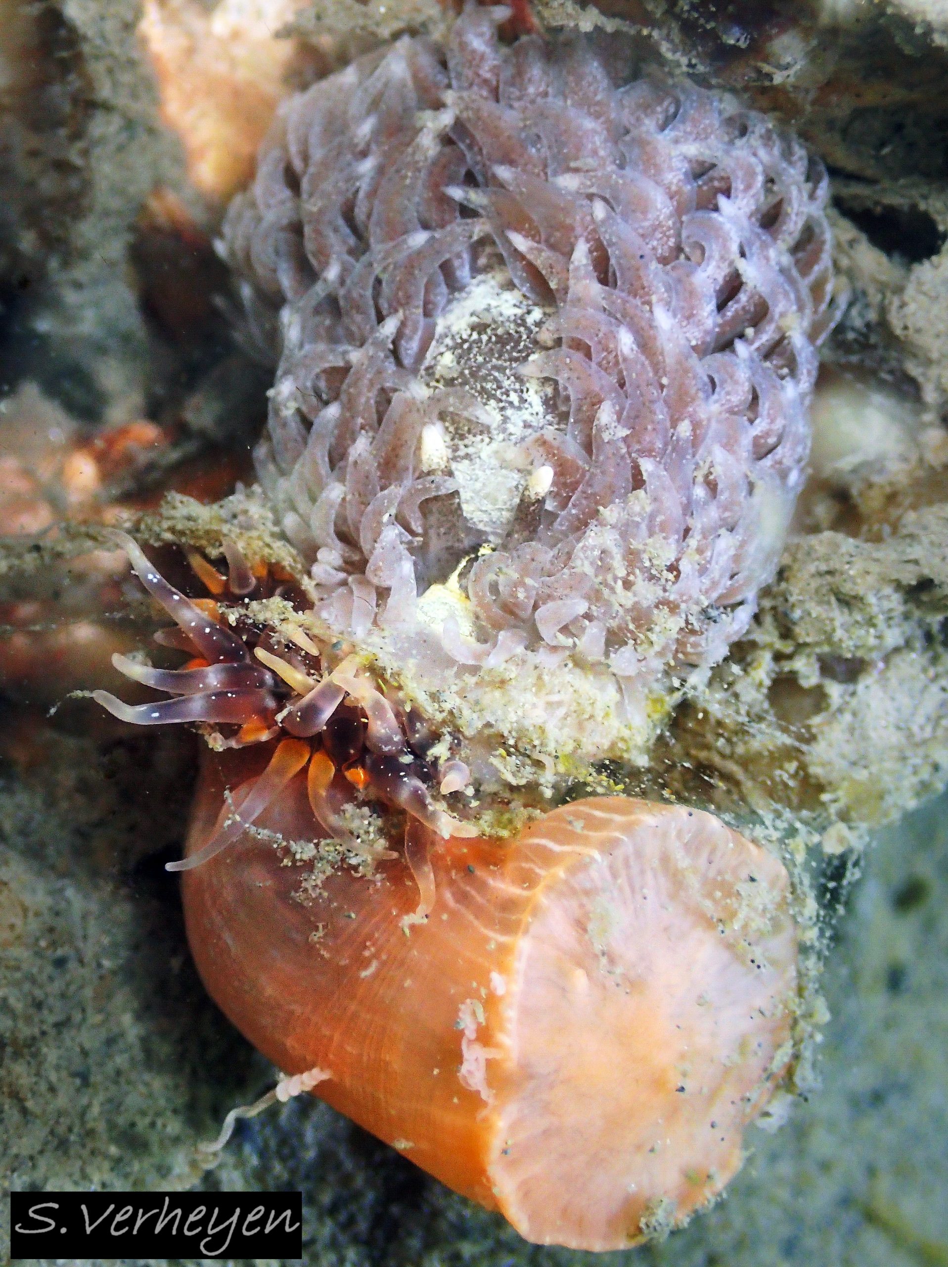
It lives sublittorally and on lower shores where there is some hard substrate, including estuaries 04 Aeolidia filomenae & 27 Aeolidia filomenae down to 20‰ salinity, and on muddy sand with isolated stones coated with sediment, where its widely extended foot enables it to traverse the soft substrate 28 Aeolidia filomenae .
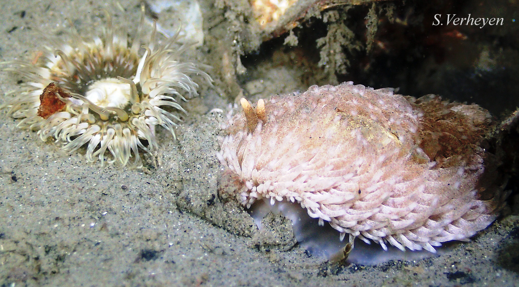
It will attack and eat sea anemones, including Cylista elegans (Dalyell, 1848) 28 Aeolidia filomenae & 29 Aeolidia filomenae and Actinia equina (Linnaeus, 1758) 30 Aeolidia filomenae . It is immune to the toxic nematocysts in their tentacles and exuded acontia, and it ingests them and stores them in cnidosacs at the tips of its cerata for release when it is attacked. The effect is powerful enough in a dog’s mouth to make it drop the slug (IFS pers. obs.) but it does not prevent haddock, Gadus aeglefinus, from consuming it (Thompson & Brown, 1984). Other enemies include copepod parasites and Nucella lapillus which occasionally eats its spawn.
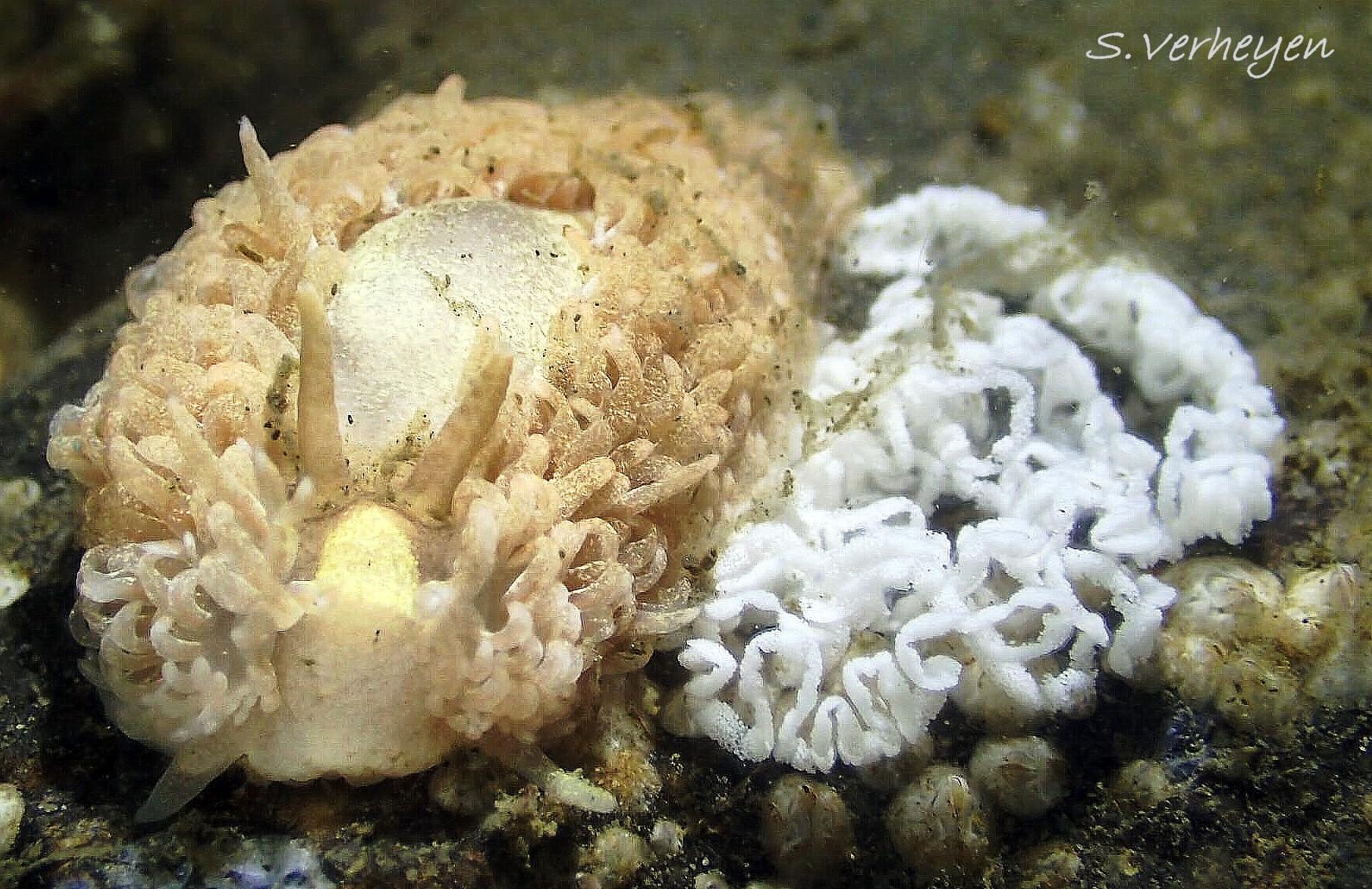
A. filomenae is a simultaneous hermaphrodite. Its convoluted white, cord of spawn, somewhat resembling a coiled spring, is attached spirally to hard substrate 31 Aeolidia filomenae from January to August in Britain. It is very similar to the spawn of A. papillosa so its identification requires presence of an adult, ideally in the process of spawning.
Veliger larvae hatch from the spawn and live in the plankton before metamorphosis. Small juveniles are not often found on shore; they may spend their early life sublittorally (Thompson & Brown, 1984).
Distribution and status
A. filomenae lives on Atlantic coasts of Europe and is sympatric with A. papillosa in the Netherlands and the British Isles. Kienberger et al. (2016) verified with molecular sequencing specimens from the Netherlands, Atlantic France, north-west Spain and Portugal. GBIF map www.gbif.org/species/8763248 . UK NBN map species.nbnatlas.org/species/NHMSYS0021168707
Acknowledgements
For use of images I gratefully thank Matt Barnes, Chris Bryan, Jeff Goddard, , Guillaume Lemonnier, Sutherland Maciver, Erin McKittrick, Stefan Verheyen and Alex Wilson. I thank Simon Taylor for specimens to photograph. For information and advice I thank Leila Carmona and J. Lucas Cervera, but any errors or omissions are my (IFS) responsibility.
Appendix
Kienberger et al. (2016) stated of A. papillosa, “A white Y–shaped or triangular mark extending from the oral tentacles to the pericardial area between the rhinophores may be present”. But the common practice of European recorders on iNaturalist and Facebook is to record all specimens with a white Y as A. filomenae.
As substantiation for their statement, Kienberger et al. refer to their Fig. 5B which shows a sequenced A. papillosa from the White Sea with a whitish mark on the head resembling a wine glass with the stem passing back between the rhinophores. Apart from it, the only European Aeolidia image with a wine glass mark found by IFS is one from the Mersey Estuary 32 Aeolidia filomenae. Whether the wine glass mark should be regarded as a form of white Y is an open question. Sometimes it is centrally faded with small lateral branches to resemble a Y, but the branches bend to meet at an obtuse angle of nearly 180° instead of the acute junction of the arms of a Y 18 Aeolidia filomenae and 33 Aeolidia filomenae .
Judging from the many images posted as A. papillosa on iNaturalist, and ‘Fig. 5F’ from Alaska in Kienberger et al. (2016), the wine glass mark is frequent on it in North America both in the Atlantic and Pacific. In the Pacific, it is often misidentified as A. loui Kienberger, Carmona, Pola, Padula, Gosliner & Cervera, 2016, especially in Washington and further north where there is no molecular evidence for A. loui ( L. Carmona, pers. comm. 7 October 2022).
Kienberger et al. differentiated A. papillosa and A. filomenae on the basis of molecular sequencing, and they described morphological features observed on the specimens studied which correlated with each species. It is possible that subsequent experience and photography of larger numbers have revealed more morphological variations but they need molecular sequencing for substantiation.
In June 2016, J. Lucas Cervera, co author with Kienberger, posted on Facebook Group N. E. Atlantic Nudibranchs to say that an in depth, focused study of UK and Irish specimens is still required to understand much better the diversity of this complex in that area, but that without material it is not possible. He would like collections to be made from different localities and then sent to him together in one or two packs, as if he receives the material scattered over time it is difficult to arrange students and material for a study focused on the issue. If you wish to help, please first email leila.carmona@uca.es for detail of how to preserve and send specimens.
Without further linked morphological and molecular study, morphological identifications of British and Irish Aeolidia spp., including those illustrated in this account, are uncertain.
References and links
Alder, J. & Hancock, A. 1845-1855. A monograph of the British nudibranchiate mollusca. London, Ray Society. Family 3 Plate 9 [A. papillosa sensu lato includes A. filomenae ] www.biodiversitylibrary.org/item/131598#page/314/mode/1up
Carmona, L., Pola, M., Gosliner, T. M. and Cervera, J. L. (2013) A tale that morphology fails to tell: a molecular phylogeny of Aeolidiidae (Aeolidida, Nudibranchia, Gastropoda). PLoS ONE 8(5): e63000. journals.plos.org/plosone/article?id=10.1371/journal.pone…
iNaturalist www.inaturalist.org/observations
Kienberger, K., Carmona, L., Pola, M., Padula, V., Gosliner, T.M. and Cervera, J.L. 2016. Aeolidia papillosa (Linnaeus, 1761) (Mollusca: Heterobranchia: Nudibranchia), single species or a cryptic species complex? A morphological and molecular study. Zool. J. Linn. Soc., 177: 481–506. www.researchgate.net/publication/303953645_Aeolidia_papil…
Thompson, T.E. & Brown, G.H. 1984. Biology of opisthobranch molluscs 2. London, Ray Society. [A. papillosa sensu lato includes A. filomenae. ]
Glossary
acontia = thread-like tissue containing numerous stinging nematocysts which are released by some sea anemones.
amphiboreal = living in the cold temperate boreal zone to south of Arctic on more than one side of the Atlantic and/or Pacific.
cerata = (sing. ceras, adj. ceratal) lobes on notum of some nudibranchs.
cleioproctic = (of anus) located on the notum to the right of the midline.
Cnidaria = hydroids, jellyfish, sea anemones etc. which possess cnidocytes.
cnidocytes = explosive stinging cells of Cnidaria. en.wikipedia.org/wiki/Cnidocyte
cnidosac = storage capsule at tips of cerata of Aeolidiidae for ingested cnidocytes.
digestive gland = large organ in gastropods which acts like the liver and pancreas in mammals to absorb food.
distal = away from centre of body or from point of attachment.
hermaphrodite, simultaneous = individual acts as both male and female at the same time with similar partner.
molecular sequencing = technique for determining the sequence of the bases adenine, guanine, cytosine, and thymine (A, G, C and T) in a DNA molecule.
notum = (of sea slugs) the dorsal surface of the body; the back.
partim = in part (indicates taxon which has been split and a new name used for part of what used to be included in it).
pericardium = sac containing heart, sometimes visible as a raised pericardial mound behind rhinophores in sea slugs.
pleuroproctic = (of anus) located on side of body below the notal edge or cerata.
propodial tentacles = tentacular, lateral extensions on anterior of the foot.
rhinophore = chemo-receptor tentacle; many sea slugs have a pair on top of the head.
sensu lato = (abbreviation s.l.) in the wide sense, possibly an aggregate of more than one species.
sensu stricto = (abbreviation s.s.) in the strict sense, excluding species that have been aggregated or confused with it.
veliger = shelled larva which moves by action of cilia on a velum (bilobed flap). Stage may be passed in plankton or within liquid-filled egg-capsule.