Click image to enlarge with full caption. Main text below slider.
01 Leptochiton cancellatus
02 Leptochiton cancellatus
03 Leptochiton cancellatus
04 Leptochiton cancellatus
05 Leptochiton cancellatus
06 Leptochiton cancellatus
07 Leptochiton cancellatus
08 Leptochiton cancellatus
09 Leptochiton cancellatus
10 Leptochiton cancellatus
11 Leptochiton cancellatus
12 Leptochiton cancellatus album COMPARISON Lepidochitona cinerea.
13 Leptochiton cancellatus album COMPARISON Lepidochitona cinerea.
14 Leptochiton cancellatus album COMPARISON Lepidochitona cinerea
15 Leptochiton cancellatus album. COMPARISON L. scabridus & Lepidochitona cinerea.
16 Leptochiton cancellatus album COMPARISON L. asellus.
17 Leptochiton cancellatus album COMPARISON L. asellus. .
18 Leptochiton cancellatus album COMPARISON L. asellus.
18.1 Leptochiton cancellatus album COMPARISON Leptochiton scabridus
18.2 Leptochiton cancellatus album COMPARISON L. scabridus.
19 Leptochiton cancellatus album COMPARISON L. sarsi.
20 Leptochiton cancellatus album COMPARISON L. sarsi.
Leptochiton cancellatus (Sowerby, 1840)
Current taxonomy: World Register of Marine Species (WoRMS)
www.marinespecies.org/aphia.php?p=taxdetails&id=138675
Synonyms: Chiton cancellatus G.B.Sowerby ii, 1840; Lepidopleurus cancellatus (Sowerby, 1840);
Vernacular: Arctic cancellate chiton.
Revised, 2020, PDF version at www.researchgate.net/profile/Ian_Smith19/research
GLOSSARY BELOW This account uses the standardised terminology for chitons proposed by Schwabe (2010). Some of Jones & Baxter (1987) alternatives are indicated in the glossary as a.k.a..
Preface
This small species may be overlooked as a juvenile pale Lepidochitona cinerea. The single specimen used to illustrate this account was discovered during close examination of several L. cinerea. It shows some deviation from the features of Leptochiton cancellatus described in Kaas (1981) and Kaas & Belle (1985). On balance, and until molecular studies of the Leptochiton species can be compared, its features, location and habitat appear to support an identification as L. cancellatus. The text is intended to conform with Kaas & Belle’s description and figures, and deviations are mentioned in the image captions. Other specimens of L. cancellatus are on Nature22 website of marine life from Brittany, whence Kaas and Belle took specimens for their figures. nature22.com/estran22/estran.html
Shell Description
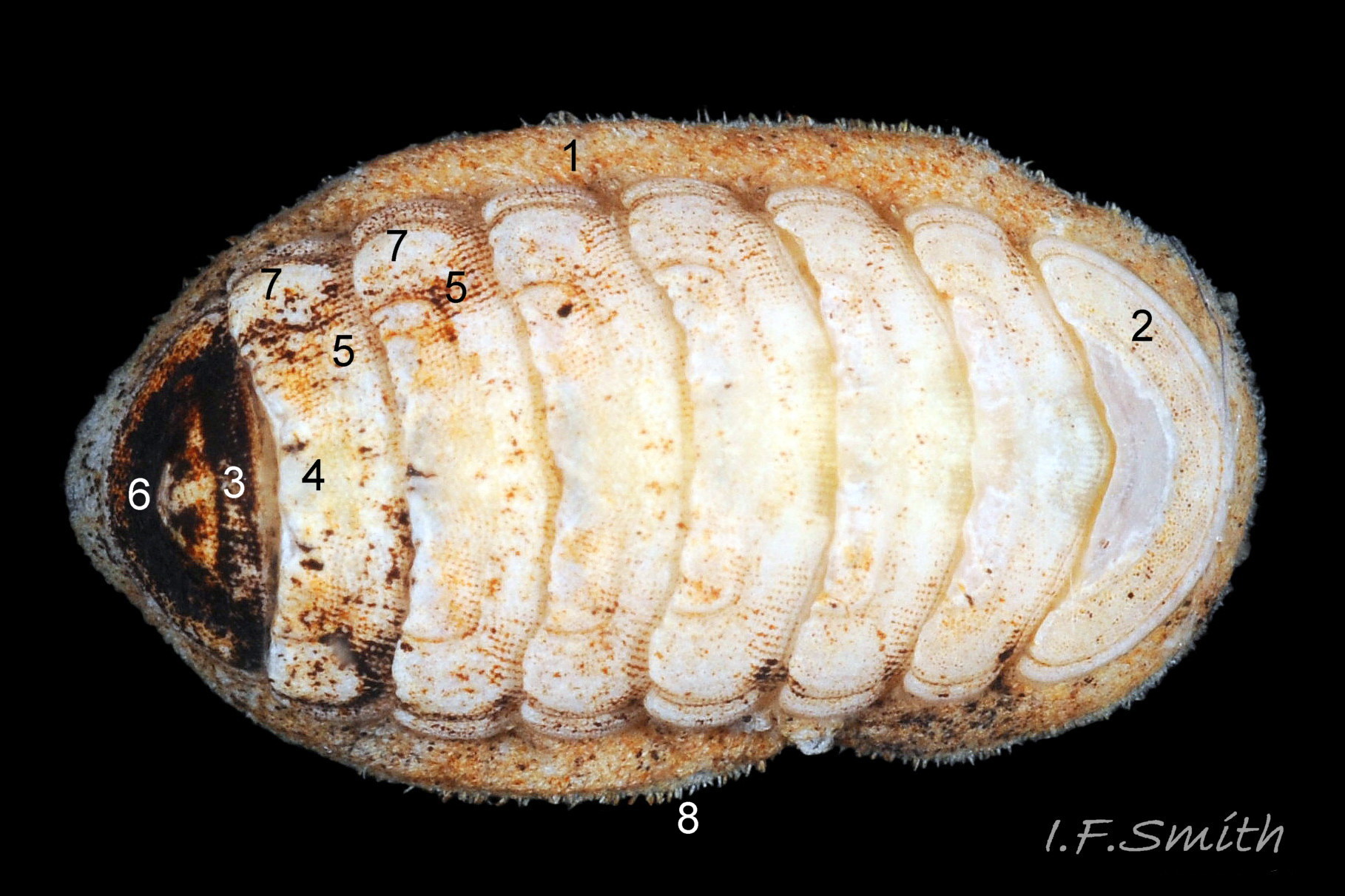
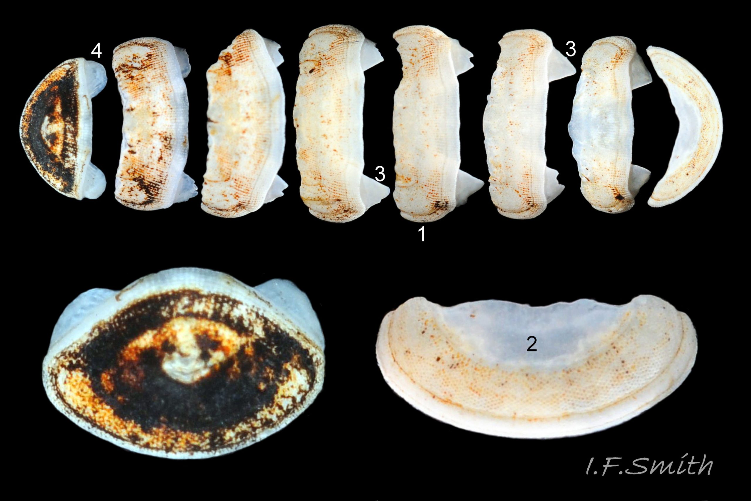
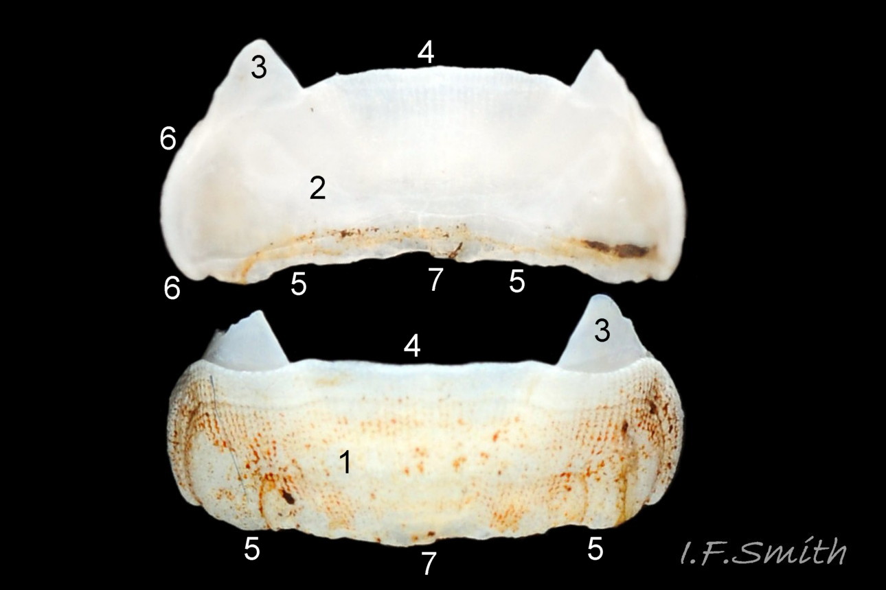
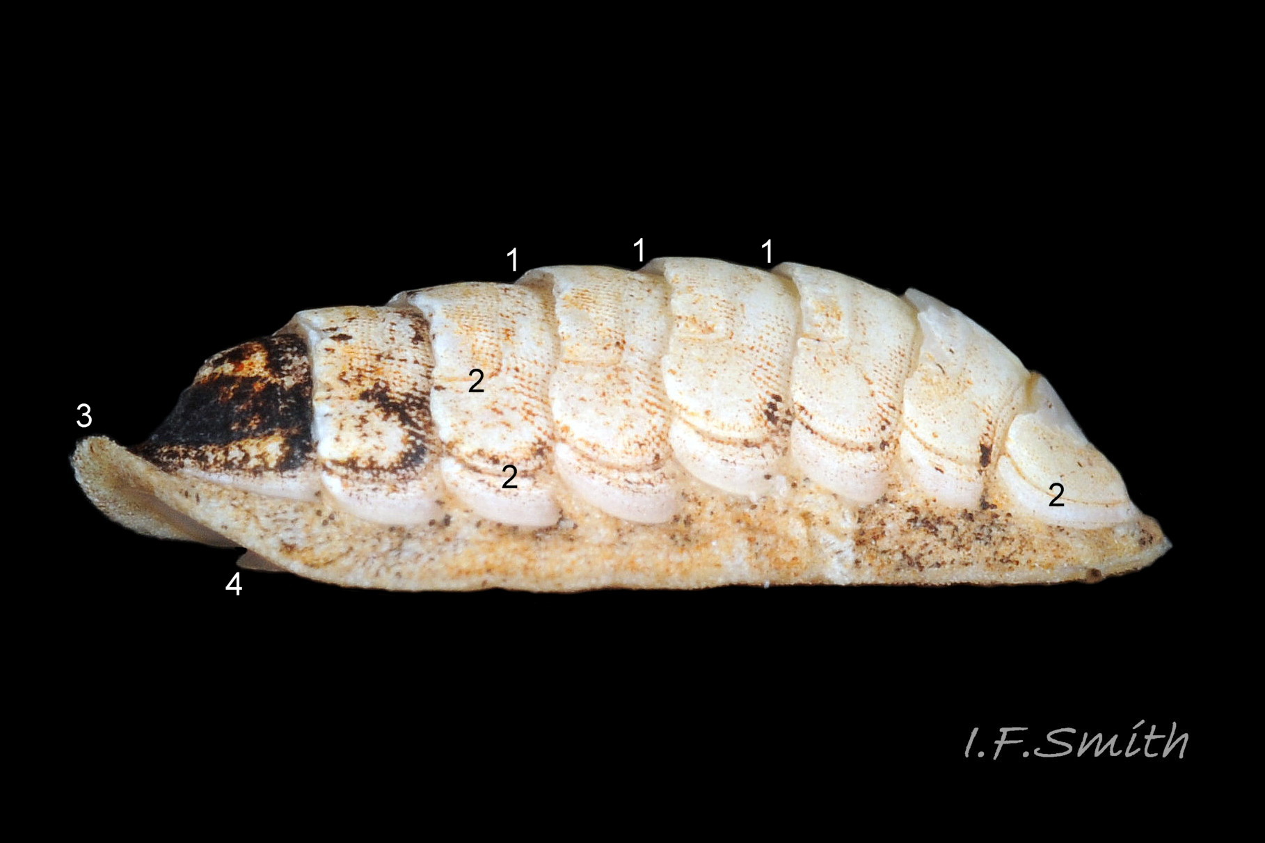
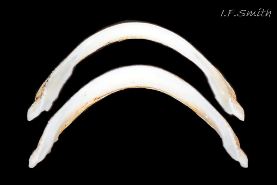
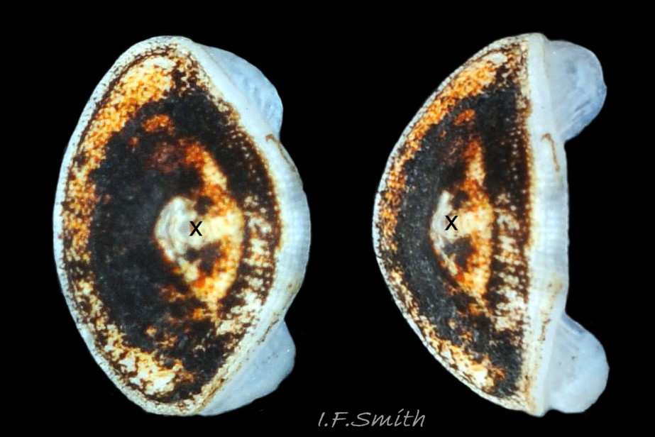
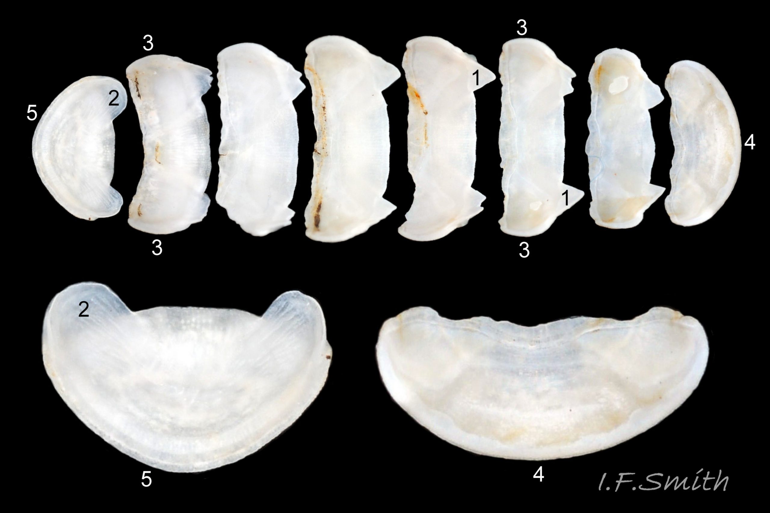
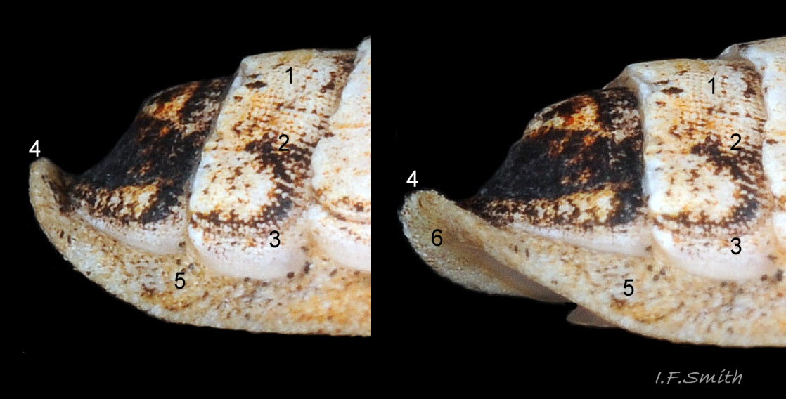
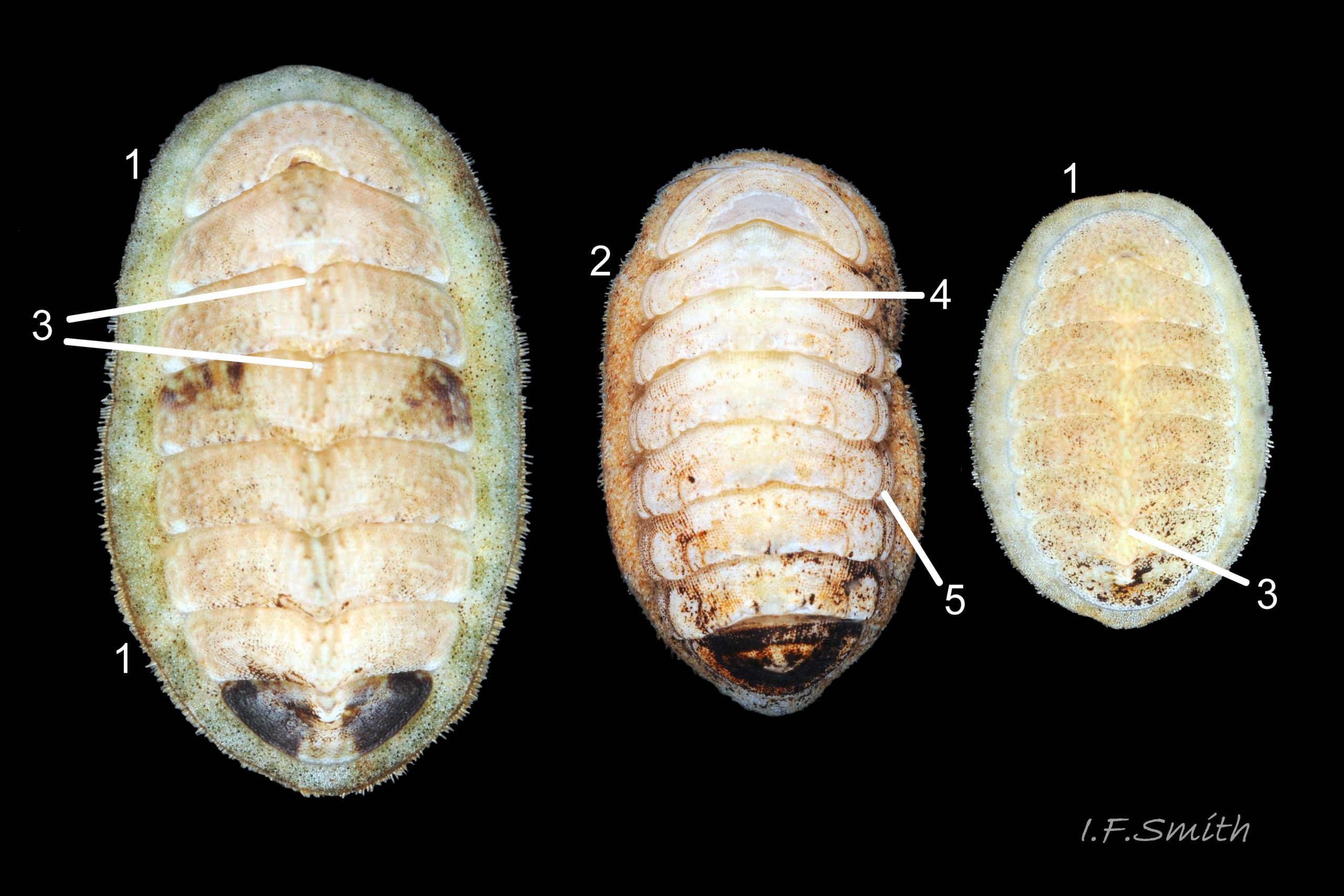
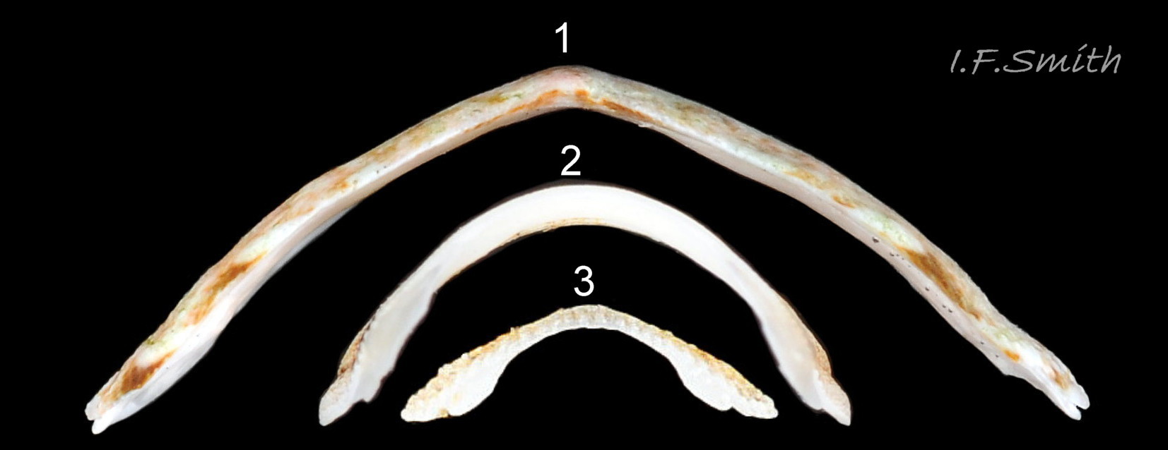
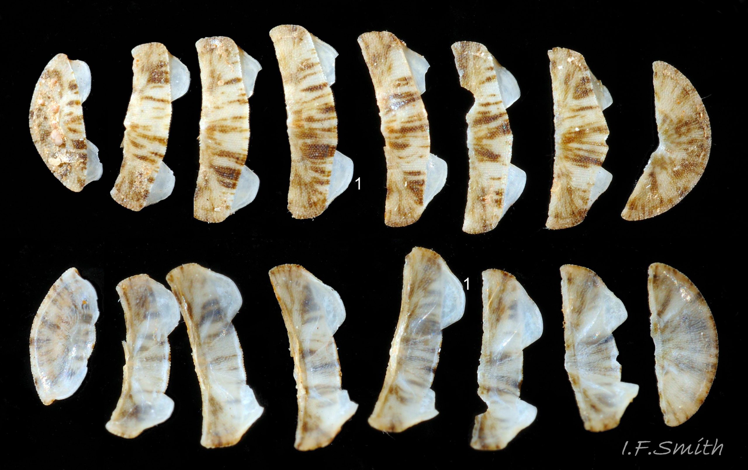
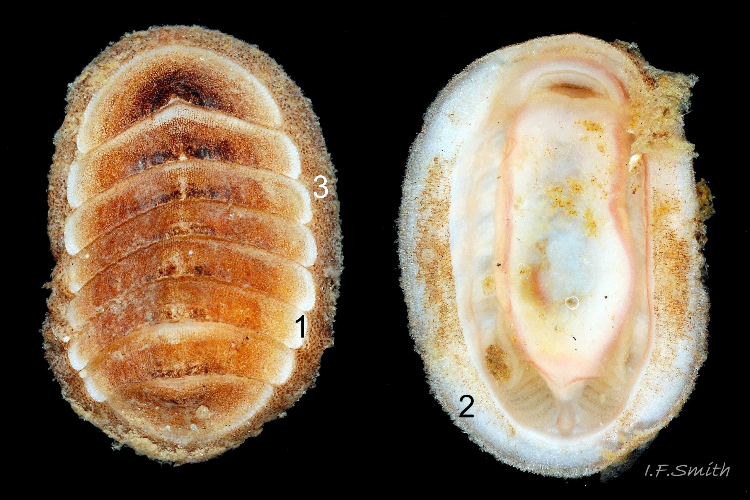
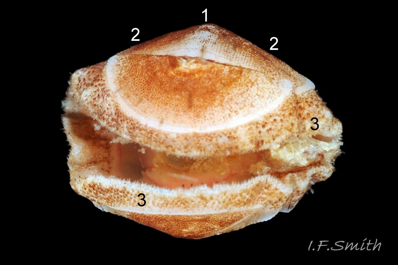
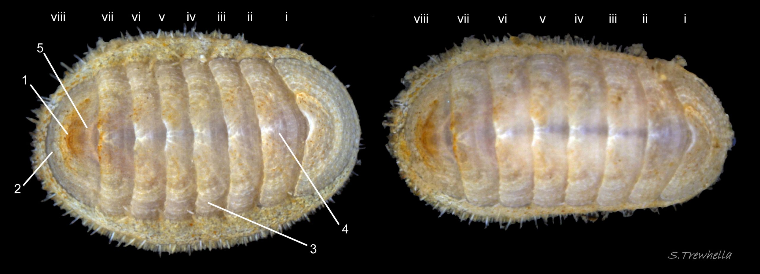
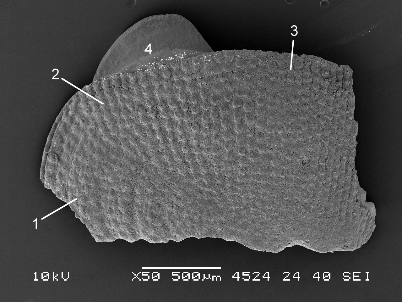
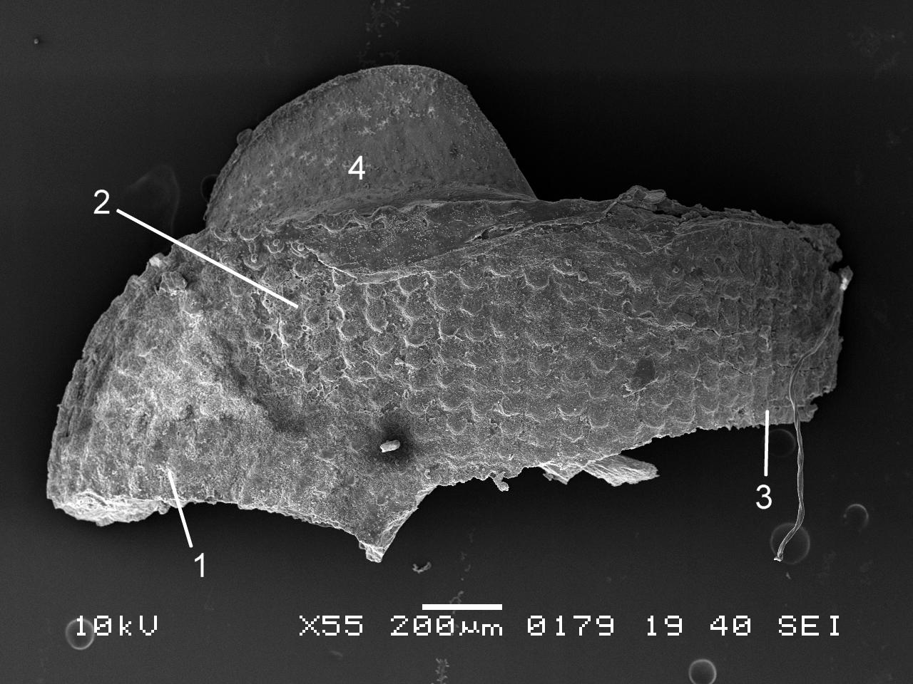
Up to 9mm long and 4mm wide; smallest chiton species in British waters apart from L. scabridus. In dorsal view, outline elliptical; width about 50% of length when extended 01 Leptochiton cancellatus . Girdle narrow, so intermediate shell valves (ii to vii) occupy 80 to 90% of chiton’s width. Eight overlapping, thin, fragile, white or cream valves 02 Leptochiton cancellatus principally made of 1) tegmentum: dorsal layer of aragonite 03 Leptochiton cancellatus permeated and weakened by canals, and 2) articulamentum: ventral layer of aragonite also 3) properiostracum: outermost proteinaceous layer and 4) myostracum: microscopically thin discontinuous innermost layer. Dorsal surface of valves often stained in patches or streaks with black manganese and/or rust coloured ferrous deposits 01 Leptochiton cancellatus . Head valve (i) less than semicircle, posterior margin widely V shaped; approximately crescentic on live animal 01 Leptochiton cancellatus . Intermediate valves (ii to vii) have gently curving, almost parallel, anterior and posterior edges, with somewhat obliquely truncated anterior corners, and they lack any sign of a beak 03 Leptochiton cancellatus & 04 Leptochiton cancellatus . Smooth curved profile with no sign of a keel when intermediate valve viewed from posterior 05 Leptochiton cancellatus . Tail valve (viii) quite large, roughly triangular 06 Leptochiton cancellatus , and slightly concave to posterior 07 Leptochiton cancellatus of slightly swollen mucro which is positioned to posterior of centre when extracted valve viewed from above 06 Leptochiton cancellatus .
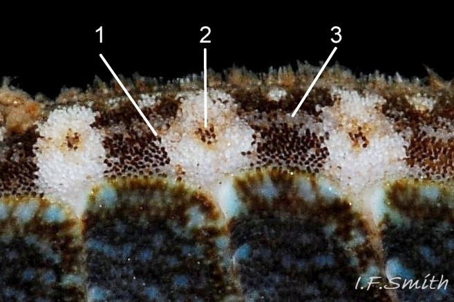
Canals permeate tegmentum and terminate on its dorsal surface in small oval granules with a cap (megalaesthete) at anterior, and a few smaller subsidiary caps (micraesthetes) in posterior part of granule (Jones & Baxter, 1987).
Intermediate valves ii to vii each have two widely separated, small, triangular apophyses 03 Leptochiton cancellatus on anterior edge that extend under next valve forwards 08 Leptochiton cancellatus . Tail valve viii has rounded trapezoidal apophyses. L. cancellatus, like others in family Leptochitonidae, lacks insertion plates that most chitons have at ends of intermediate valves, at anterior of head valve and posterior of tail valve.
Dorsal surface cancellated by closely packed, almost touching, oval granules in chainlike lines running a) longitudinally in front of mucro on tail valve and in the jugal and pleural areas of valves ii to vii, and b) radially on head valve, behind mucro on tail valve and on lateral areas of valves ii to vii 01 Leptochiton cancellatus & 09 Leptochiton cancellatus . Lines of granules may be eroded on large old specimens. Lateral and pleural areas slightly differentiated by slight elevation of lateral area and difference in orientation of granules. Usually a few strong growth lines clearly visible on all valves 04 Leptochiton cancellatus .
Body Description
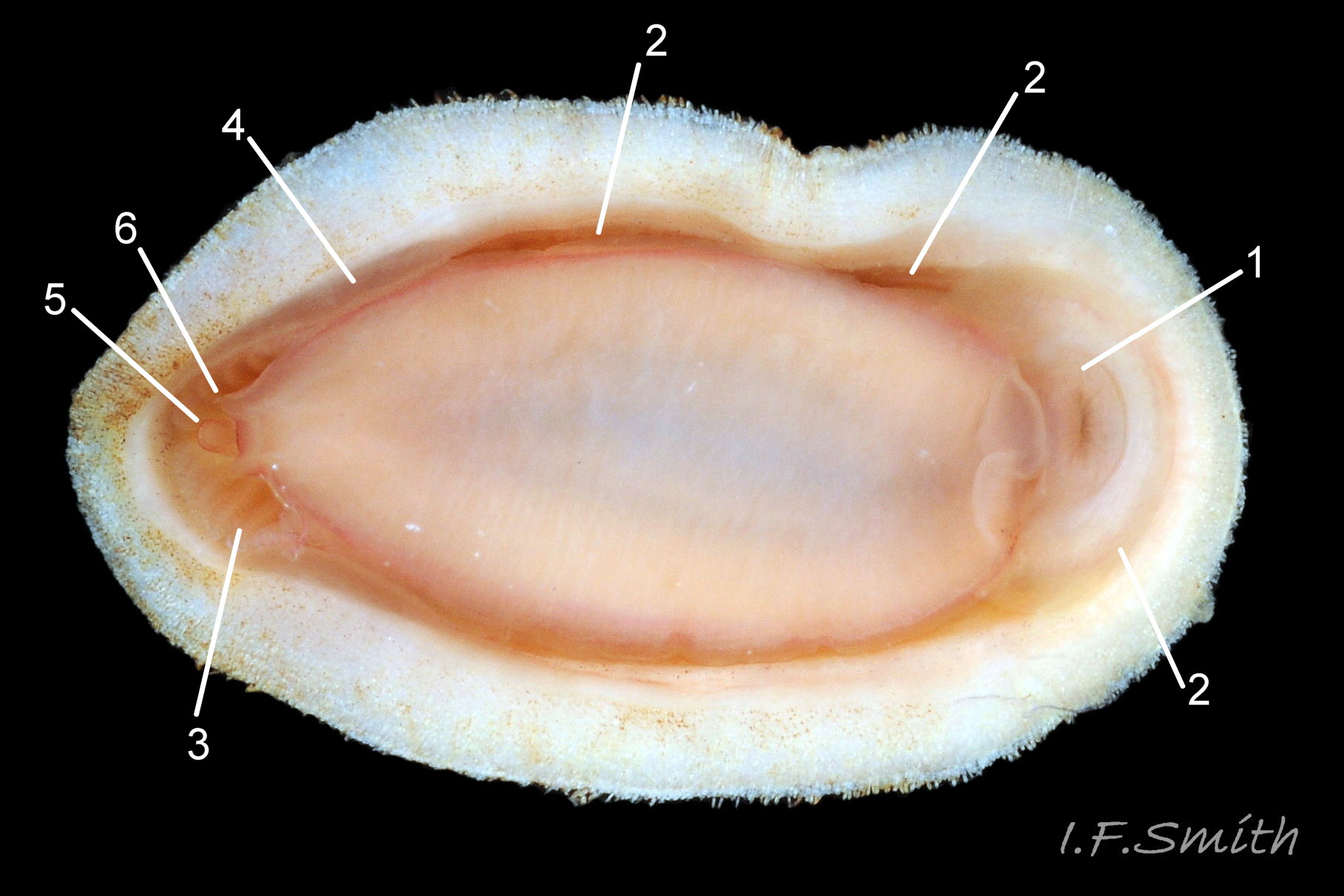
Head and foot rarely protrude into view naturally on live animal 04 Leptochiton cancellatus , can only be examined if animal removed from substrate, or placed on glass. Most of head occupied by large transverse slit-mouth with wrinkled lips , surrounded by white hood 10 Leptochiton cancellatus ; no eyes or sensory tentacles.
Aesthetes (sensory tissue) fill canals that permeate the tegmentum and parts of articulamentum; terminate in oval granules as sense organs on dorsal surface of valves. Perinotum (dorsal surface) of girdle, whitish but often stained rusty colour with mineral deposits; densely packed, squarish scales with curled tips give a rough appearance 09 Leptochiton cancellatus . Hyponotum (ventral surface of girdle), whitish variably stained with mineral deposits; has imbricated, bluntly pointed, flat scales (microscope needed to see detail on perinotum and hyponotum). Peripheral margin of girdle has fringe of ribbed pointed spicules about 0.1mm long 01 Leptochiton cancellatus . Girdle can be flexed up at posterior to allow release of faeces, excreta, ova or sperm 04 Leptochiton cancellatus .
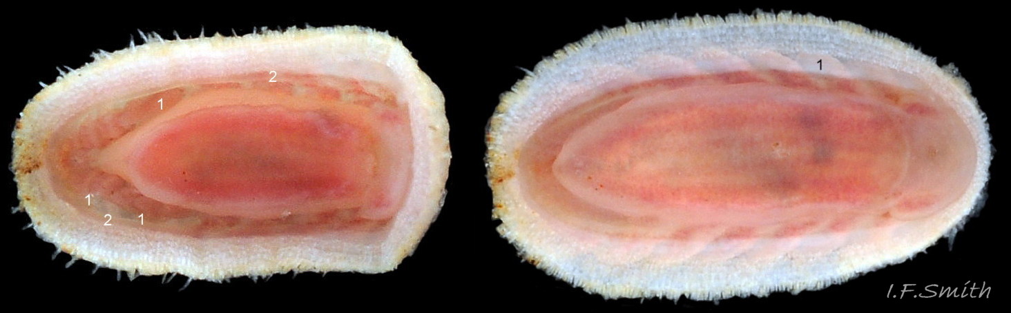
Open, narrow mantle cavity runs around whole animal; contains ctenidia close to anus at posterior (merobranch arrangement) 10 Leptochiton cancellatus . Between foot and hyponotum, the mantle fold is unobtrusive but can seal the mantle cavity and conceal ctenidia when in place. Anus on papilla opens into mantle cavity at posterior where girdle deflected dorsally for expulsion of faeces. Nephridiopores and gonopores open laterally into posterior quarter of cavity. No penis as external fertilization. Foot, elongate ellipse with small projection either side of anal papilla, sole whitish pink centrally, becoming reddish pink towards periphery, no medial dividing line 10 Leptochiton cancellatus . Foot spreads widely, concealing pallial cavity with help of mantle fold when gripping substrate.
Key identification features
Leptochiton cancellatus
1: Small; maximum length 8mm (illustrated specimen slightly larger). Valves whitish or cream, often stained in patches or streaks with black manganese and/or rust coloured ferrous deposits. No lateral insertion plates or slits. 02 Leptochiton cancellatus
2: Only at LWS and sublittorally. All round Britain. Probably overlooked.
3: Narrow whitish girdle has no pattern, may be stained with rust coloured ferrous, and/or blackish manganese, deposits. Dorsal girdle scales squarish with rolled tip. 09 Leptochiton cancellatus .
4: Arch of valves is moderately high smooth curve with no sign of a keel. Elevation of arch valves iv & v; height/width 39% (Kaas&Belle), but illustrated specimen 46%. 05 Leptochiton cancellatus .
5: No posterior beak on intermediate valves. 04 Leptochiton cancellatus .
6: Cancellated by closely packed, almost touching, oval granules in chainlike lines running radially on valves i & viii, and on lateral areas of valves ii to vii (sometimes obsolete on lateral areas) . Lines longitudinal on central areas of valves ii to vii. 09 Leptochiton cancellatus .
7: Sole whitish pink centrally, becoming reddish pink towards periphery. 10 Leptochiton cancellatus .
8: Ctenidia close to anus at posterior (merobranch) 10 Leptochiton cancellatus .
9: Tail valve shorter than on L. sarsi; length 45% of width in life position ( 51% on illustrated specimen) 06 Leptochiton cancellatus .
10: Mucro slightly swollen and to posterior of centre of extracted valve viii (part of anterior concealed on live specimens) 06 Leptochiton cancellatus . Postmucronal slope entirely concave 07 Leptochiton cancellatus .
Similar species
*or ** indicate principal distinguishing features.
Lepidochitona cinerea (Linnaeus, 1767) Leptochitona cinerea
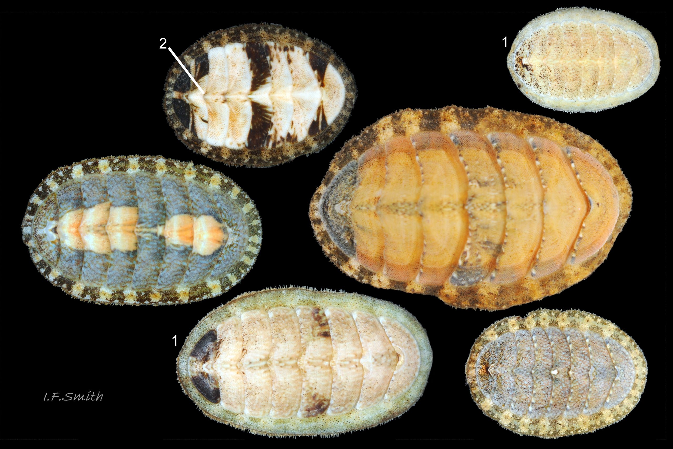
1*: Maximum length 28mm. Valves diversly coloured and patterned, including whitish resembling Leptochiton cancellatus 12 Leptochiton cancellatus. Lateral insertion plates separated by single slit on each end of valves ii to vii.
2: Midshore level and below. All round Britain. Commonest littoral chiton.
3**: Girdle has a unique (in N.W. Europe) lozenge pattern 13 Leptochiton cancellatus, but sometimes indistinct, especially on whitish specimens which are easily confused with Leptochiton cancellatus 14 Leptochiton cancellatus .
4*: Arch of valves is keeled 15 Leptochiton cancellatus.
5*: Distinct posterior beak on intermediate valves 12 Leptochiton cancellatus.
6: Dorsal surface of valves has densely packed rounded granules, not in straight lines.
7: Sole pinkish white to orange pink, usually with grey viscera showing centrally.
8*: Usually 16 to19 ctenidia each side for whole length of foot (holobranch).
Leptochiton asellus (Gmelin, 1791)
1*: Maximum size 18mm X 10mm. Valves whitish, sometimes with distinct black longitudinal lines 16 Leptochiton cancellatus or general staining by black or rust coloured mineral deposits 17 Leptochiton cancellatus. No lateral insertion plates or slits.
2: Only at LWS and sublittorally. All round Britain, probably commonest sublittoral chiton.
3: Whitish girdle has no pattern, may be stained with rust coloured ferrous, and/or blackish manganese, deposits. Dorsal girdle scales elongate, bluntly pointed 18 Leptochiton cancellatus.
4**: Arch of valves is quite high with distinct keel* and straight* side slopes 18 Leptochiton cancellatus. (Elevation of arch valves iv & v; height/width 36%)
5: Often a slight beak on intermediate valves, especially valves ii & iii.
6: Roughly oval granules in slightly disjointed lines, oriented as on L. cancellatus.
7: Sole whitish pink centrally, becoming reddish pink towards periphery.
8: Eight to thirteen ctenidia in posterior half of pallial groove (merobranch).
9: Tail valve length less than 50% of width in life position.
10: Mucro not swollen and slightly to anterior of centre of valve viii.
Leptochiton scabridus (Jeffreys, 1880).
18.2 Leptochiton cancellatus
1: Small; maximum size 8mm X 4mm. Valves whitish to dull orange, sometimes with staining by black or rust coloured mineral deposits. No lateral insertion plates or slit.
2*: Only at LWS and sublittorally. Rare, only SW England, Channel Islands & Brittany.
3: Narrow whitish girdle has no pattern, may be stained with rust coloured ferrous, and/or blackish manganese, deposits.
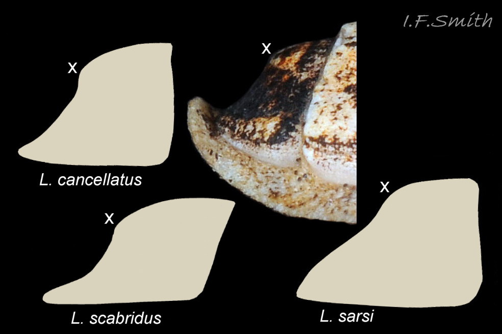
4: Arch of valves is a smooth curve with very slight indication of carina, lower in profile than on L. cancellatus.
5: No posterior beak on intermediate valves, but anterior of valve ii tends to be angulated.
6*: Rows of raised, round granules on valves, oriented as on L. cancellatus, but coarser. Compare at nature22.com/estran22/mollusques/polyplacodentale/chitons…
7**: Whole sole strikingly bright red. 18.1 Leptochiton cancellatus
8: Ctenidia confined to posterior half (merobranch).
9: Tail valve length about 55% of width in life position.
10: Mucro swollen and near centre of valve viii. Postmucronal slope entirely concave.
Leptochiton sarsi Kaas, 1981
Very similar and closely related to L. cancellatus. All Scandinavian specimens previously recorded as L. cancellatus that were examined by Kaas (1981) were considered by him to be L. sarsi. No British records on NBN, but one from Guernsey (1922) and others from Mediterranean (Dell’Angelo et al, 2009), so, if these correctly identified, possibilty that L. sarsi may be in British waters unrecognised.
1: Maximum size 10mm X 5mm.
2*: Western Sweden to northern Norway at depths 40m to 700m.
3: Narrow girdle. Dorsal scales elongate, conical.
4: Arch of valves is high and rounded to slightly keeled. Elevation of arch of valves iv & v; height/width 48%.
6*: Valve granules coarser than on L. cancellatus with wider gaps between them. Granule lines not radial on valves i & viii or lateral areas of valves ii to vii. Lines longitudinal on central areas of valves ii to vii; spaced wider than on L. cancellatus so fewer rows (12 rows match width of an apophysis, about 16 on L. cancellatus). 19 Leptochiton & 20 Leptochiton cancellatus.
9: Tail valve long, length 60% of width.
10*: Mucro on tail valve viii not swollen and postioned slightly to anterior of centre. Postmucronal slope concave only to immediate posterior of mucro; changes to straight or slightly convex further to posterior 07 Leptochiton cancellatus .
Leptochiton rugatus Carpenter in Pilsbury, 1892
All Pacific specimens previously recorded as L. cancellatus that were examined by Ferreira (1979) were considered by him to be L. rugatus. It has a quite different radula.
Habits and ecology
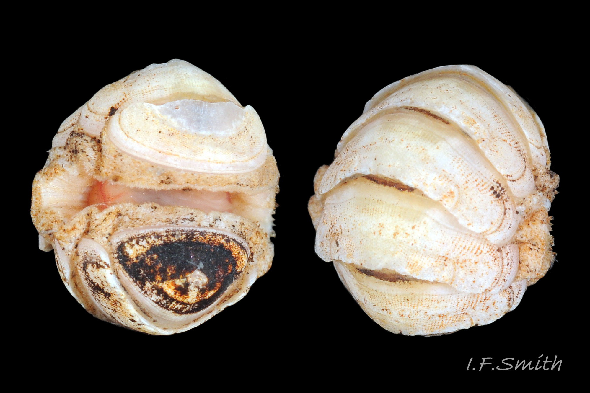
Lives on and under stones and dead shells on silty sand at LWS and sublittorally to beyond 900 metres. When alarmed, can increase grip suctorially by raising part of hyponotum to form partial vacuum, and if displaced from substrate, can roll into a ball 11 Leptochiton cancellatus . Respiration: cilia on ctenidia and mantle create inhalent water-current entering pallial cavity wherever girdle is raised at anterior. Water current passes through ctenidia and then along cavity as exhalent current to exit at posterior under raised girdle.
In absence of eyes or of tactile or chemoreceptor tentacles on head, senses environment through aesthetes exposed on surface of shell.
Feeds by scraping micro algae and associated organisms from the rock surface using its hard radula of chitin mineralized with magnetite. Water current in pallial cavity carries excreta from lateral nephridiopores to posterior, where faecal pellets from anus join the flow; all expelled at posterior under raised girdle 09 Leptochiton cancellatus .
Travels by monotaxic retrograde compression waves on sole of foot.
Breeding: dioecious. Water current in mantle cavity carries sperm or ova from lateral gonopores to posterior and out through channel in deflected girdle 10 Leptochiton cancellatus . As fertilization is external, synchronised emission of sperm and ova needed to ensure success; trigger in many chiton species is moon-phase/ state of tides. Planktonic trochophore larvae hatch and metamorphose into small adult-form young without intervening veliger stage.
Distribution and status
Shetland to Spain, Madeira and Mediterranean. Not Baltic. GBIF map www.gbif.org/species/5193797 (shows disputed Scandinavian and Pacific records). All around Britain and Ireland on suitable substrate, except scarce/absent in North Sea; NBN UK map species.nbnatlas.org/species/NHMSYS0021056576
Acknowledgements
I am most grateful to Dr Julia Sigwart for valuable advice, images and access to literature, but I (IFS) am liable for any errors or omissions.
Links and references
Botelho, A. 2013. Zoologger: mollusc grows hardest teeth in the world New Scientist 3rd October 2013 www.newscientist.com/article/dn24329-zoologger-mollusc-gr…
Dell’Angelo, B., Giusti, F., Paolini, P., Sosso, M. and Bonfitto, A. (2009) New data on Polyplacophora from Tuscan Archipelago. I. Leptochiton sarsi Kaas, 1981 and Leptochiton pepezamorai Carmona Zalvide, Urgorri & Garcia, 2004, two species new to the Mediterranean Sea. Italian Journal of Zoology,76:1,76 to 82.
Fernandez, C.Z., Vendrasco, M.J. & Runnegar,B. 2007. Aesthete canal morphology in twelve species of chiton (Polyplacophora). The Veliger 49(2) 51 – 69 www.researchgate.net/publication/246548141_Aesthete_Canal…
Forbes, E. & Hanley S. 1849-53. A history of the British mollusca and their shells. vol. 2 (1853) London, van Voorst. Free pdf at archive.org/stream/historyofbritish02forbe#page/410/mode/2up (Use slide at base of page to select pp.410 to 411.)
Fox, R. 2007. Invertebrate Anatomy On Line. Katharina tunicata lanwebs.lander.edu/faculty/rsfox/invertebrates/katharina….
Hayward, P.J. & Ryland, J.S. 1995. Handbook of the marine fauna of north-west Europe. Oxford, Oxford University Press.
Jardim, J.A. & Simone, L.R.L. 2010. Redescription of Hanleya brachyplax (Polyplacophora, Hanleyidae) from the south-southeastern Brazilian coast. Pap. Avulsos Zool. (São Paulo) 50 no.40
Jeffreys, J.G. 1862-69. British conchology. vol. 3 (1865). London, van Voorst. (As Chiton cancellatus); Free pdf at archive.org/stream/britishconcholog03jeffr#page/216/mode/2up . (Use slide at base of page to select pp.217-218.)
Jones, A.M. & Baxter, J.M. 1987. Molluscs: Caudofoveata, Solenogastres, Polyplacophora and Scaphopoda London, Linnean Society, and Estuarine and Brackish-water Sciences Association.
Kaas, P. & van Belle, R.A. 1985. Monograph of living chitons Vol 1: 43 and 44, figure on p.42 (no clear demarcation between fig. and adjacent text of L. asellus). Leiden-London-Köln-København: E. J. Brill/W. Backuys. Preview at
books.google.co.uk/books?id=CKautf2EUPkC&pg=PA7&s…
Kaas, P. 1981. Scandinavian species of Leptochiton Gray, 1847 (Mollusca, Polyplacophora). Sarsia 66 :217-229. Bergen.
Abstract at
www.researchgate.net/publication/233359994_Scandinavian_s…
Matthews, G. circa1954. The identification of British chitons. Papers for Students No.9. London, Conchological Society of Great Britain and Ireland.
Nature22 website, chiton page. (Good images of several chiton species.) nature22.com/estran22/mollusques/polyplacodentale/chitons…
Schwabe, E. 2010. Illustrated summary of chiton terminology. Spixiana 33(2):171-194.
Free pdf at verlag-pfeil.de/04biol/pdf/spix33_2_02.pdf
Glossary
a = image 3 in Leptochiton cancellatus album.
aesthete (in chitons) = one of complex of canals filled with sensory tissue that permeate tegmentum and parts of articulamentum. Occur in bundles of a large megalaesthete surrounded by several smaller radiating micraesthetes that open as sensory macropores and micropores on dorsal surface of valves. Some are photoreceptors; other function(s) uncertain, may include chemoreception, mechanoreception, properiostracum replenishment and/or secretion of protective substances.
a.k.a. = also known as.
antemucronal area = area situated to anterior of mucro.
apophysis (pl. apophyses) = natural protruberance within a shell for attachment of muscles. On chitons, anterior extension of articulamentum which underlies preceding valve; on all valves except head valve.
aragonite = orthorhombic crystalline mineral form of calcium carbonate www.minerals.net/mineral/aragonite.aspx . Less common on land than calcite, but, currently, the more frequent mineral-form in oceans and living mollusc shells.
articulamentum = ventral shell-layer of chiton valves, usually hard, white, porcelaneous aragonite and often differently coloured in central part. (Partially overlain ventrally by inconspicuous myostracum layer.)
cancellated = lattice like pattern.
chemoreception = sensing of chemicals; “smell / taste”.
chitin = semitransparent flexible horny protein.
chitinous = (adj.) made of chitin.
ctenidium (pl. ctenidia) = comb-like molluscan gill; usually an axis with a row of filaments either side.
dioecious = having separate male and female individuals, not hermaphrodite.
ELWS = extreme low water spring tide (usually near March and September equinoxes).
girdle (on chiton) = peripheral band of thickened, reflexed mantle that encloses ends of valves.
gonopore = opening through which eggs or sperm are released.
haemoglobin = oxygen-carrying substance in blood; scarlet when oxygenated.
holobranch (of chitons) = arrangement of gills in pallial groove that extends full length of foot.
hyponotum = ventral surface of chiton’s girdle.
insertion plate (on most chitons) = extension of articulamentum on lateral margin of intermediate valves, anterior margin of head valve and posterior margin of tail valve. Inserts into, and anchors valve to, the girdle muscle block.
intermediate valve = (of chiton) any valve (ii to vii), except head valve (i) and tail valve (viii).
imbricated = overlapping like roof tiles.
jugal area = triangular middle section of central area of intermediate valves, with apex pointing to posterior; discernible when defined by differences of colour and/or sculpture (dorsal surface).
jugal tract = triangular middle section of central area of intermediate valves, with apex pointing to posterior; discernible when defined by densely arranged aesthete pores (ventral surface).
jugum = triangular middle section of central area of intermediate valves. (See jugal area and jugal tract.)
lateral area (on intermediate valve of chiton) = triangular area with its base along lateral edge of valve and its apex near the centre of the posterior edge. a.k.a. lateral triangle.
LWS = low water spring tide, two periods of a few days each month when tide falls lowest.
magnetite = mineral of iron oxide, hardest material made by any living organism (Botelho, 2013).
mantle = sheet of tissue covering visceral mass of molluscs. Secretes shell of shelled species, and forms part or all of dorsal body surface (notum) of those without shells. On chitons, forms mantle/pallial cavity and is toughened to form the girdle surrounding the shell valves.
megalaesthete = (see aesthete).
merobranch = (of chitons) gills in pallial groove only in posterior two-thirds of animal.
micraesthete = (see aesthete).
monotaxic = (of locomotion waves on foot) single series of waves across complete width of foot.
mucro = (on chiton) projection on tail valve (viii) demarking posterior from rest of valve. Varies in prominence and position.
myoglobin = red oxygen-binding protein in muscle tissue; often in buccal-mass muscles of gastropods. Similar to red haemoglobin in vertebrate blood, but green haemocyanin is usual oxygen-carrier in mollusc blood. See www.researchgate.net/publication/251227038_Radular_myoglo…
myostracum = microscopically thin discontinuous innermost layer of chiton valve.
nephridium (pl. nephridia) = cilia-lined excretory/osmoregulatory tubule (kidney).
nephridiopore = opening of nephridium for excretion. a.k.a. nephropore, or renal pore.
odontophore = firm, approximately ellipsoid, structure of cartilage supporting radula. Protruded like a tongue to operate radula. Often reddish from myoglobin, and medially grooved.
papilla (pl. papillae) = small nipplelike protuberance.
perinotum = dorsal surface of chiton’s girdle.
plankton = animals and plants that drift in pelagic zone (main body of water).
pleural area (on intermediate valve of chiton) = triangular area with its base along anterior edge of valve and its apex near the centre of the posterior edge. a.k.a. median triangle.
porcelaneous = resembling vitreous glazed ceramic material.
postmucronal = situated to posterior of mucro on tail valve.
properiostracum (on chitons) = proteinaceous material covering the shell. Different composition from periostracum of most other molluscs.
quincunx = pattern of five as on playing card.
radula = ribbon of chitin bearing chitinous teeth that is extruded on a tongue-like odontophore of cartilage to rasp food. On chitons and limpets, teeth are usually impregnated with magnetite, a hard magnetic mineral of iron.
en.wikipedia.org/wiki/Radula
radular sac = tube, ending in a caecum, where radula is created and stored.
retrograde (of locomotion waves on foot) = waves travel from anterior to posterior.
SEM = scanning electron microscope.
sublittoral = below level of low water spring tide
trochophore = spherical or pear-shaped larvae that move with aid of girdle of cilia. Stage preceding veliger, passed within gastropod egg in most spp. but free in plankton for limpets, Trochidae, Tricolia pullus and (with no veligers) chitons.
tegmentum = outer shell-layer of chiton valves, usually porous and relatively soft. (Covered by properiostracum when live.)
valves (on chitons) = the eight dorsal, articulated shell plates.
veliger = shelled larva of marine gastropod or bivalve mollusc which swims by beating cilia of a velum (bilobed flap).