Click image to enlarge with full caption. Main text below slider.
01 Myosotella myosotis 6.2 mm high. Same specimen in air (left) and in water (right). Salting on tidal River Dee, Flintshire, Wales. December 2018.
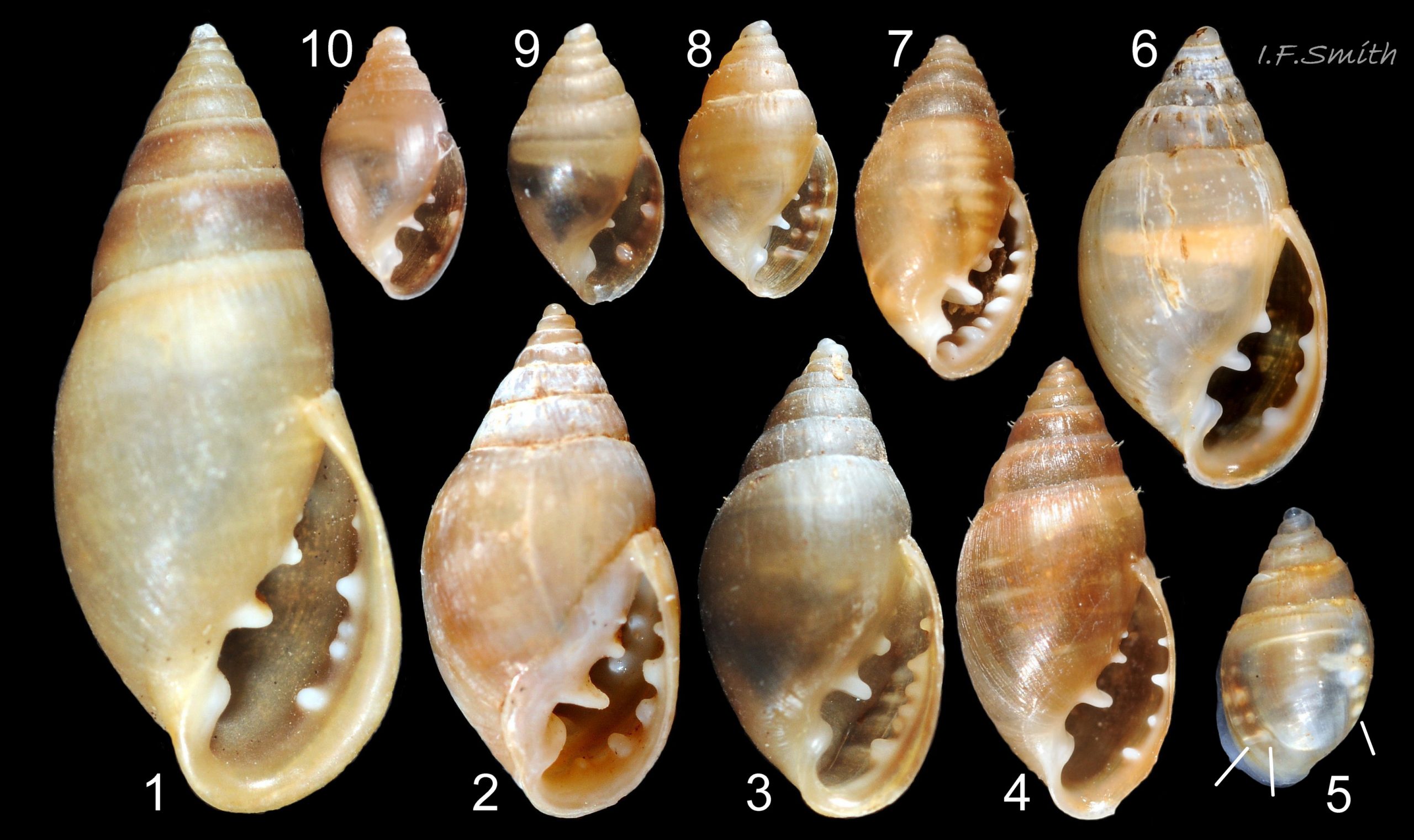
02 Myosotella myosotis 7.5 mm high adult & 4.5 mm high juvenile. Salting on Severn Estuary, Gloucestershire, England. August 2011.
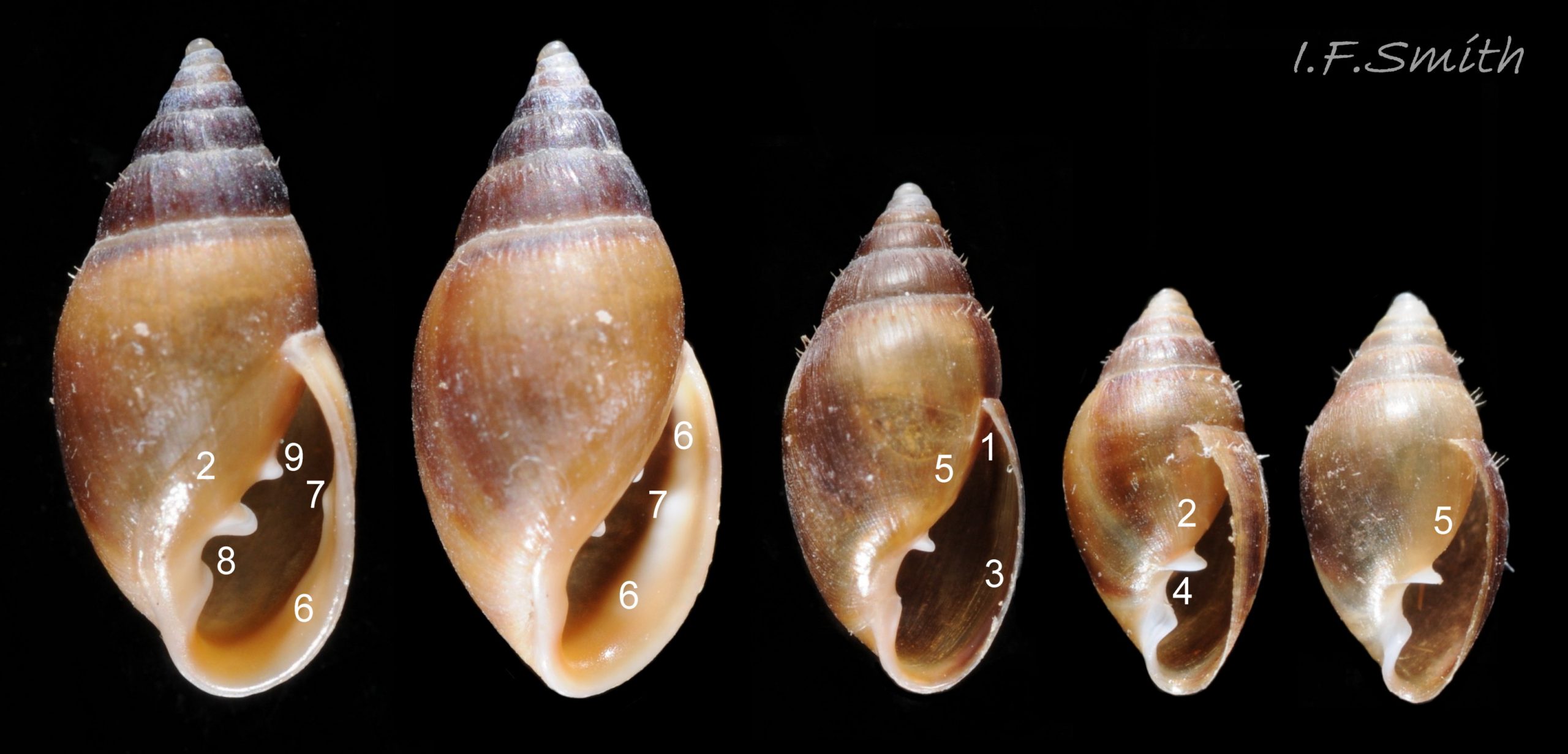
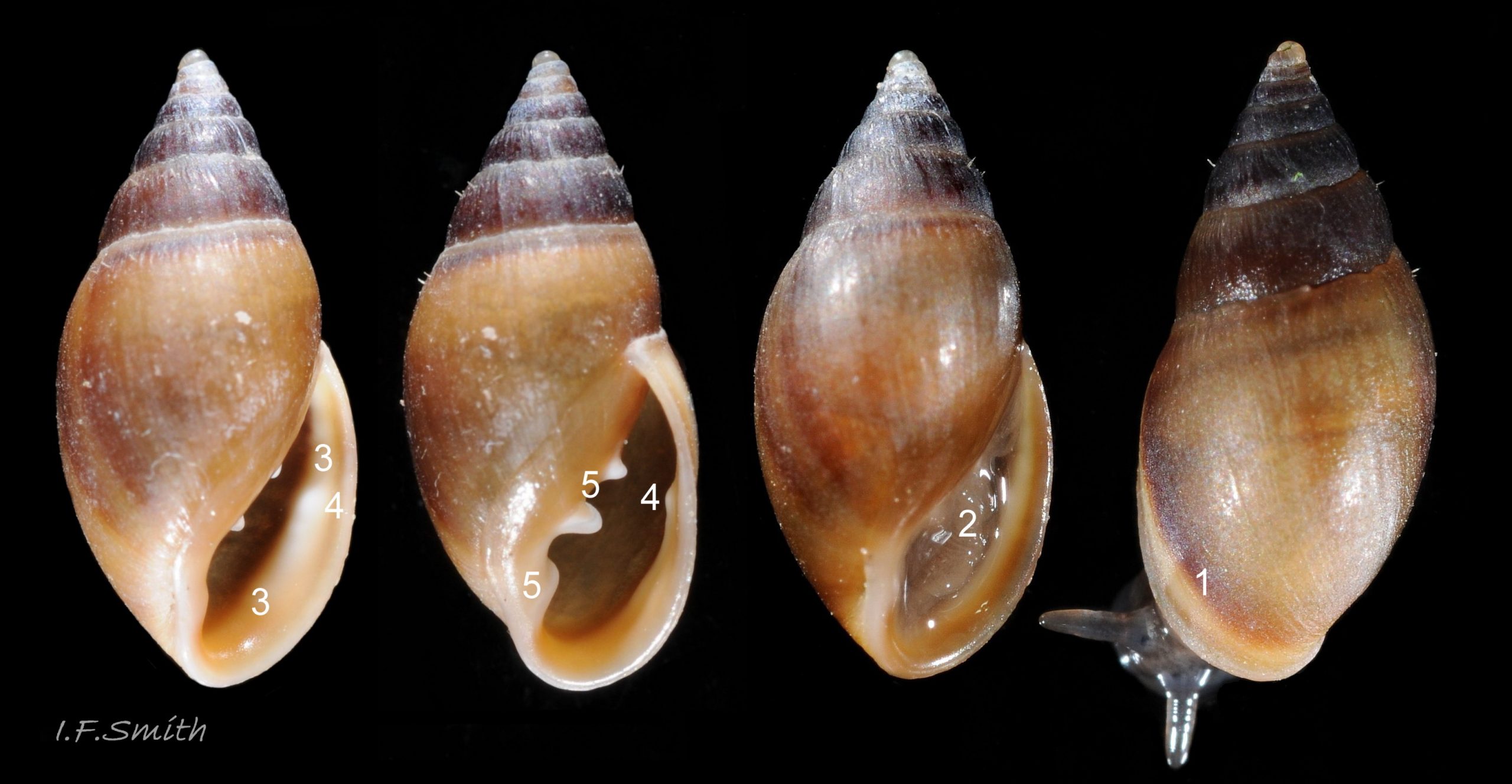
03 Myosotella myosotis apertural views. Salting on Severn Estuary, Gloucestershire, England. August 2011.
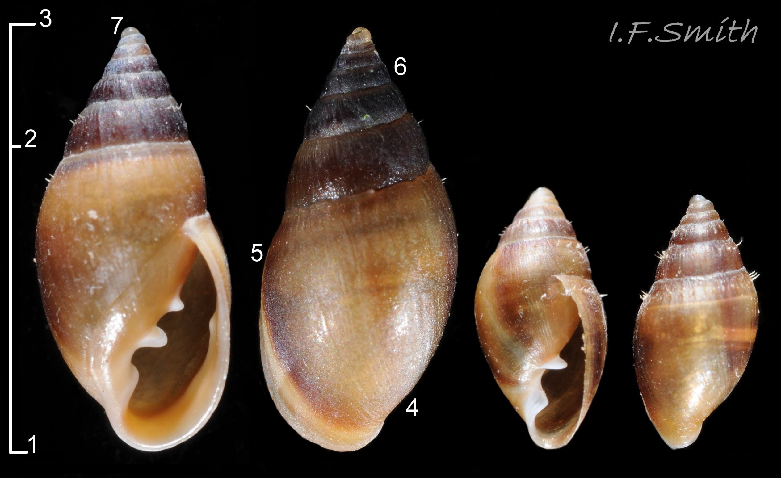
04 Myosotella myosotis. 4.5 mm high juvenile & 7.5 mm high adult. Salting on Severn Estuary, Gloucestershire, England. August 2011.
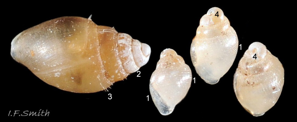
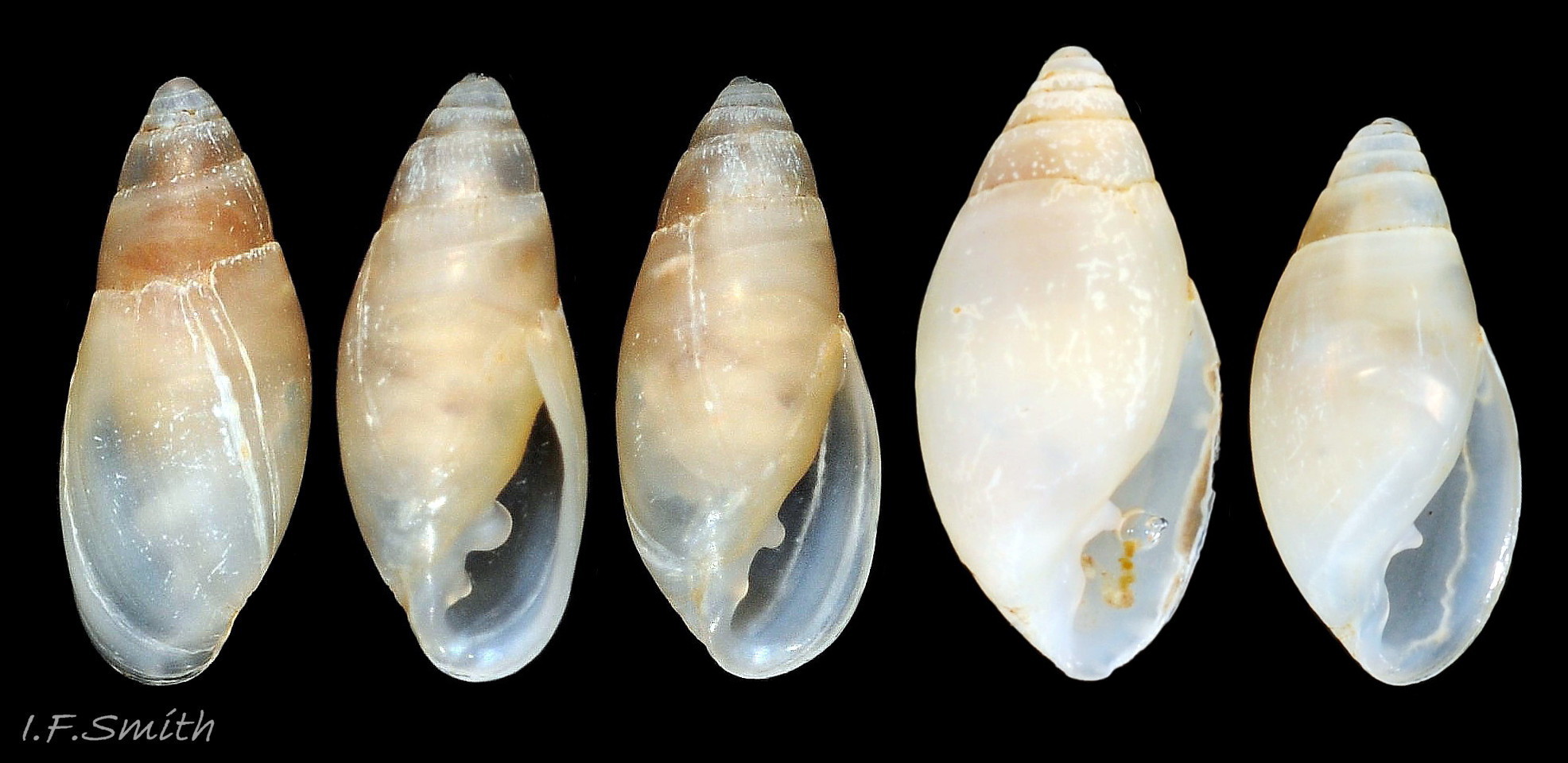
05 Myosotella myosotis. Juveniles from 1.2 mm to 2.4 mm high. Salting on tidal River Dee, Flintshire, Wales. December 2018.
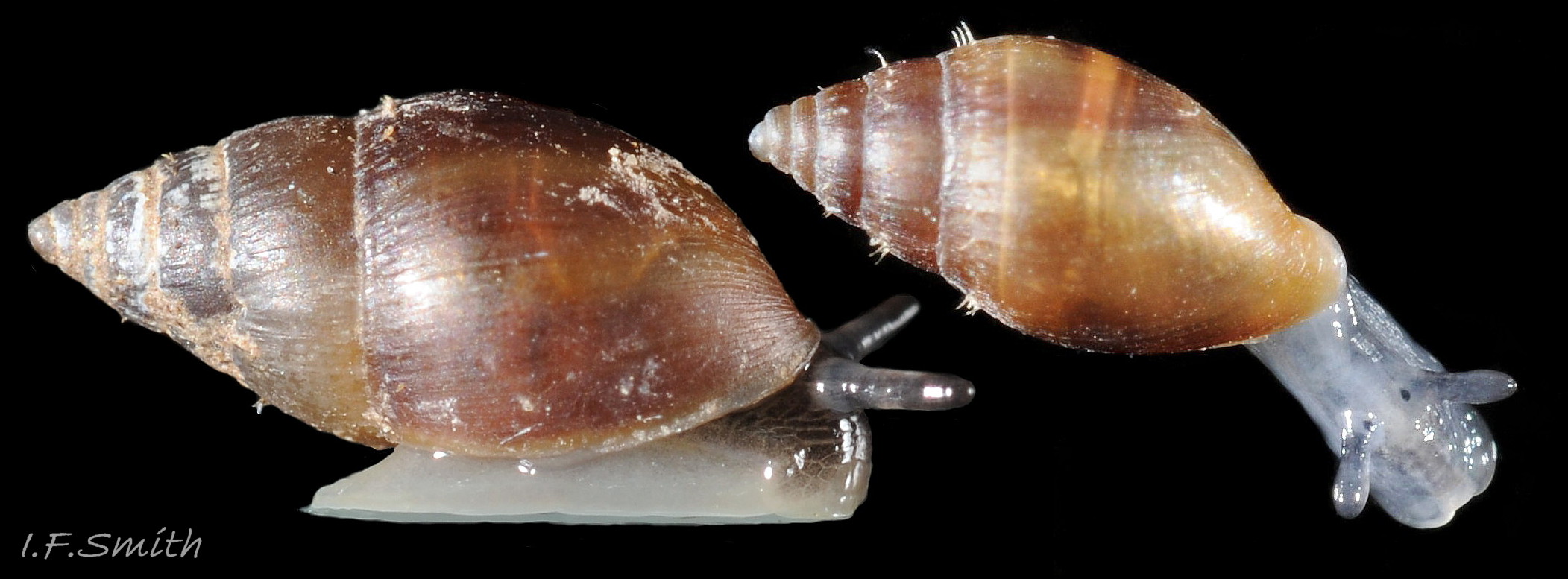
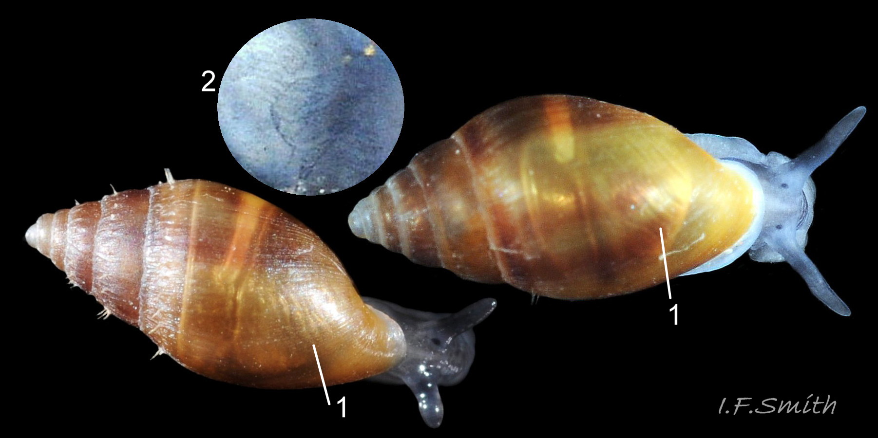
06 Myosotella myosotis. (Left) 5.7mm high. Salting on Dee Estuary, Cheshire, England. May 2018. (Right) 4.5mm high. Salting on Severn Estuary, Gloucestershire, England. August 2011. Both photographed in air.
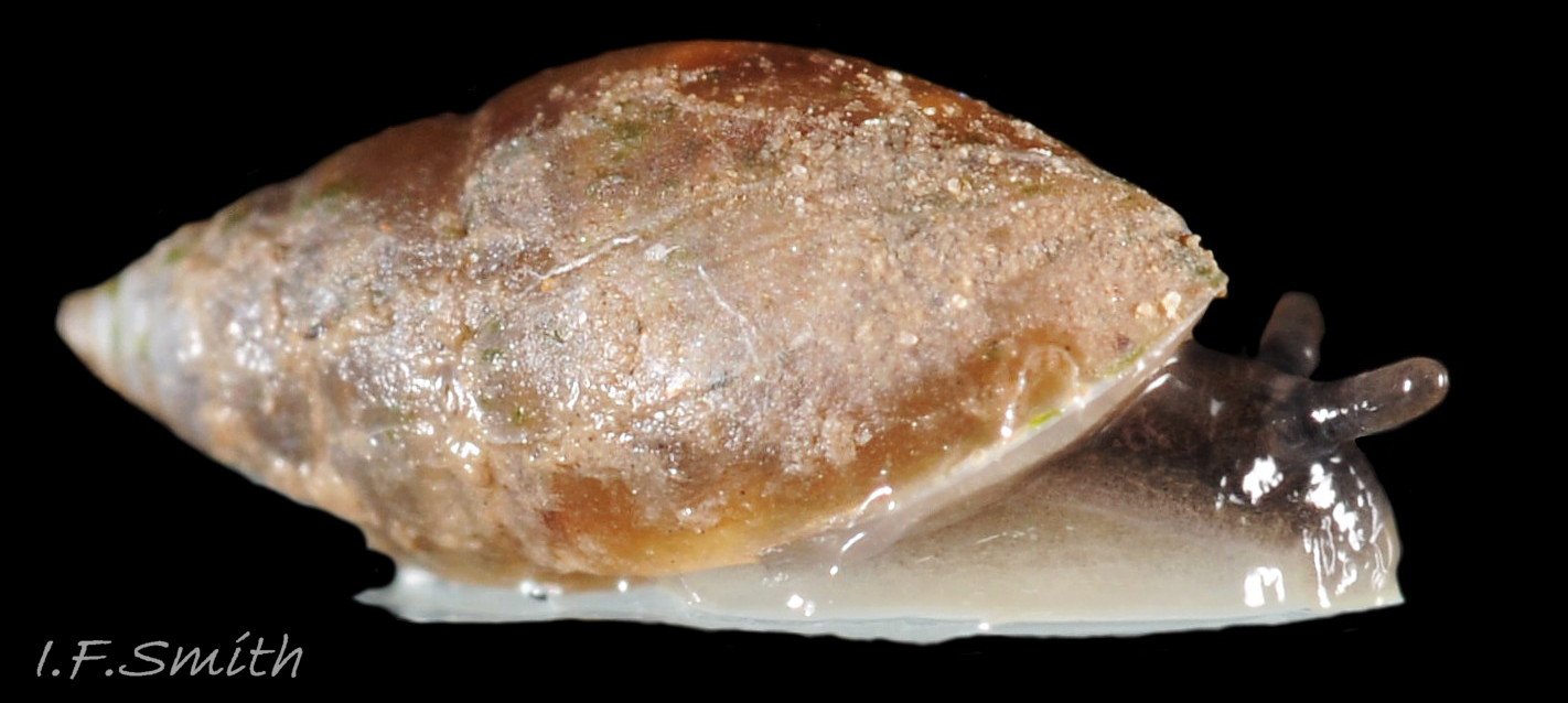
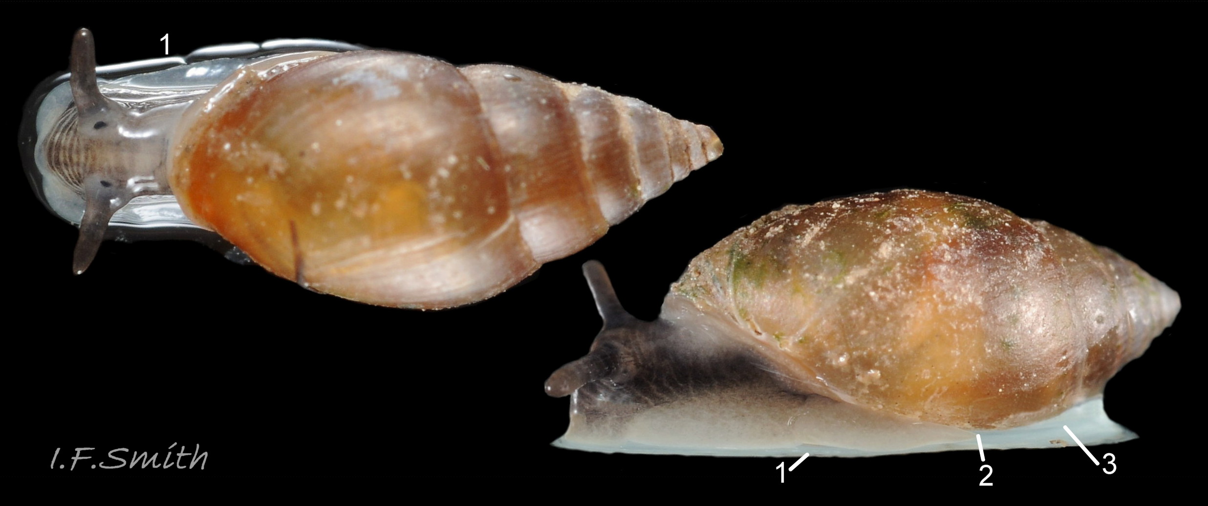
07 Myosotella myosotis. Heights 6mm (in air) & 7.5 mm (in water). Salting on Dee Estuary, Cheshire, England. May 2018.
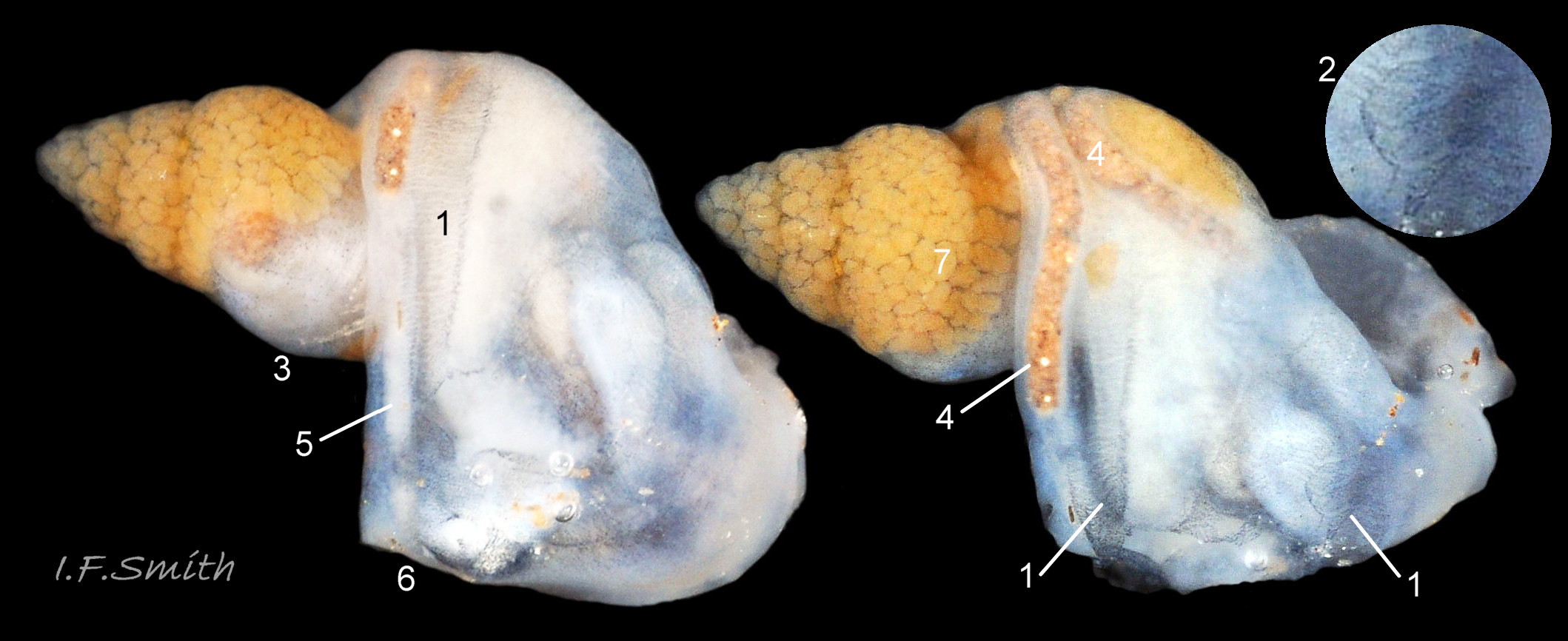
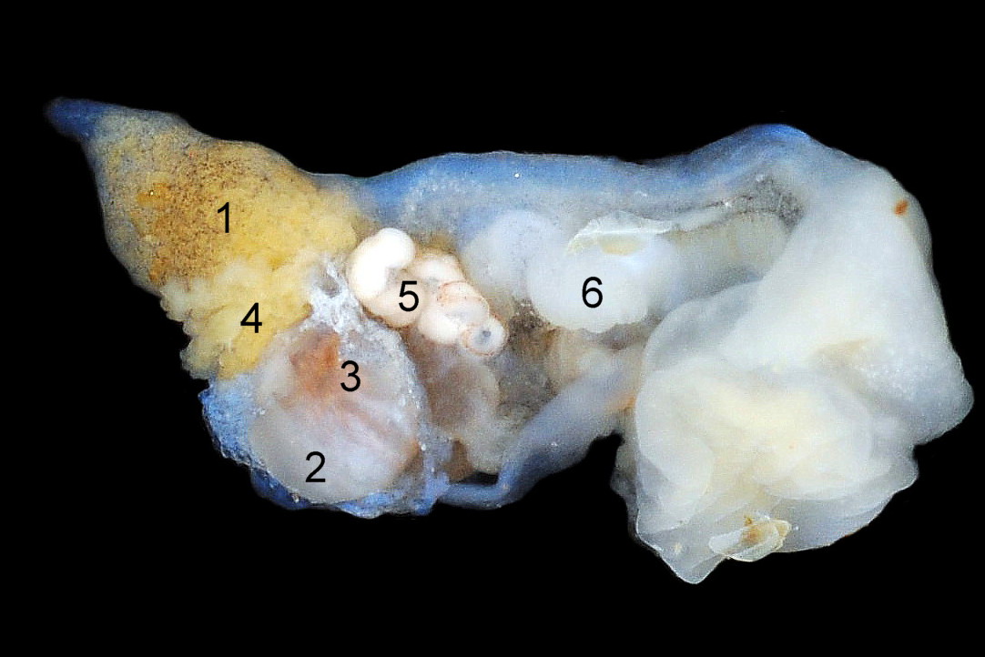
08 Myosotella myosotis. Height 8.8 mm. Shell-wall removed to show interior. Salting on Dee Estuary, Cheshire, England. June 1997.
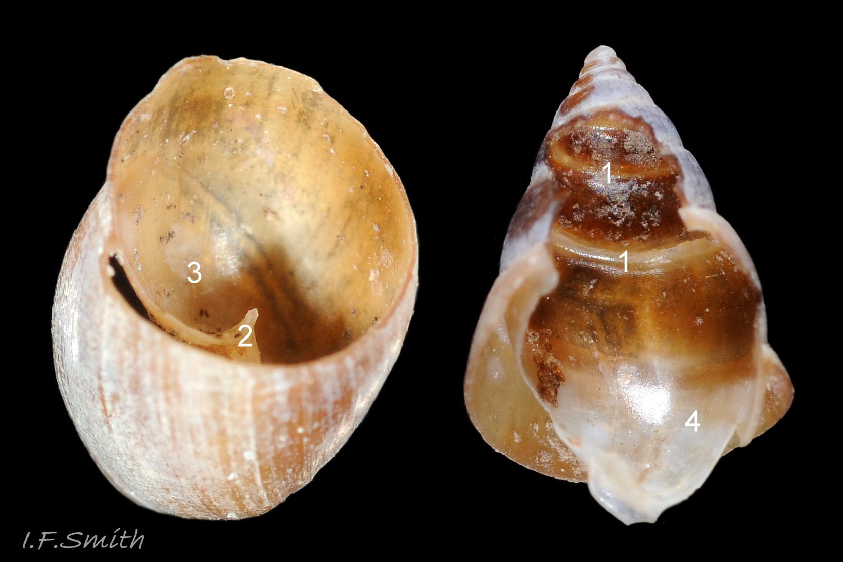
09 Myosotella myosotis. 8mm high. Shell cut transversely, and part of spire wall removed to show interior. Salting on Dee Estuary, Cheshire, England. June 1997.
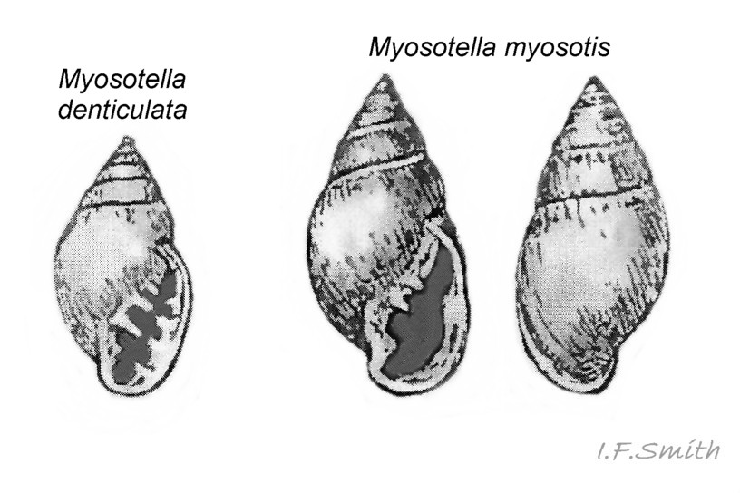
10 Myosotella myosotis album. Images of M. myosotis and M. denticulata from Forbes & Hanley (1853).
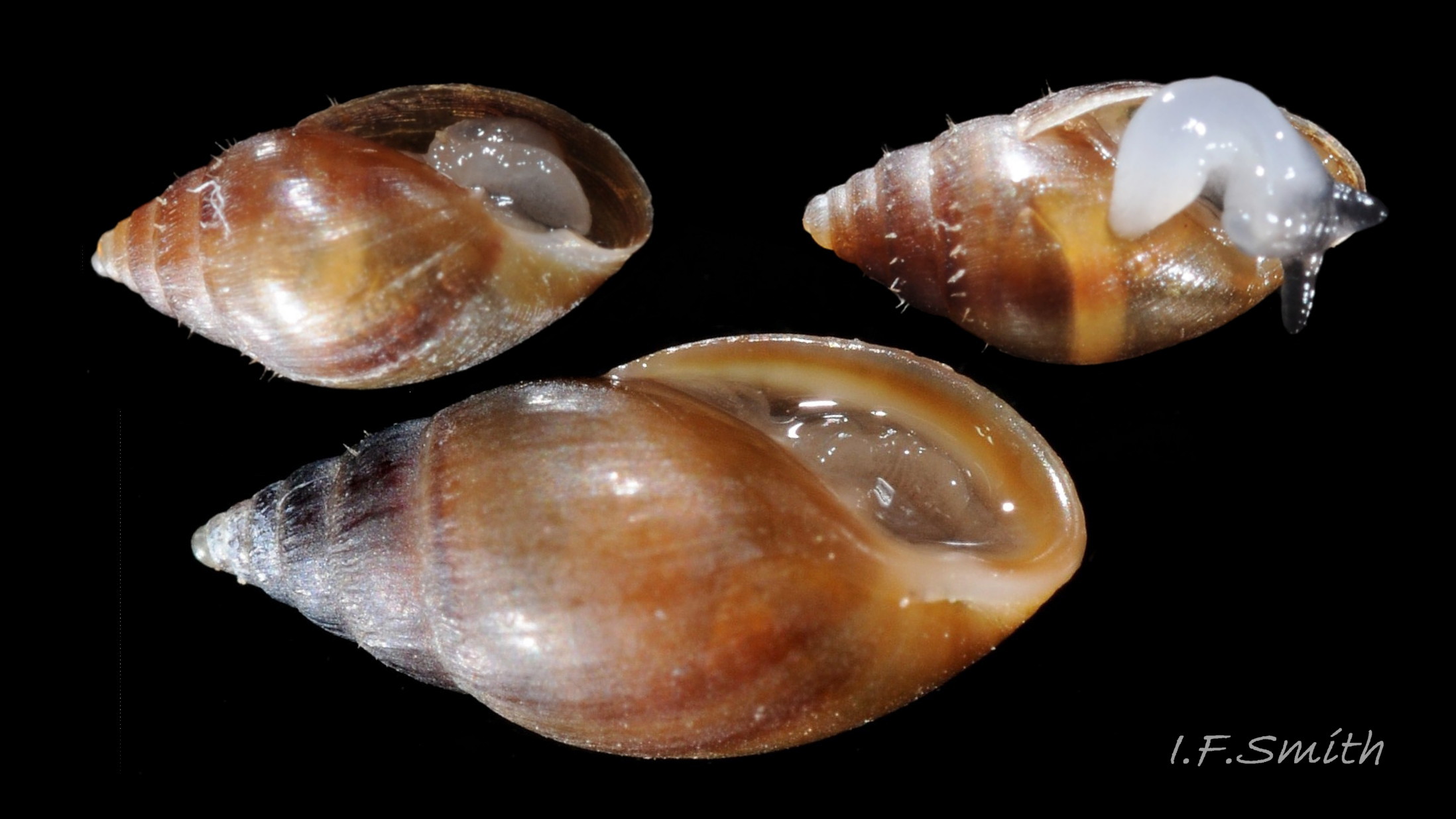
11 Myosotella myosotis. Adult. Height 7.5 mm. Salting on Severn Estuary, Gloucestershire, England. August 2011.
12 Myosotella myosotis. Heights 4.5mm, 4.5 mm and 7.5 mm. Salting on Severn Estuary, Gloucestershire, England. August 2011.
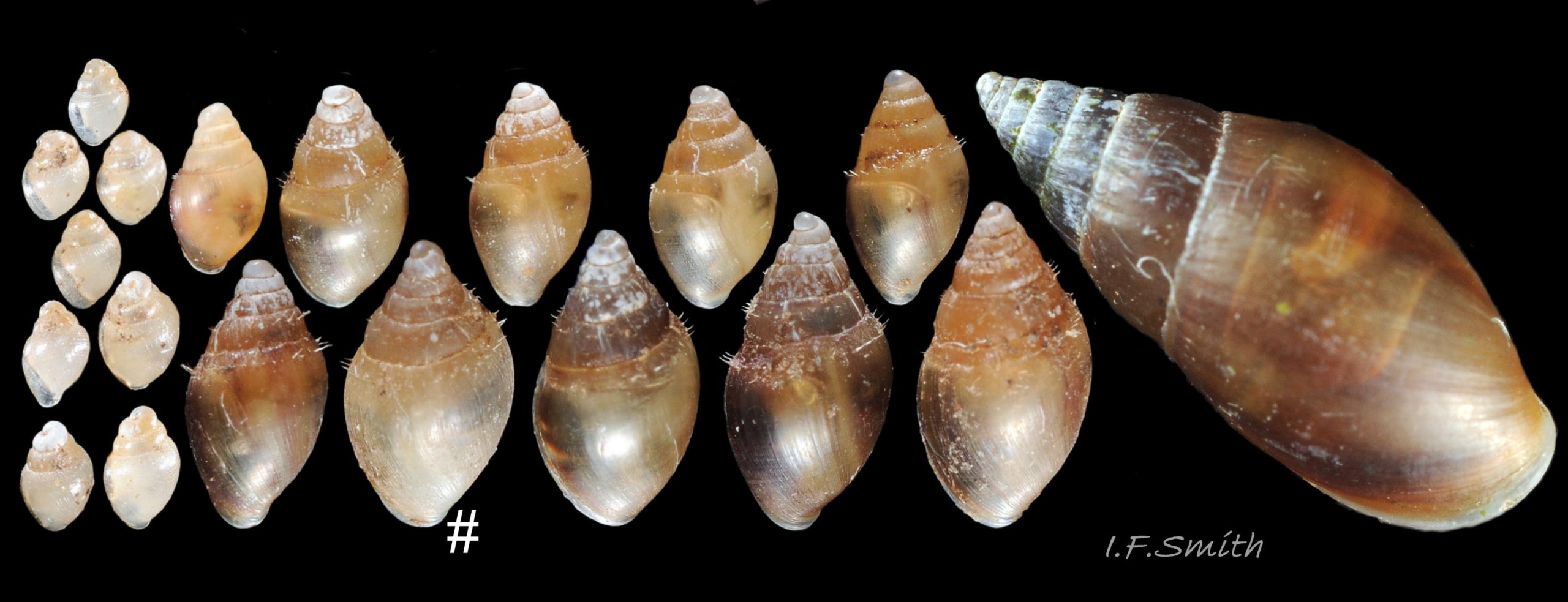
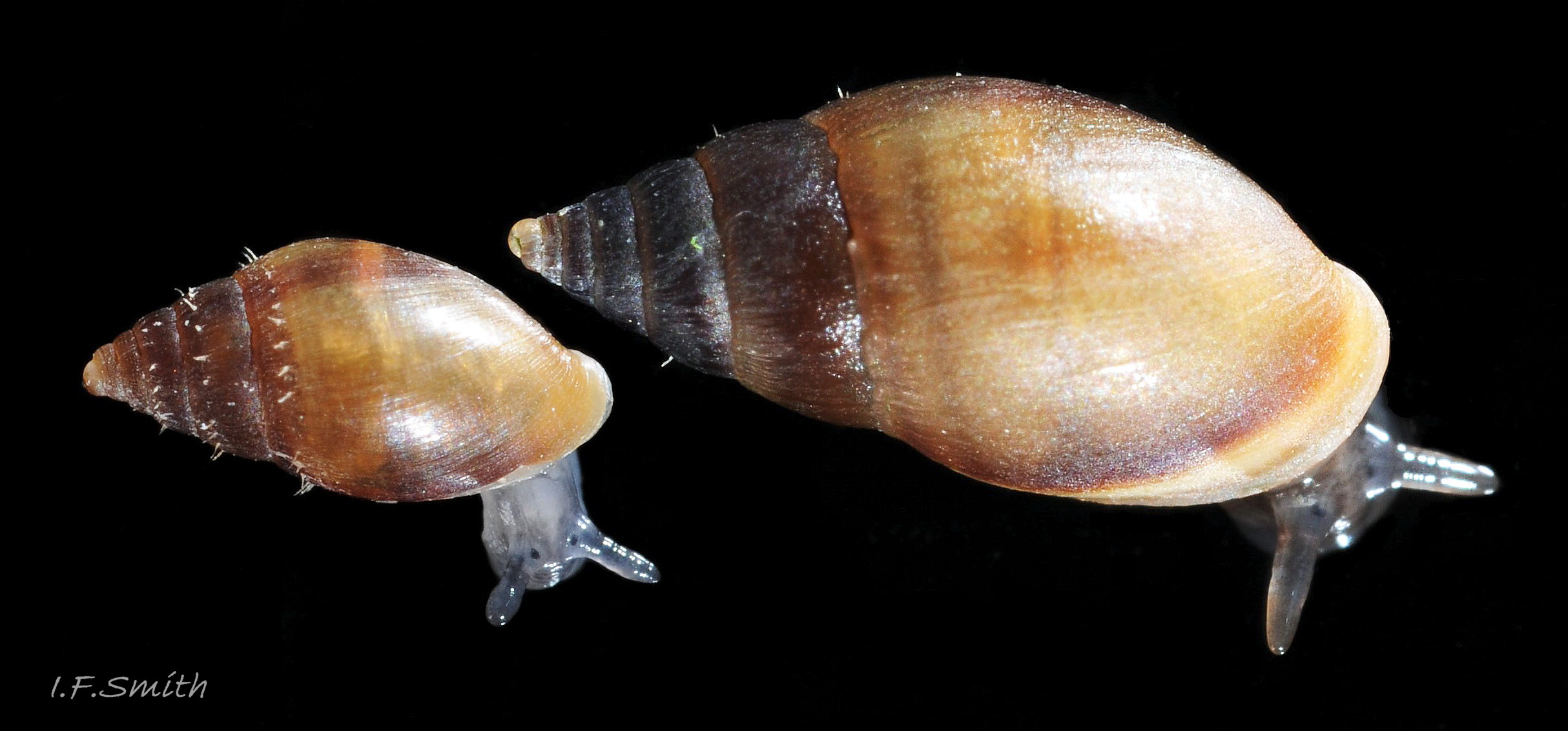
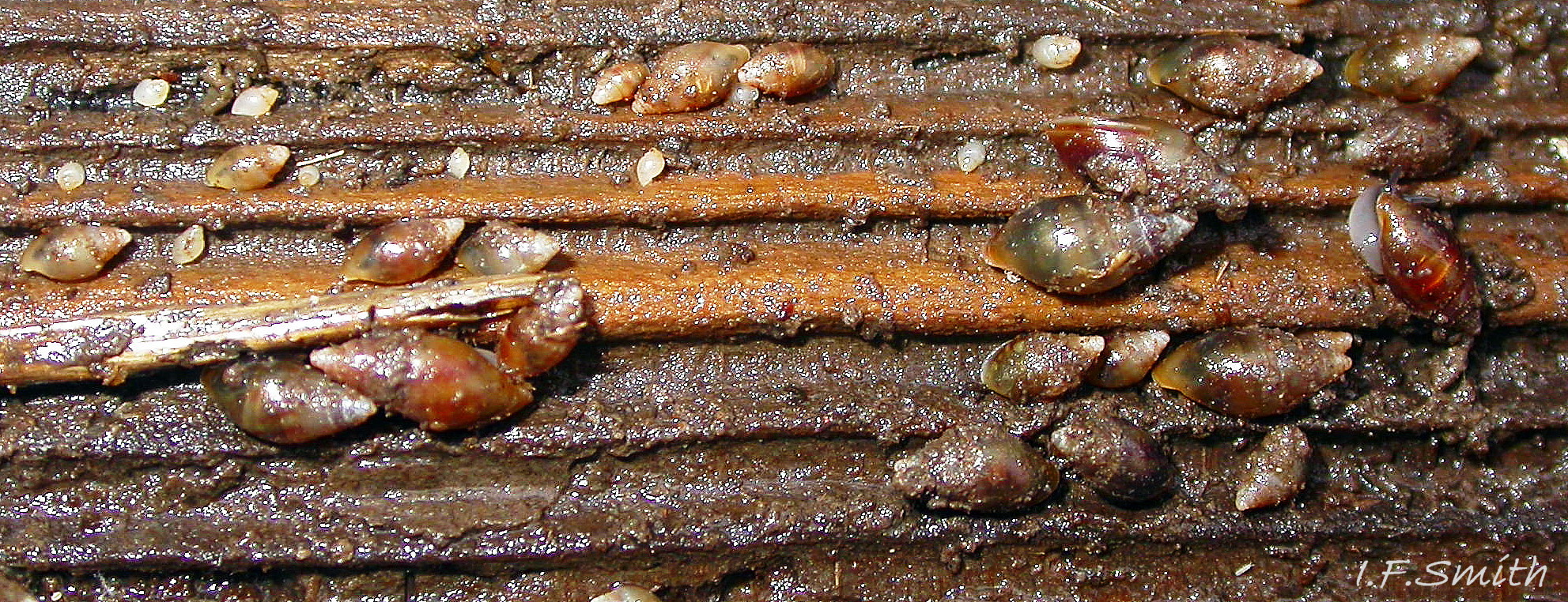
13 Myosotella myosotis. Juveniles 1mm high and above. Adult on right 7.6mm high. (Single M. denticulata marked with # .) All shells occupied by live animal. Saltmarsh-grass sward on tidal River Dee, Flintshire, Wales. December 2018.
14 Myosotella myosotis. Height 5.7 mm. Salting on Dee Estuary, Cheshire, England. May 2018.
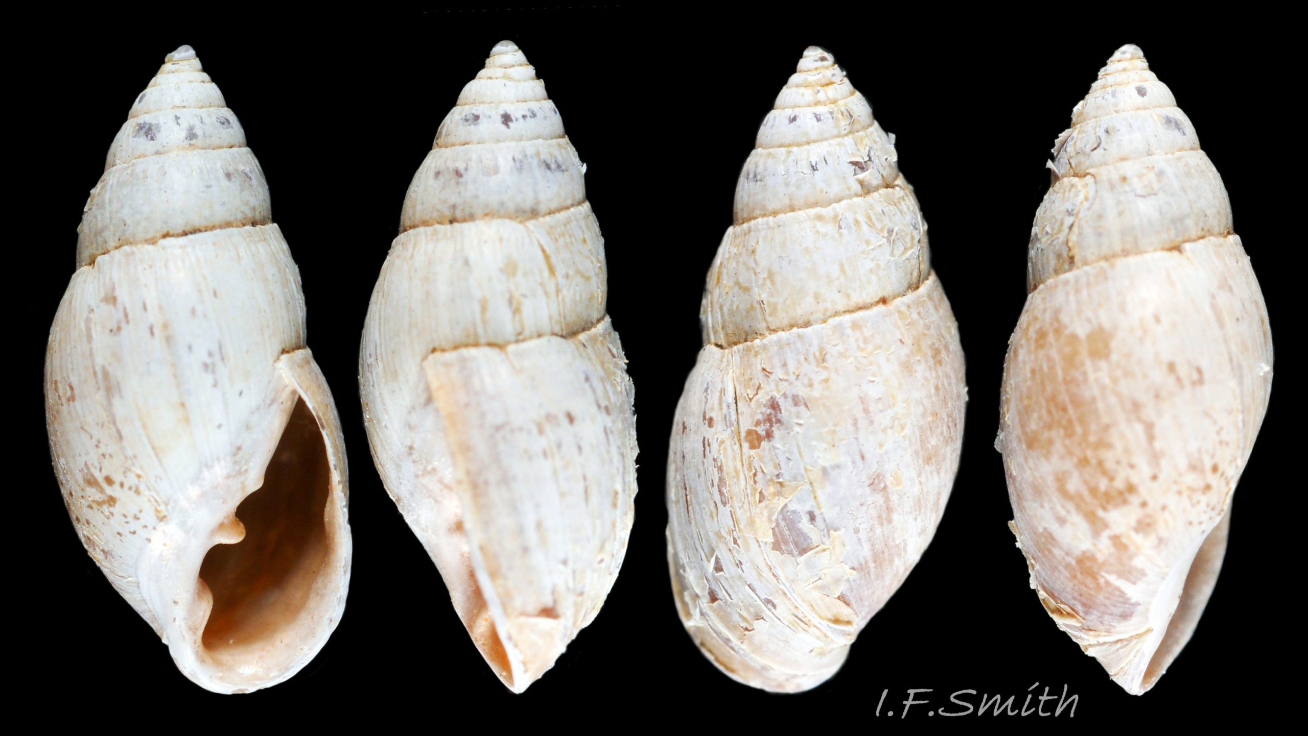
15 Myosotella myosotis. Height 8.2 mm. Salting on Dee Estuary, Cheshire, England. May 2018.
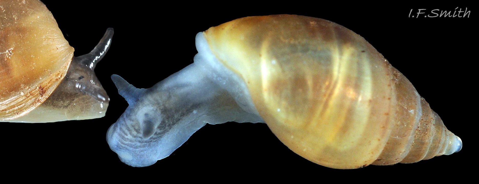
16 Myosotella myosotis. Height 7.2 mm. Salting on Dee Estuary, Cheshire, England. May 2018. Left: in air. Right: same specimen in water.
17 Myosotella myosotis. Shell heights 6mm and 5.6 mm. Salting on Dee Estuary, Cheshire, England. May 2018. Both in air.
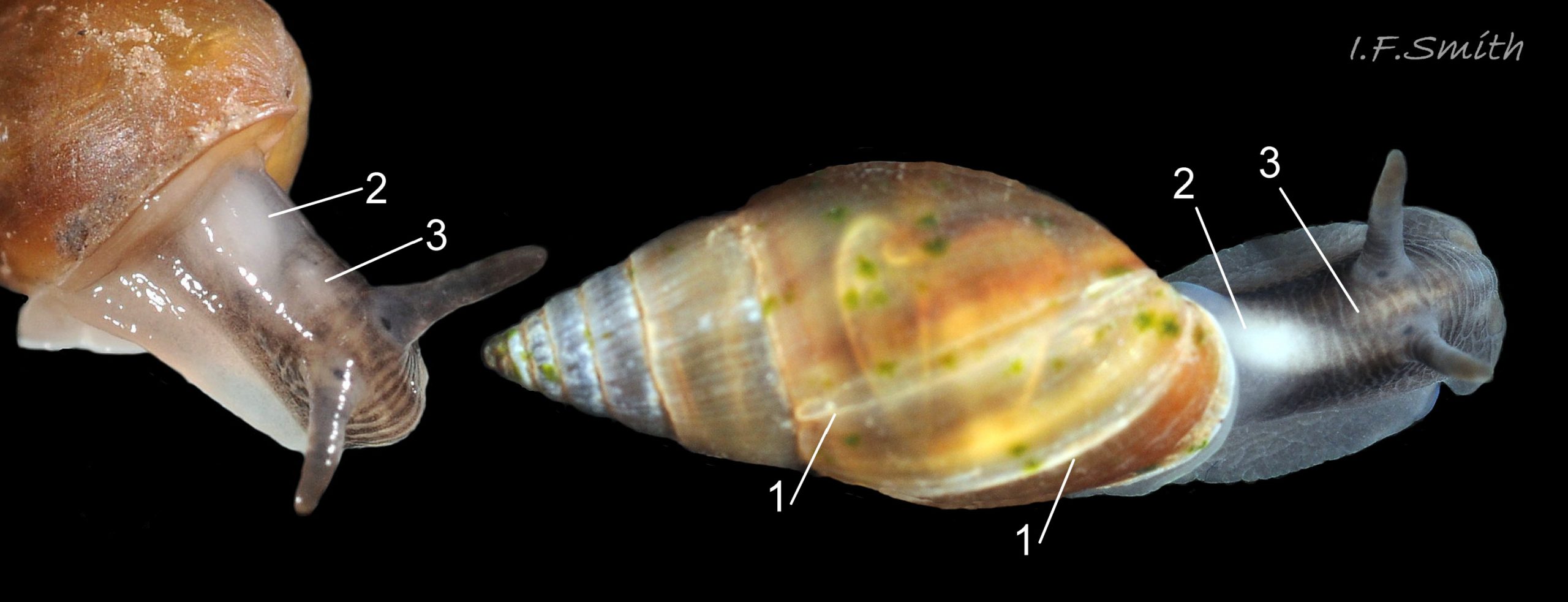
18 Myosotella myosotis. Shell heights 6.4 mm and 7.4 mm. Salting on Dee Estuary, Cheshire, England. May 2018.
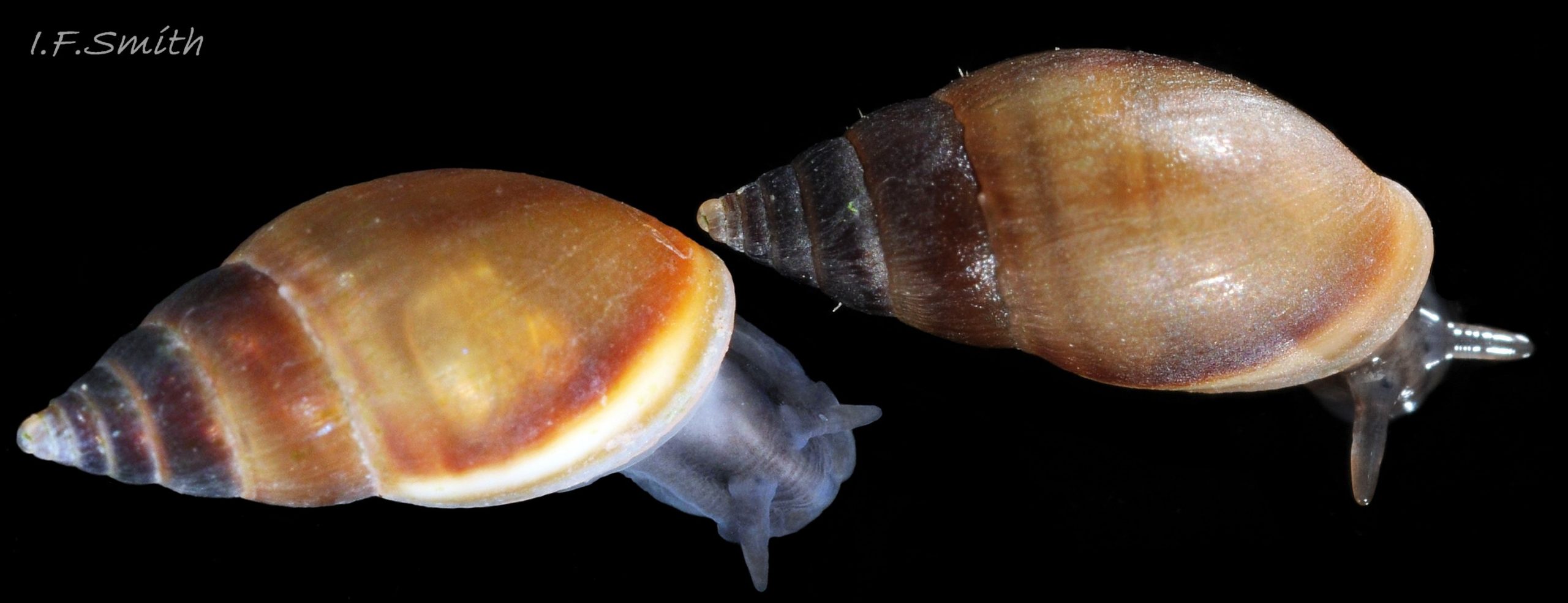
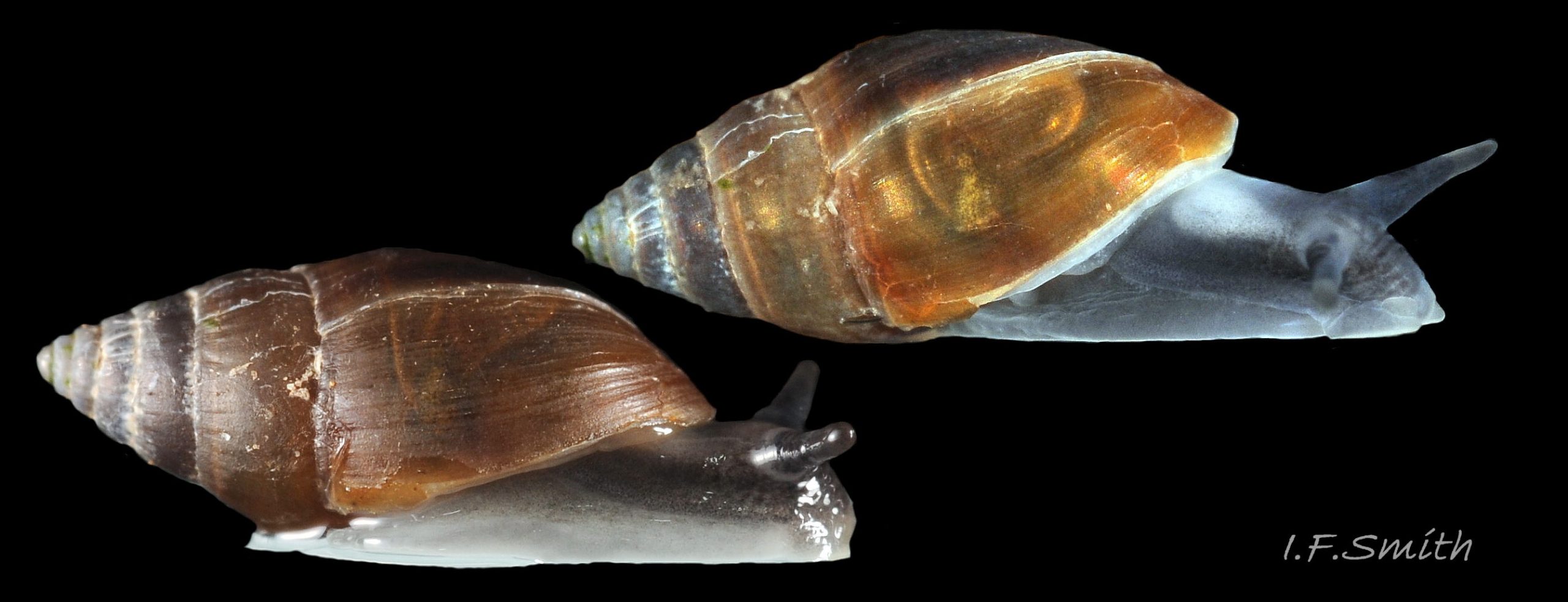
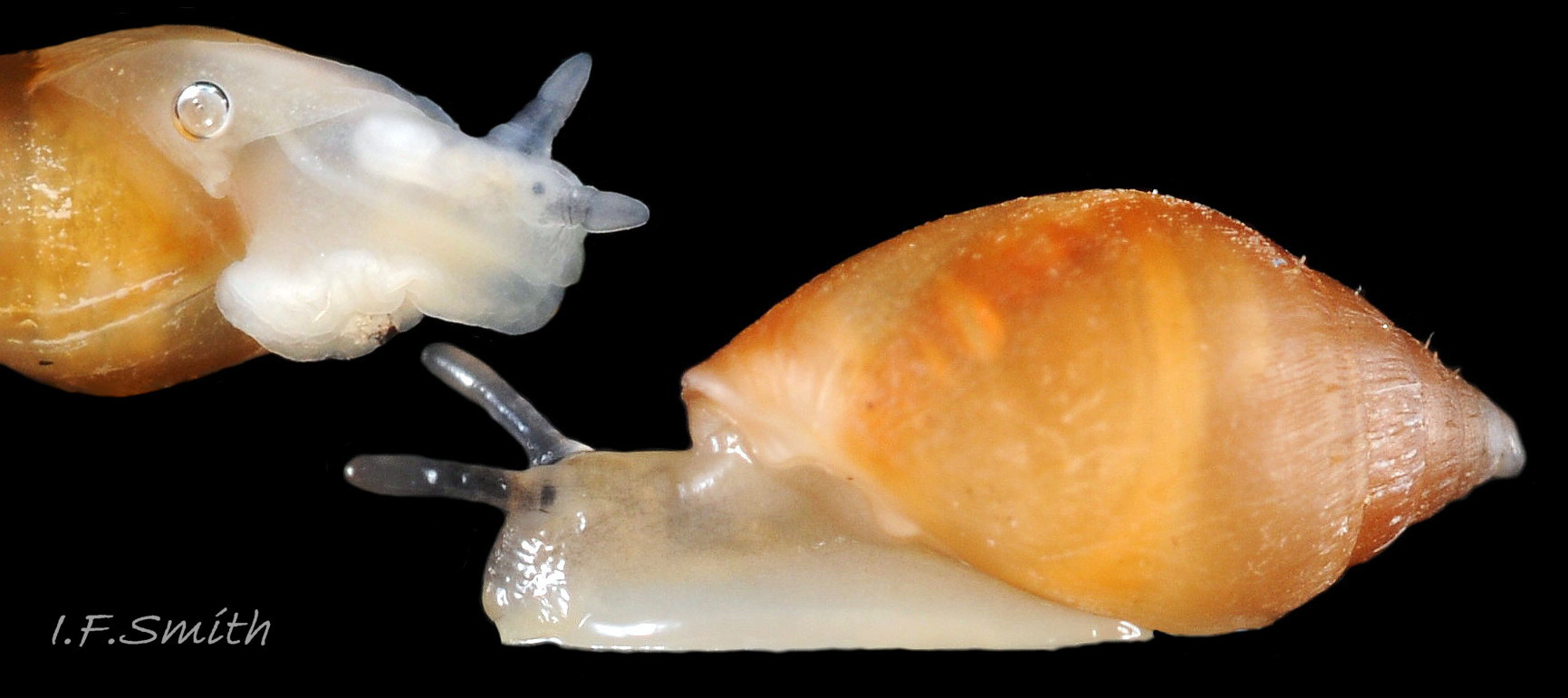
19 Myosotella myosotis. Adult. Height 7.5 mm. Salting on Severn Estuary, Gloucestershire, England. August 2011. Left: in water. Right: same specimen in air.
20 Myosotella myosotis. Shell heights 4.5 mm & 7.4 mm. Salting on Dee Estuary, Cheshire, England. May 2018.
21 Myosotella myosotis removed from shell. Salting on tidal River Dee, Flintshire, Wales. December 2018.
22 Myosotella myosotis. Left in air, right in water. Salting on Severn Estuary, Gloucestershire, England. August 2011.
23 Myosotella myosotis. Shell height 6 mm. Salting on Dee Estuary, Cheshire, England. May 2018.
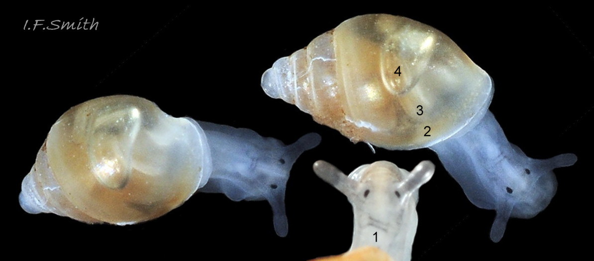
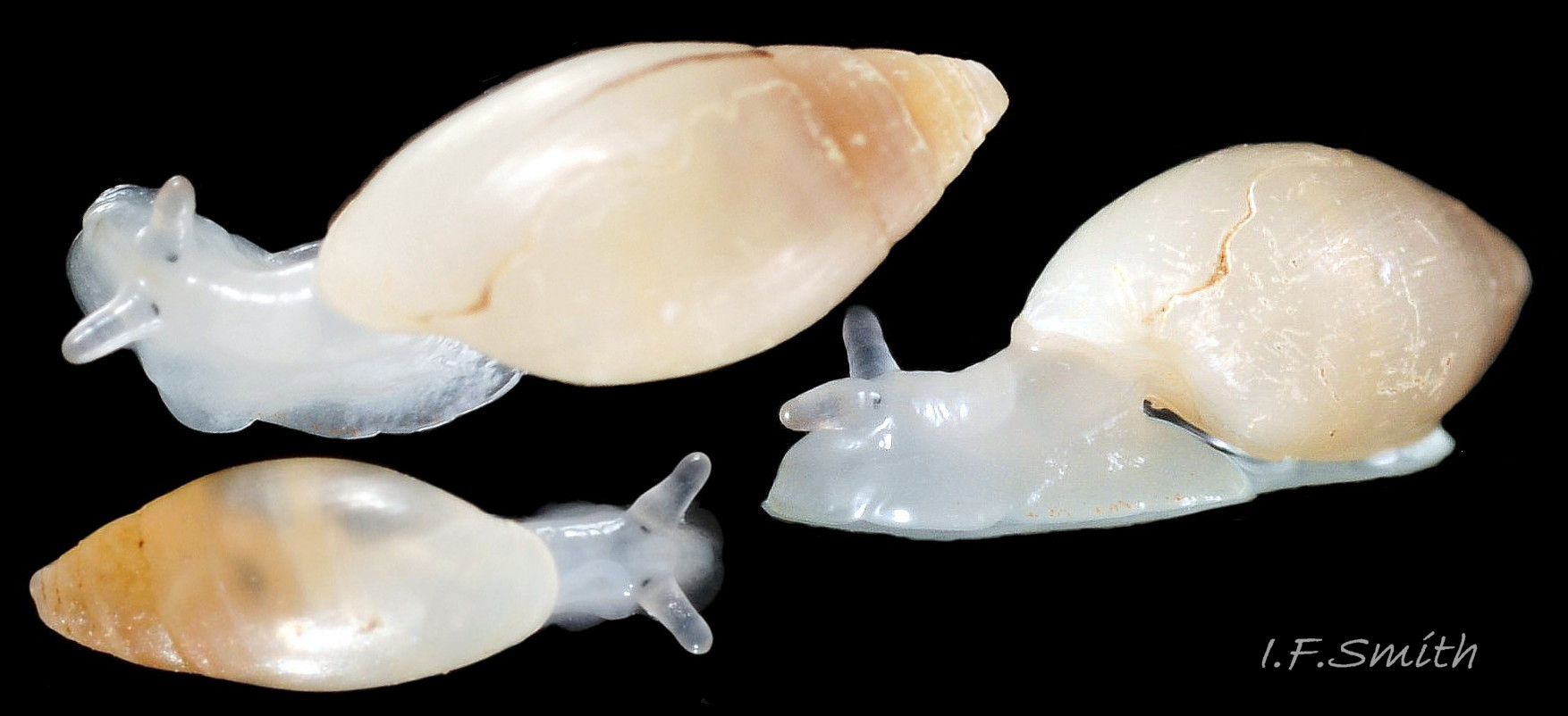
24 Myosotella myosotis. Juveniles with transparent shells. Shell heights 2.5mm (top in water),3mm (bottom in air; less translucent). Salting on tidal River Dee, Flintshire, Wales. December 2018.
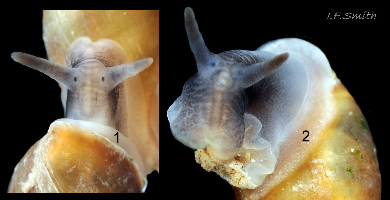
25 Myosotella myosotis. Shell height 6 mm. Salting on Dee Estuary, Cheshire, England. May 2018. Photographed in air.
26 Myosotella myosotis. Shell height 6.2 mm. Salting on Dee Estuary, Cheshire, England. May 2018. Photographed in water.
27 Myosotella myosotis. Adapical angle of aperture. Salting on Dee Estuary, Cheshire, England. May 2018. Photographed in air.
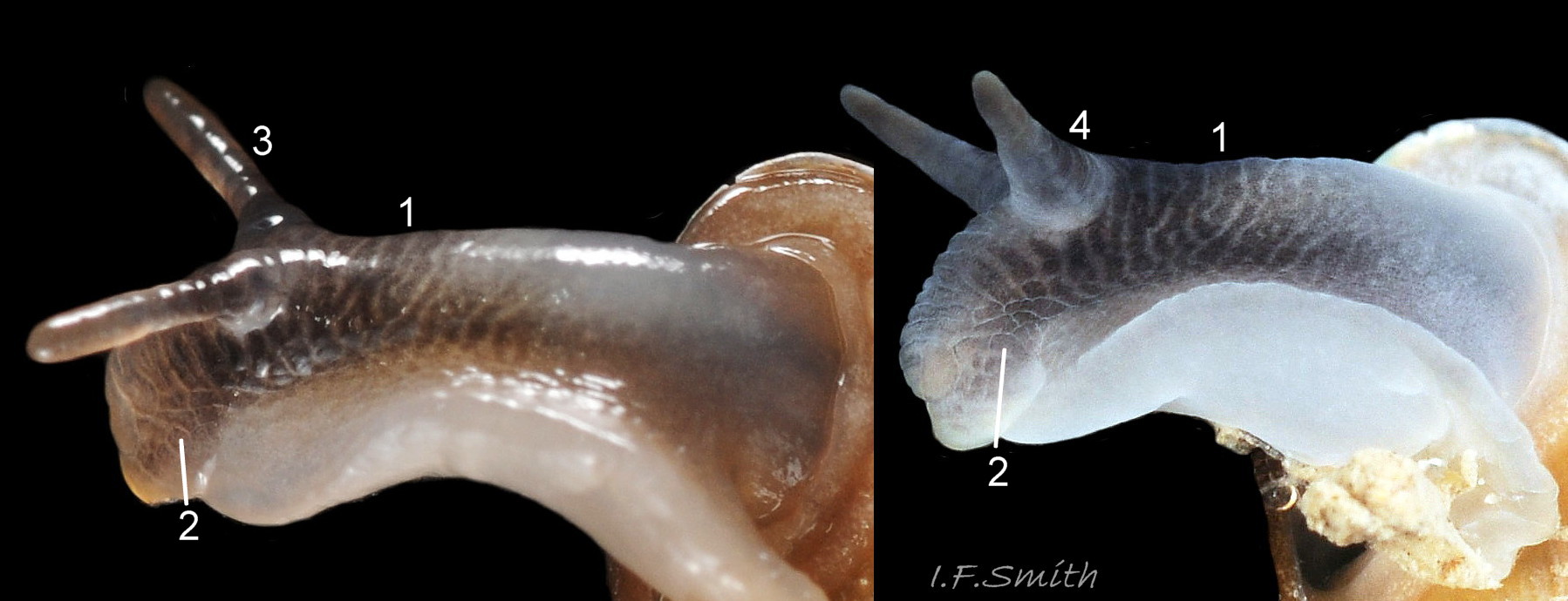
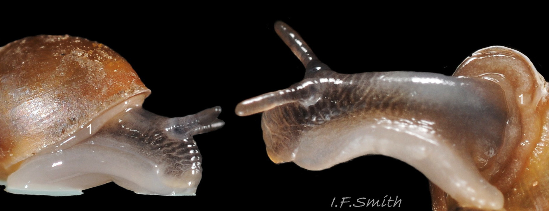
28 Myosotella myosotis. Shell height 7.4 mm. Salting on Dee Estuary, Cheshire, England. May 2018. Photographed in water.
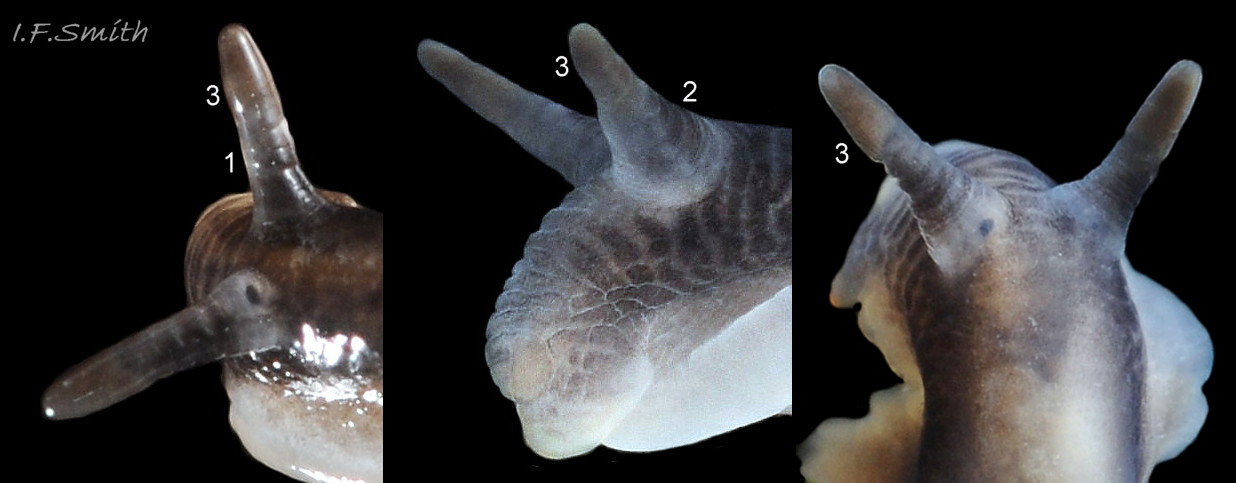
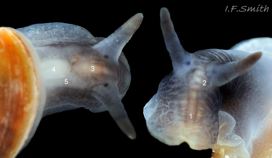
29 Myosotella myosotis. Shell heights left: 7.2 mm, in air; centre & right: 7.4 mm, in water. Salting on Dee Estuary, Cheshire, England. May 2018.
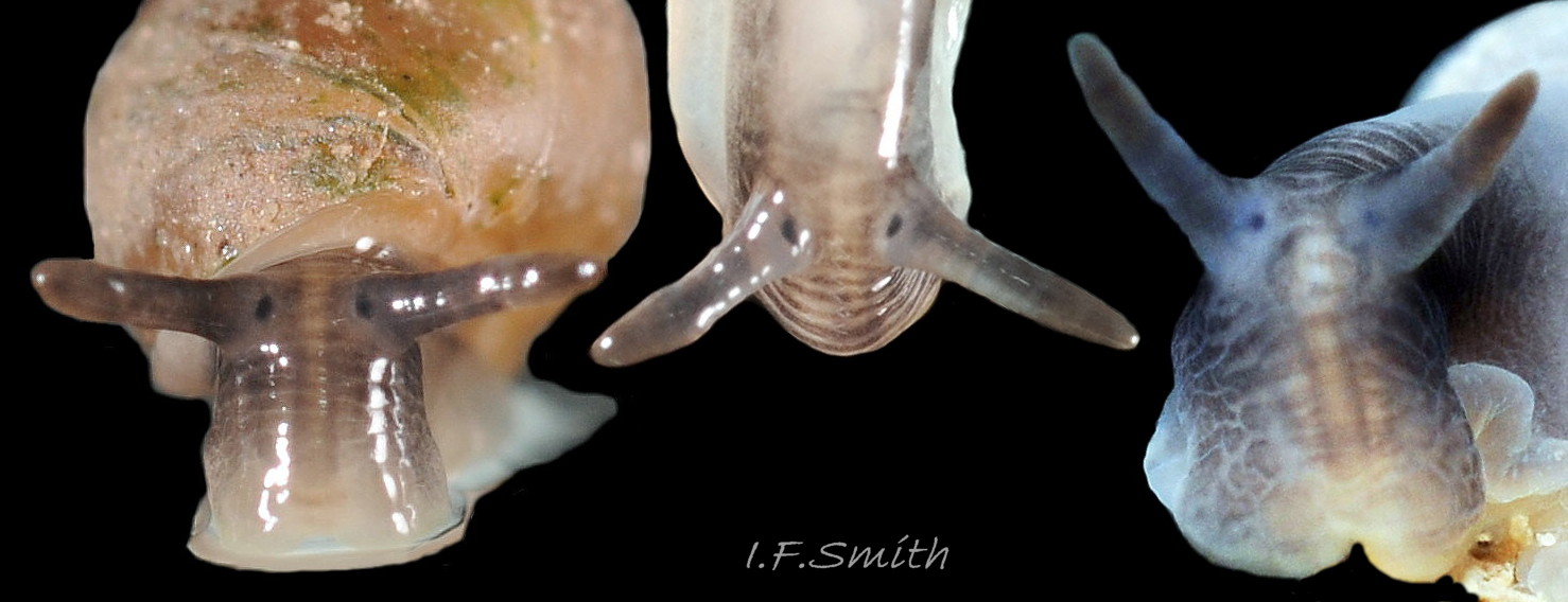
30 Myosotella myosotis. Shell heights 5.6 mm, 6 mm, in air, & 7.4mm in water (on right). Salting on Dee Estuary, Cheshire, England. May 2018.
31 Myosotella myosotis. Shell height 6 mm. Salting on Dee Estuary, Cheshire, England. May 2018.
32 Myosotella myosotis. Body removed from shell; left side. Salting on tidal River Dee, Flintshire, Wales. December 2018.
33 Myosotella myosotis. Body removed from shell and viscera exposed; right side. Salting on tidal River Dee, Flintshire, Wales. December 2018.
34 Myosotella myosotis. Stomach. Salting on tidal River Dee, Flintshire, Wales. December 2018.
35 Myosotella myosotis. Salting on tidal River Dee, Flintshire, Wales. December 2018.
36 Myosotella myosotis. Head and anterior of body with mantle removed. Salting on tidal River Dee, Flintshire, Wales. December 2018.
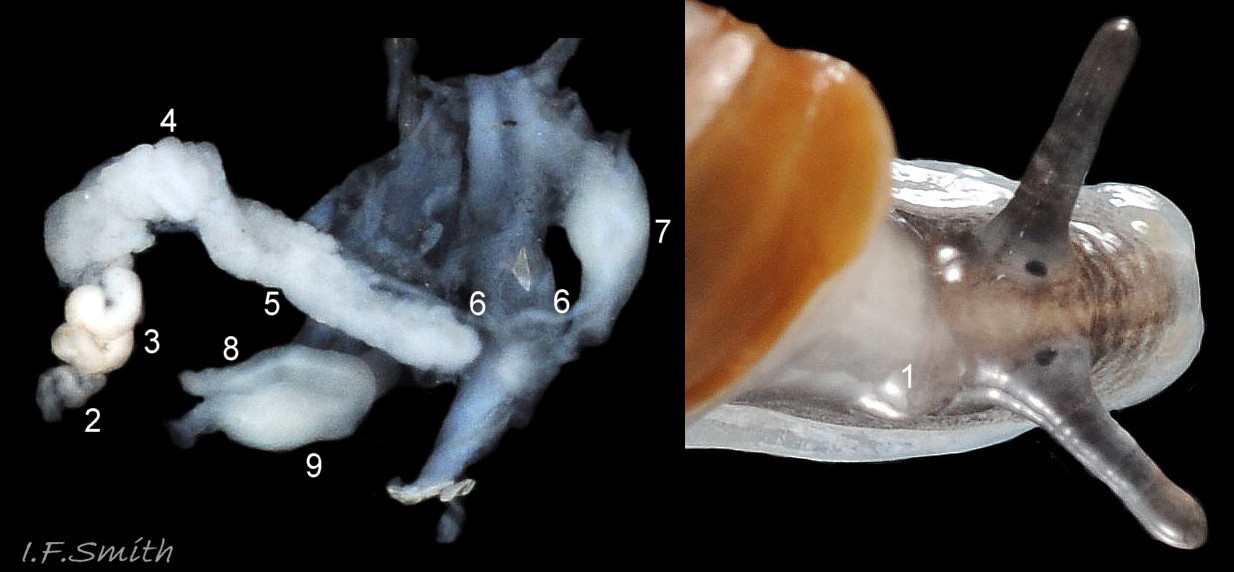
37 Myosotella myosotis. Part of reproductive organs. Salting on tidal River Dee, Flintshire, Wales. December 2018.
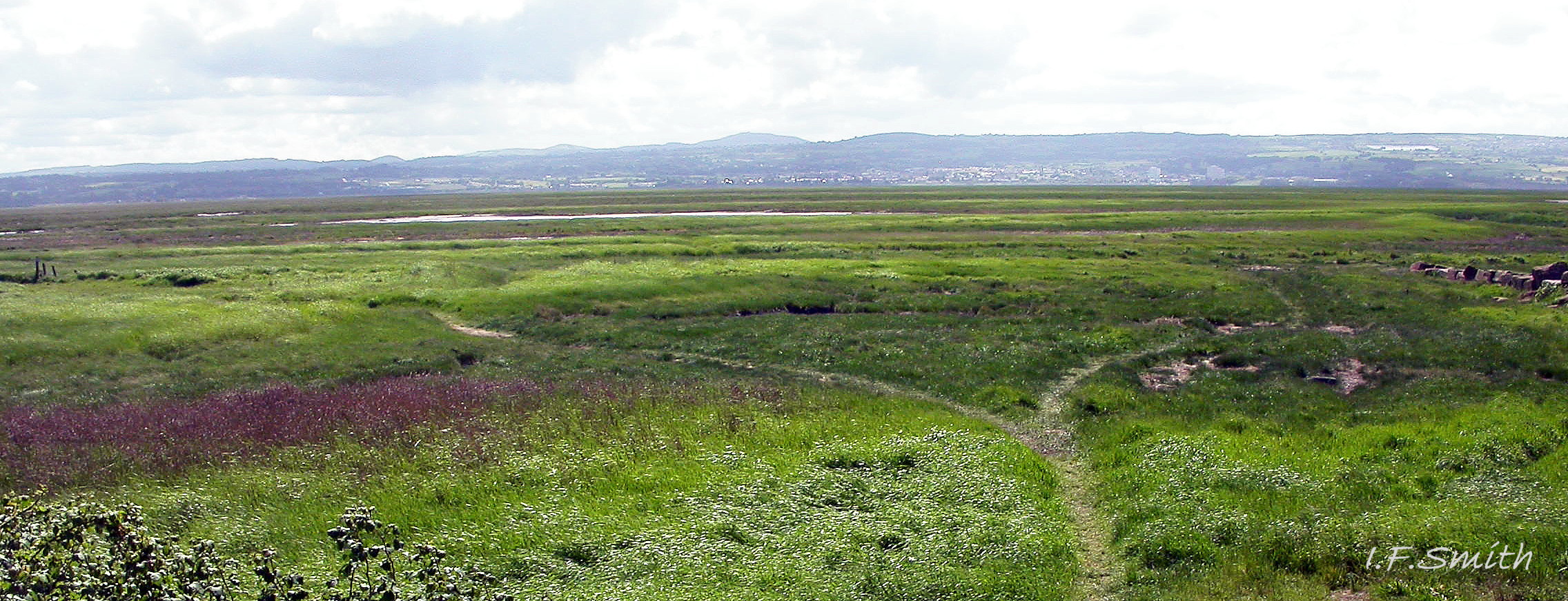
38 Myosotella myosotis habitat. Salting on Dee Estuary, Cheshire, England (Welsh hills in background). May 2018.
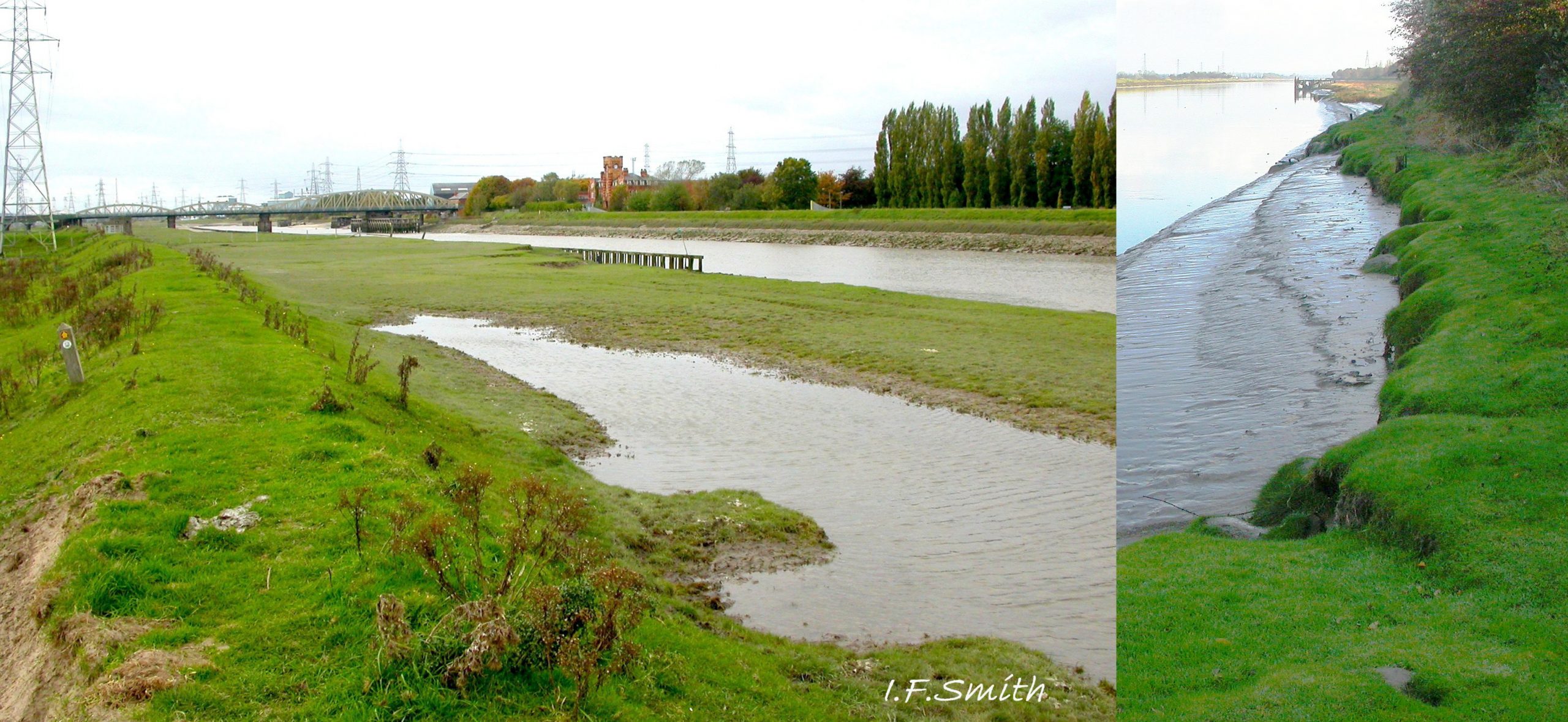
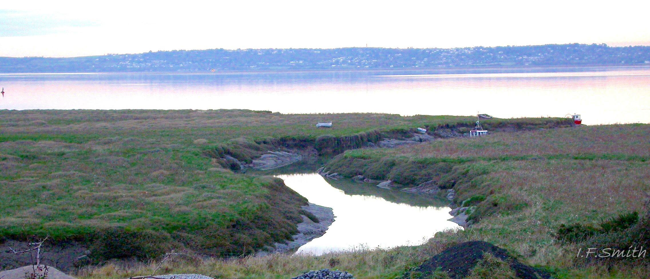
39 Myosotella myosotis habitat. Tidal River Dee, Flintshire, Wales.
40 Myosotella myosotis habitat. Tidal River Dee, Flintshire, Wales. December 2018.
41 Myosotella myosotis habitat. Estuarine salting, Flintshire, Wales.
42 Myosotella myosotis habitat. Tidal River Dee, Flintshire, Wales. December 2018.
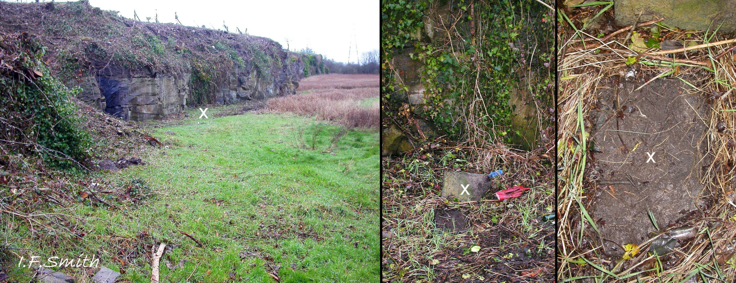
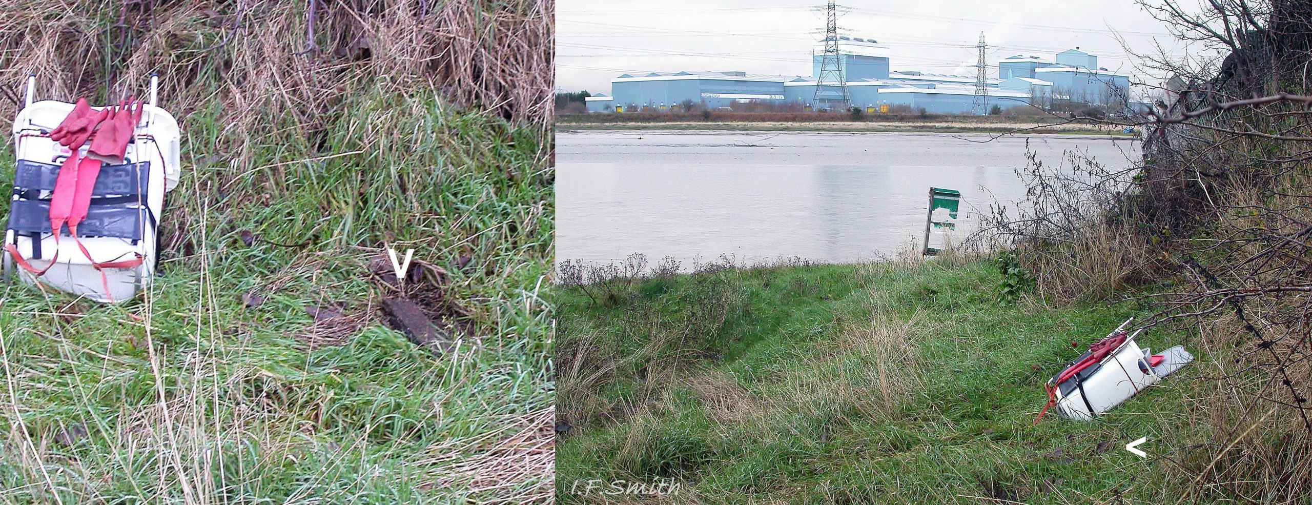
43 Myosotella myosotis habitat. Tidal River Dee, Flintshire, Wales. December 2018.
44 Myosotella myosotis faeces. Tidal River Dee, Flintshire, Wales. December 2018.
45 Myosotella myosotis album. COMPARISON images of M. denticulata. All at same scale, largest 10mm high.
46 Myosotella myosotis album. COMPARISON images of M. denticulata. Shell heights 4.5mm & 5mm. Menai Strait, Wales. April 2018.
47 Myosotella myosotis album. COMPARISON images of M. denticulata. Shell height 6.1mm. Tidal River Dee, Flintshire, Wales. December 2018.
48 Myosotella myosotis album. COMPARISON images of Leucophytia bidentata. Menai Strait, Wales. April 2018 (shell height 4.3mm) & September 2011 (shell height 3.2mm).
49 Myosotella myosotis album. COMPARISON images of Leucophytia bidentata. Menai Strait, Wales. April 2018.
Myosotella myosotis (Draparnaud, 1801)
Preface
[EDIT July 2021: specimens illustrated in this account which were supplied to Amgueddfa Cymru (the Natural History Museum, Wales) were sequenced by Ben Rowson who found no difference in the DNA of M. myosotis and M. denticulata and concluded that they were a single species; Myosotella myosotis. This has now been accepted by WoRMS; see www.marinespecies.org/aphia.php?p=taxdetails&id=139672 ]
The World Register of Marine Species (WoRMS) accepts Myosotella myosotis and M. denticulata as valid species, but those identified as such in Britain may be distinct ecotypes of a single species. A possibility, raised by Martins (2013), is that the true M. myosotis (Draparnaud, 1801) occurs in the Mediterranean and that both British shell forms are ecotypes of M. denticulata (Montagu, 1803). This account treats them separately as, whichever status is determined by planned DNA sequencing, they have distinct apertural sculptures associated with different habitats.
Because of its special habitat intermediate between terrestrial and marine, this species, and its Myosotella and Leucophytia relatives in the family Ellobiidae, are omitted from some identification guides, while variously appearing in others devoted solely to either terrestrial, marine or even freshwater mollusca.
Synonyms: Auricula myosotis Draparnoud, 1801; Ovatella myosotis (Draparnaud, 1801); Alexia myosotis (Draparnaud, 1801); Phytia myosotis (Draparnaud, 1801); Conovulus denticulatus var. myosotis in Forbes & Hanley (1853); Melampus myosotis in Jeffreys (1869);
Vernacular Probably also applied to M. denticulata: Mouse-eared Alexia, Mouse ear(ed) snail (English); Clust llygoden (Welsh); Evesnegl (Danish); Muizenoortje (Dutch); Ovatelle naine des vases (French); Stranddvärgsnäcka (Swedish); Mäuseöhrchen (German);
Applied to just this species/ecotype: Estuarine mouse-ear (English); Gewoon muizenoortje (Dutch);
Description
When in water, the body is more swollen, and both shell and body are more translucent, lighter and brighter in colour, and less reflective, than when in air 01 Myosotella myosoti. The following shell description is of specimens in air.
Shell
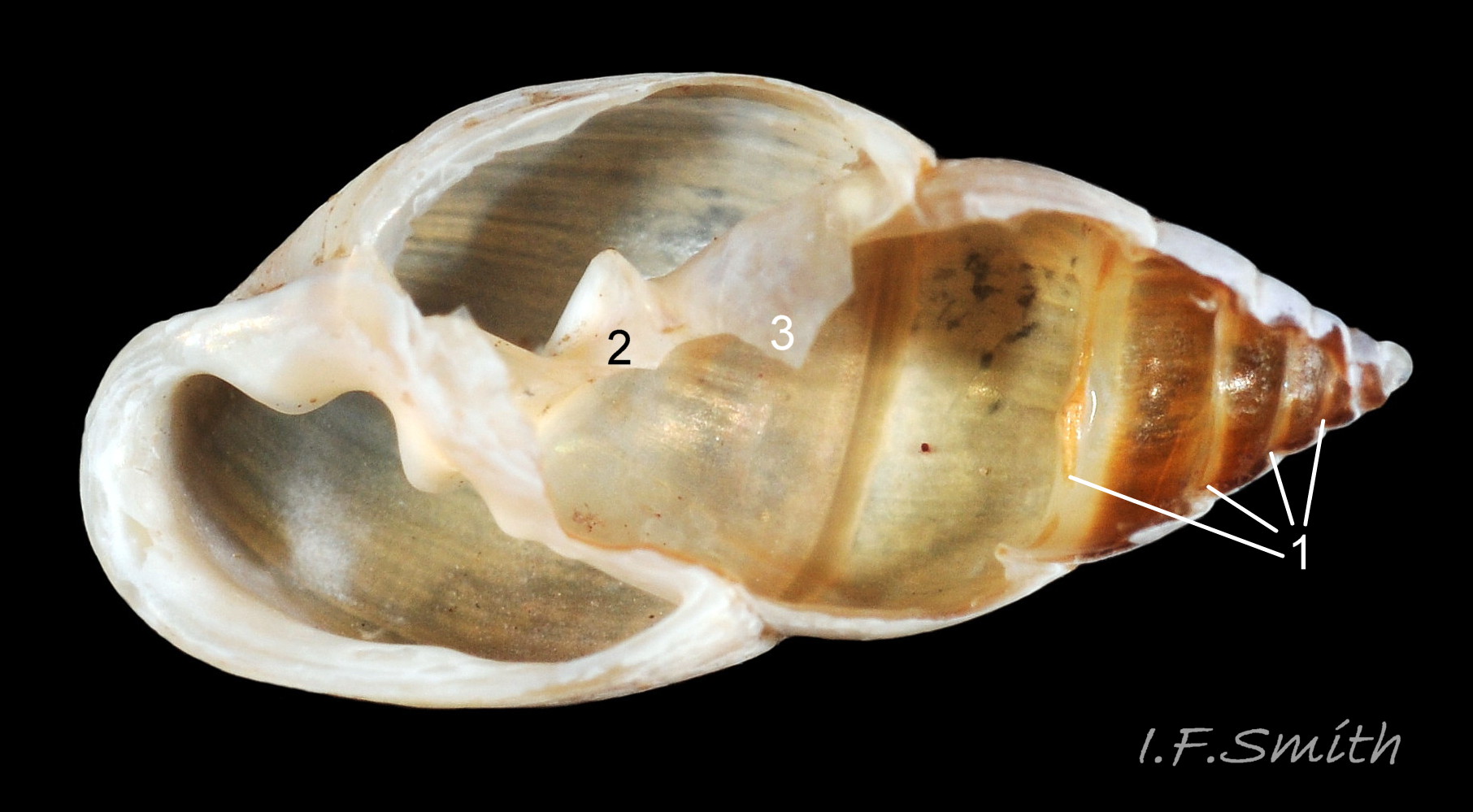
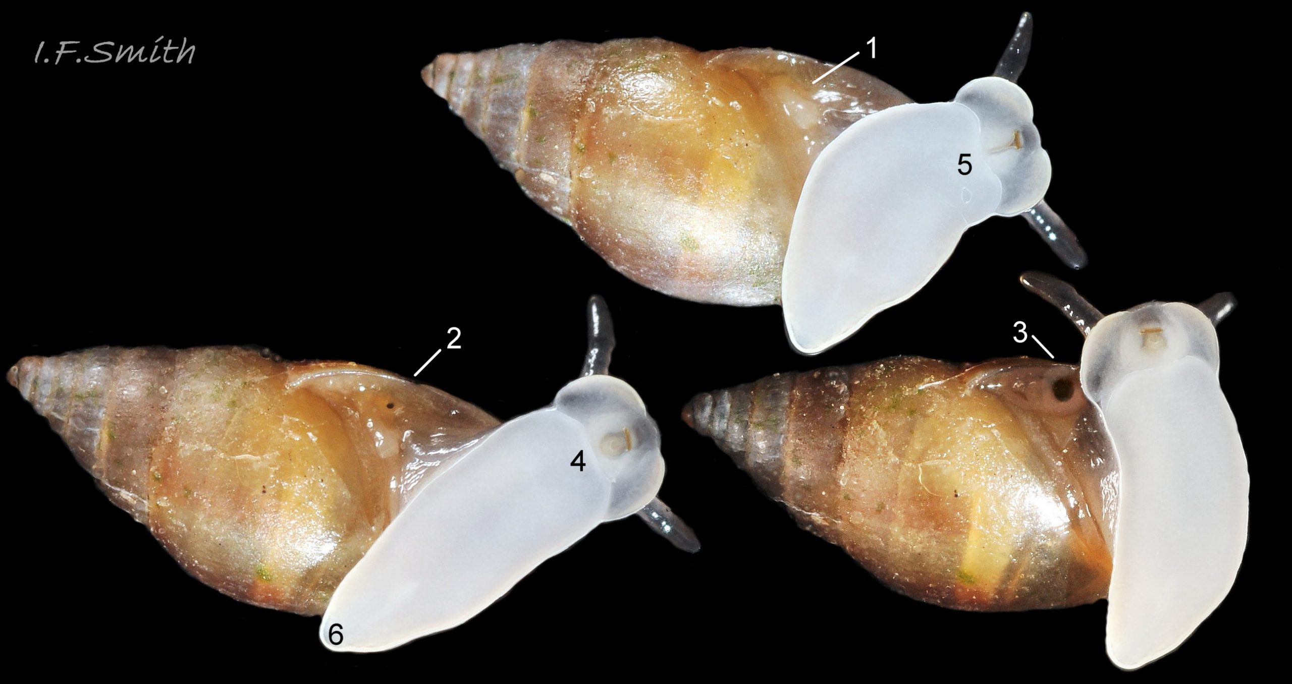
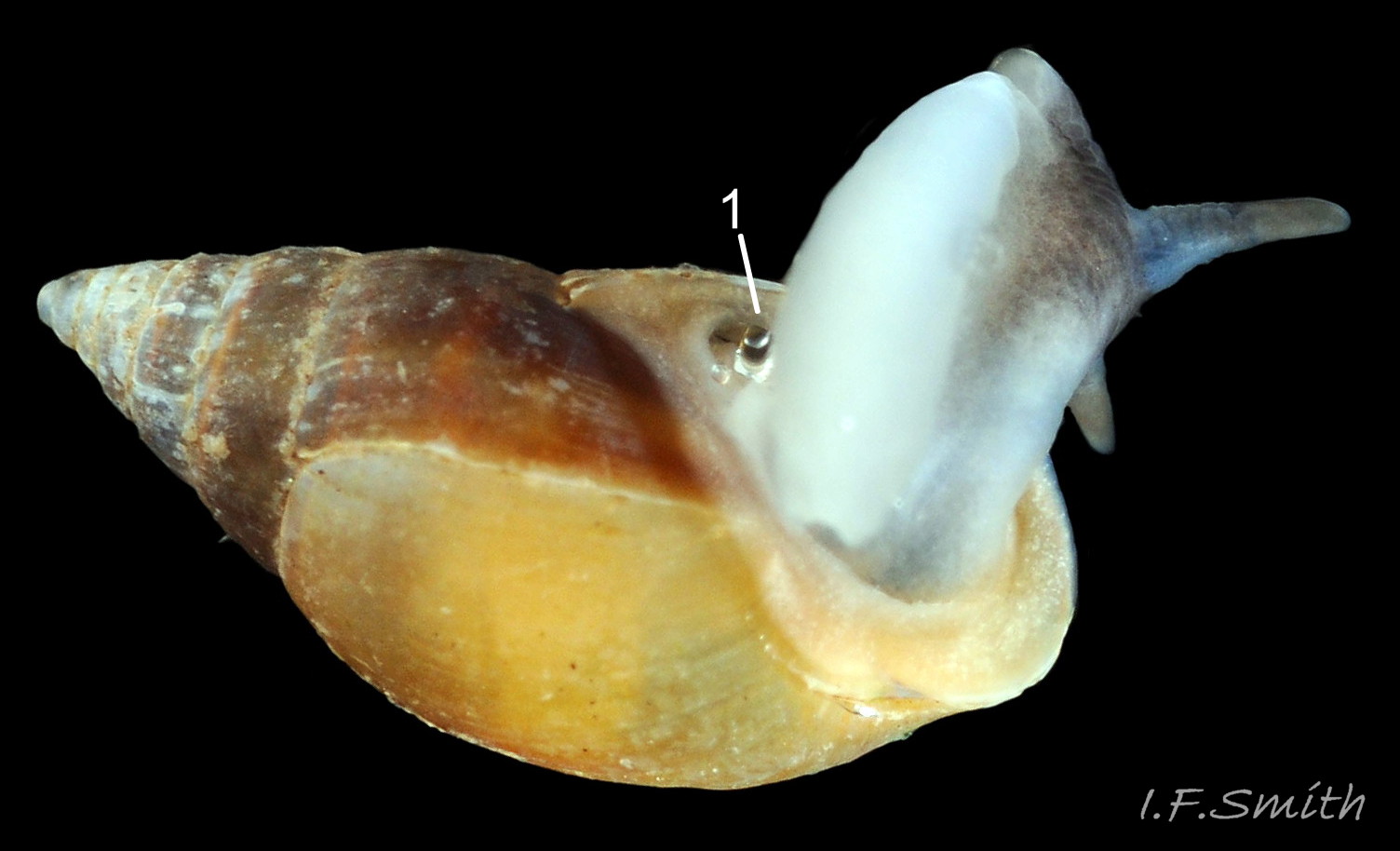
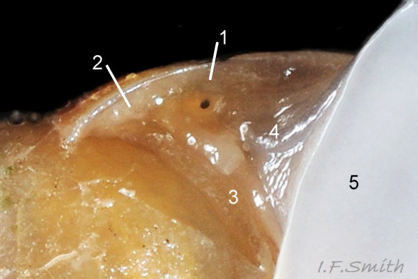
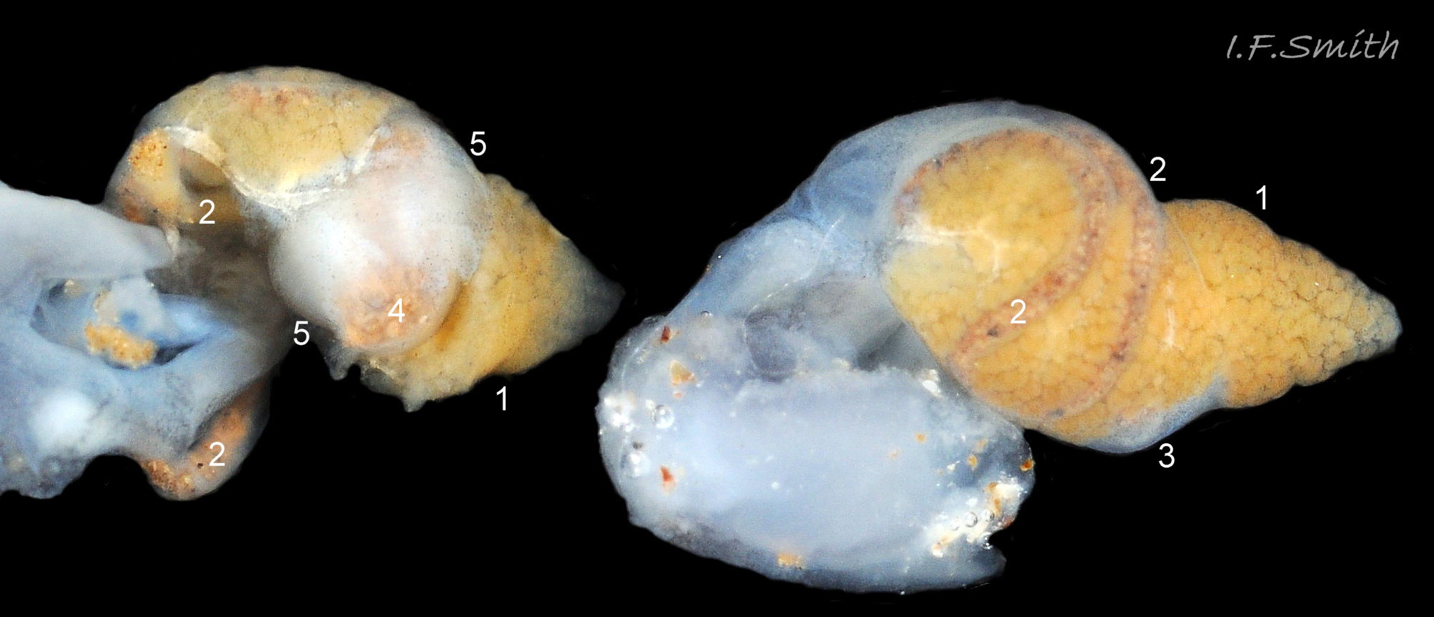
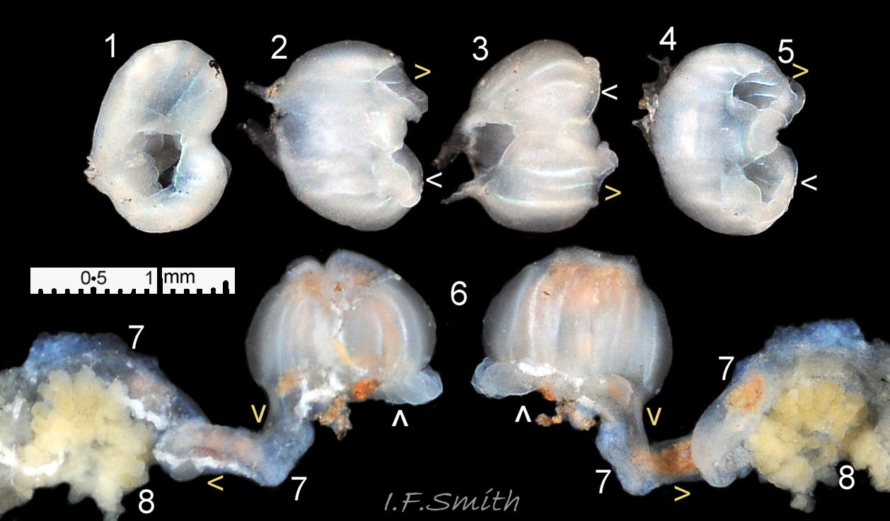
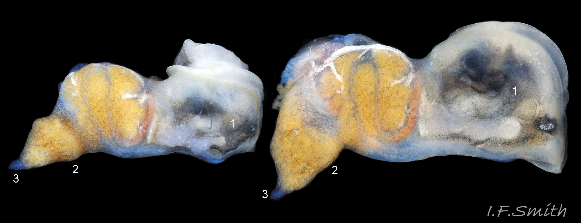
Juvenile shell usually less than 6.5mm high. Adult shell usually up to 8mm high and 3.5mm broad, exceptionally 10mm high and 5mm broad; ridge often within palatal (outer) lip 02 Myosotella myosotis. Fusiform shell, width 45% to 50% of height 03 Myosotella myosotis. Small spire with sharp apex; body whorl c. 73% of height of 7.5mm adult; 77% of 5.8mm juvenile; 80% of 4.4mm juvenile. Apex slightly twisted due to change from sinistral protoconch to dextral teleoconch. Shell-wall thin, opaque or slightly translucent, with a silky sheen when clean 02 Myosotella myosotis. Up to 8 moderately convex whorls separated by distinct shallow sutures. On juveniles, the periostracum is drawn into a row of bristles below the sutures 04 Myosotella myosotis, but they are worn off over time; a few bristles may survive on adult shells. Earliest juveniles with three or fewer whorls lack periostracum and bristles; their shells are white-translucent with punctate spiral lines which may persist for a time as the shell grows 05 Myosotella myosotis; other later whorls may have them concealed under the periostracum. Very fine, closely spaced, costal lines sometimes visible on adults, especially on spire whorls 06 Myosotella myosotis . Growth lines sometimes emphasised by change of shell colour 07 Myosotella myosotis. Usually no umbilicus except for an umbilicus-like slit in the apex caused by the change from the sinistral larval shell (protoconch) to a dextral shell 05 Myosotella myosotis. Within the shell, when it reaches 2½ whorls, the columella and septa between the spire whorls are resorbed by the mantle, leaving an open space except for the columella and septum of the body whorl 08 Myosotella myosotis & 09 Myosotella myosotis. Aperture about 50% of adult shell height, 65% of 4.6mm juvenile; shaped like a narrow ear with a rounded base and a sharp adapical angle 03 Myosotella myosotis & 10 Myosotella myosotis . Palatal (outer) lip of juveniles (under c.6mm shell-height) is thin without protrusions (folds/teeth/denticles); lip sometimes weakly reflected 04 Myosotella myosotis on adults (over c.6mm high) often with a pale calcareous ridge within the aperture near the palatal rim. The ridge sometimes contains a single raised white denticle that is often only weakly developed. The columellar/parietal lip (inner lip of aperture) has two or three protrusions . The parietal lip consists of a wide glazed area on the body whorl. For a clear view of the features within the aperture, including, sometimes, a far-back palatal ridge formed at a previous pause in growth, the animal may need a prod with a small brush to make it withdraw, and the shell requires tilting at different angles 11 Myosotella myosotis. There is no operculum 12 Myosotella myosotis. Exterior colour varies from pale yellowish brown to dark reddish brown or, sometimes, purplish brown 02 Myosotella myosotis. Sometimes the shade of brown changes at growth lines 07 Myosotella myosotis , and, frequently, the spire is darker than the body whorl 03 Myosotella myosotis. The protoconch and juvenile shell up to 1.4mm height are white 13 Myosotella myosotis, and are retained as a white apex on the adult 02 Myosotella myosotis. On adults, the pale ridge within the aperture may show as a pale band on the exterior of the slightly translucent shell 11 Myosotella myosotis. Shells on saltings are often coated with mud 14 Myosotella myosotis. On dead stranded shells the periostracum often peels off and the colour bleaches to whitish 15 Myosotella myosotis.
Body
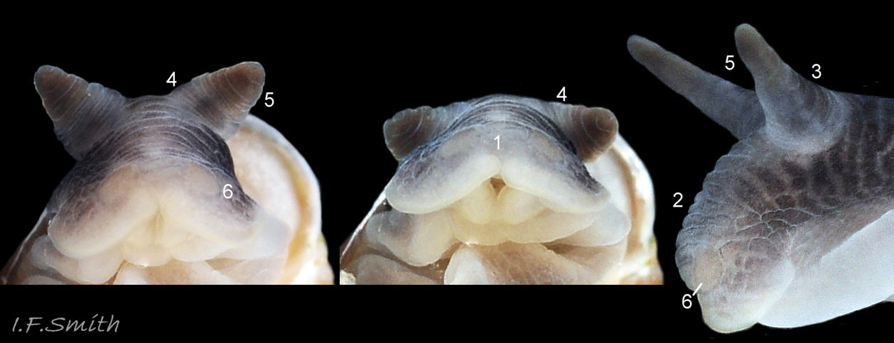
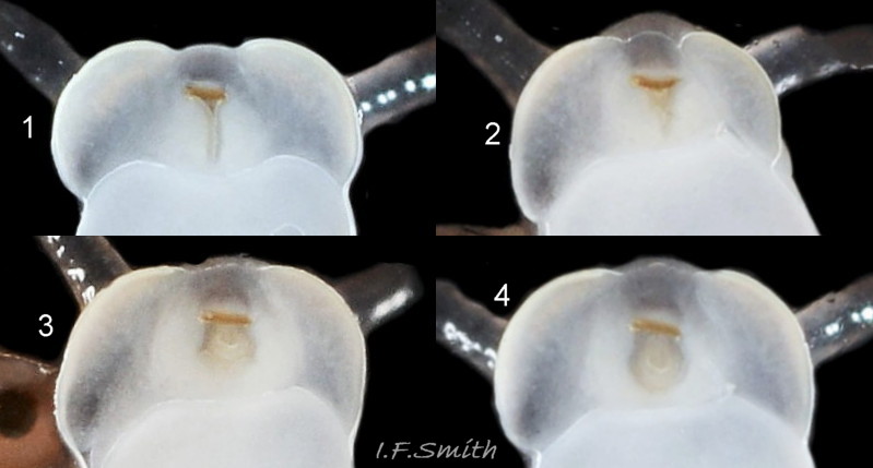
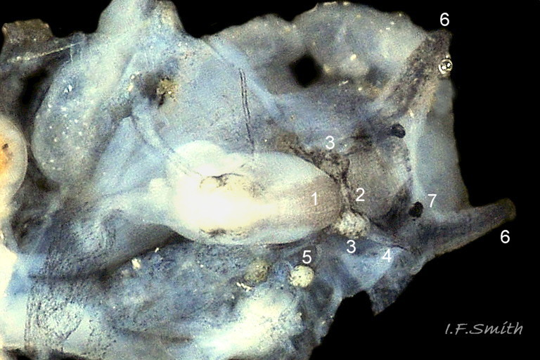
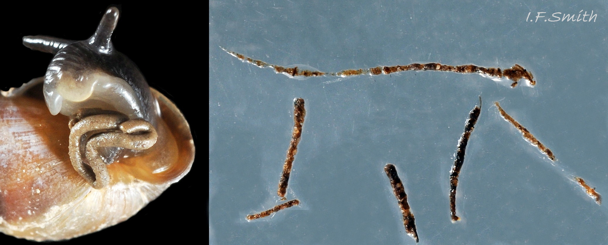
Upper parts of head and body that are exposed by normal extension are various shades of grey including whitish-grey, steel-grey and brownish-grey to nearly black 06 Myosotella myosotis & 16 Myosotella myosotis, but rarely, if ever, pure white; colour on an individual varies with degree of extension and whether in air or water, and its intensity may increase with age. The colour is arranged in transverse bands across the dorsum 07 Myosotella myosotis, and as a mosaic of tessellating blotches on the sides of the head 16 Myosotella myosotis. Sides of foot are greyish white or a paler grey than the dorsum 17 Myosotella myosotis. Body parts normally concealed by the shell have less pigment and show the white of the internal oesophagus with flanking salivary glands, reproductive organs and retractor muscles when the body is extended to its maximum 07 Myosotella myosotis & 18 Myosotella myosotis. When immersed, the body absorbs water and swells, the body colour becomes paler, and translucency of the shell and body increases 19 Myosotella myosotis. The mantle sometimes projects a short way beyond the aperture rim of the palatal lip, but is not reflected onto it 20 Myosotella myosotis. The parietal lip on the body whorl is a glaze formed by the mantle extending onto it. Within the shell, the mantle is very thin, semi-transparent and colourless apart from a faint, fine, pale-grey speckling. Where it roofs the mantle cavity 21 Myosotella myosotis, it contains many haemolymph vessels which are occasionally visible through translucent juvenile shells 22 Myosotella myosotis or on dissected mantle. The mantle cavity, which functions as a lung for respiration, is sealed off from the exterior by a thick, white or brownish-white, membranous mantle-collar which fits closely round the body as it extends or retracts 23 Myosotella myosoti & 24 Myosotella myosotis. The collar has a pneumostome which, when in air, can be opened and closed 25 Myosotella myosotis. for respiration and humidity control but, when immersed, does not effectively retain air or exclude water 26 Myosotella myosotis. The rectum and part of the intestine, visible through translucent shells in water 22 Myosotella myosotis , runs along the rear edge of the roof of the mantle cavity 21 Myosotella myosotis to the anus which opens to the exterior in a folded part of the mantle-collar 27 Myosotella myosotis in the adapical angle of the aperture close to the pneumostome. The head has two cephalic tentacles; nearly linear with a bluntly pointed tip (subulate) when dry, and conical and paler when swollen with water 16 Myosotella myosotis. When not extended, they are contracted into crumpled stumps 28 Myosotella myosotis, not retracted by inversion into the body. Both when extended or contracted, the tentacles widely diverge from their bases near the midline of the head 28 Myosotella myosotis. & 29 Myosotella myosotis. The distal half of the tentacles, sometimes slightly bulbous, is brownish and contains sensory chemoreceptor cells (Wondrak, 1984) 29 Myosotella myosotis. There is an internal black eye within the posteromesial base of each tentacle 29 Myosotella myosotis & 30 Myosotella myosotis. Distinctness of eyes varies with intensity of body pigment and amount of flesh they are viewed through. The head in front of the tentacles forms a broad, slightly bilobed “muzzle” (Forbes & Hanley,1853) which can be variably configured, but not cylindrically to form a snout like that of many marine gastropods. When not feeding, it forms a shallow curve over the mouth in anterior view, and a steep anterior in profile view 28 Myosotella myosotis. Near the anterior edge of the muzzle are two button-like, faintly-brownish grey pads (“fungiform bodies” of Wondrak, 1984) 28 Myosotella myosotis which contain sensory cells. Ventrally, the mouth is protected by white outer-lip lobes. When feeding, the ventrally translucent-white muzzle is spread out flat on the substrate and the outer lips moved aside 31 Myosotella myosotis to expose the mouth edged anteriorly by the rim of the red-brown jaw, and to allow the extension of the anterior of the radula covered in thousands of white teeth. When translucent, the muzzle may reveal dorsally the oral tube leading from the mouth to the buccal mass, and the oesophagus passing from it towards the stomach 18 Myosotella myosotis. The stomach is a large sac, partly surrounded by digestive gland. On specimens removed from the shell, the transparent lateral surface showing the stomach contents is prominently visible on the left of the visceral mass 32 Myosotella myosotis. , and the gizzard-like girdle of strong, white, folded muscle fibre surrounding it may be seen if the visceral mass is opened 33 Myosotella myosotis & 34 Myosotella myosotis. Unlike the spiral viscera of most gastropods, the viscera of M. myosotis forms a non-spiracular, approximate cone 35 Myosotella myosotis. to fill the spire after the columella and septa are resorbed 08 Myosotella myosotis & 09 Myosotella myosotis , with a small colourless tip formed in the shell’s early pre-resorption stage. On weakly pigmented, translucent specimens (most often juveniles) the dumbbell-shaped, dorsal part of the nerve ring with two cerebral ganglia may be visible 24 Myosotella myosotis. The ring encircles the oesophagus. It and its ganglia that innervate organs on the head are the nearest approximation in gastropods to a centralised brain, but other ganglia distributed on nerve cords around the body innervate other organs 36 Myosotella myosotis. The anterior edge of the translucent white sole is broad and almost straight, sometimes with a slightly indented middle, and tapers to a rounded posterior 25 Myosotella myosotis. . M. myosotis is a protandrous hermaphrodite with a penis shaped as a stout cone with the vas deferens running up the centre. The penis is normally inverted within the body, and everted for mating by hydrostatic pressure of haemolymph from an opening on the right side of the head to the rear of the tentacle 37 Myosotella myosotis.
Key identification features
Features 1 to 4, below, accord with Forbes & Hanley (1853) and Gittenberger (2004). The former aggregated M. myosotis sensu stricto with M. denticulata but “scrupulously kept apart their description.” Many currently used identification guides aggregate them and their features under M. myosotis sensu lato. Consequently, distribution maps on GBIF and NBN include many M. denticulata occurrences under “M. myosotis”, and the M. denticulata maps have under-representation of its occurrence. If making a differentiated record, ‘sensu stricto’ should be added to the record to enable it to be distinguished from lumped records.
To observe aperture sculpture the animal must be well withdrawn, and the shell tilted at different angles. Sometimes the outer (palatal) lip sculpture of an earlier growth stage is visible deep into the aperture and should be used if the sculpture on new growth has not yet developed. It is advisable to examine several specimens of different sizes from a site; sometimes both are present.
Myosotella myosotis
1. Live shell brown 02 Myosotella myosotis (beachworn shells may be dull whitish 15 Myosotella myosotis). Usual adult height 6.5mm to 8mm, exceptionally 10mm .
2. Inner (columellar/parietal) lip has only 2 or 3 apertural protrusions 03 Myosotella myosotis.
3. Outer (palatal) lip has a single apertural denticle or none 03 Myosotella myosotis. Some have a pale apertural ridge running close to the lip.
4. Flesh colour of normally extended dorsal body is grey 01 Myosotella myosotis. Shade and intensity varies with age, extension and whether in air or water, but not pure white when adult.
5. Habitat: among vegetation, often under driftwood, on low salinity estuarine saltings 38 Myosotella myosotis and Saltmarsh-grass sward by tidal rivers 39 Myosotella myosotis a little above and below EHWS. Locally abundant. (May occur with M. denticulata under stones on/near saltings 40 Myosotella myosotis.)
Similar species/ecotype
Myosotella denticulata
(full account Myosotella myosotis form denticulata)
1. Live shell brown (beachworn shells may be dull whitish). Usual adult height 3.5 mm to 7.5 mm, exceptionally 10 mm 45 Myosotella myosotis.
2. Inner (columellar/parietal) lip has 3 to 5 apertural protrusions 45 Myosotella myosotis.
3.Outer (palatal) lip has 2 to 7 (or more) apertural protrusions 45 Myosotella myosotis, sometimes set into a pale ridge which occasionally submerges them. [If no protrusions, check further back in aperture for protrusions on earlier lip position; may be visible from exterior through translucent shell , with or without connecting streaks.]
4. In its typical non-salting habitat, the flesh colour of normally extended dorsal body is white or very pale whitish grey, with darker grey tentacles 46 Myosotella myosotis. But when it occurs in muddier conditions, it may be as dark as M. myosotis 47 Myosotella myosotis.
5. Habitat: typically under slightly embedded stones at Extreme High Water Spring level and above (supralittoral) on sheltered coast without salting vegetation at fully marine salinity. Occasionally under stones on landward edge of Saltmarsh-grass sward by tidal rivers with low salinity 40 Myosotella myosotis.
Leucophytia bidentata (Montagu, 1808).
(Full account Leucophytia bidentata)
Features 1 to 4 conform with Montagu’s original description and image.
1.Live shell slightly-translucent ivory-white; yellow viscera may show through spire 48 Myosotella myosotis. Usual adult height to 5 mm, occasionally to 7 mm. Sutures shallower and whorls less rounded than on M. myosotis 49 Myosotella myosotis.
2. Inner (columellar/parietal) lip has 2 protrusions within the aperture; not more 49 Myosotella myosotis.
3. Outer (palatal) lip has no protrusions or rib (sometimes in a photo, a strong growth line might be mistaken for a rib 49 Myosotella myosotis ).
4. Flesh colour of normally extended dorsal body is amost pure white 48 Myosotella myosotis, but when contracted into body-whorl colour saturation gives it a cream appearance.
5. Lives in deep, silty, rock crevices between High Water Neap level and Low Water Spring level. Also under stones embedded into soil-like substrate at Extreme High Water Spring level and a little above on sheltered coast where it is often with M. denticulata.
Habits and ecology
M. myosotis lives in the upper littoral fringe at, and a little below, the level of EHWS tides at the base of halophyte vascular plants on estuarine saltings 41 Myosotella myosotis. and in Saltmarsh-grass sward (Puccinellia maritima) along tidal rivers 39 Myosotella myosotis. & 42 Myosotella myosotis. This zone can be extensive on large, flat, estuarine saltings 38 Myosotella myosotis or as little as a metre wide on steep river banks 39 Myosotella myosotis. At its upper limits, at or a little above EHWS, M. myosotis intermingles with some terrestrial invertebrates. It does not live in permanently submerged in pools, but can survive and be active for the short period of immersion (c. 1 hour) that occurs on 2 to 6 days per month. It lives semi-subterraneanly under driftwood 43 Myosotella myosotis or large debris, or in the groundcover matt of vegetation and debris which, for moving through, its spindle shaped shell is well adapted. When moving, the foot is cushioned on a layer of watery mucus and the shell has a thicker layer between it and the substrate 17 Myosotella myosotis. Under large pieces of wood or debris it favours the central darkest part while Assiminea grayana, if present, is found near the periphery. After immersion or during rain, it may be active in the open when its tentacles wave in the air to detect odours and, in the absence of anterior tentacles found on most pulmonates, its oral tube tests wet substances on the substrate (Wondrak, 1984). The two button-like, faintly-brownish grey pads (“fungiform bodies” of Wondrak, 1984) 28 Myosotella myosotis. near the anterior edge of the muzzle are also sensory.
It is a euryhaline species capable of surviving immersion in water from 0 p.p.t to full marine salinity or more, but individuals require time to adapt to changes in salinity and may become inactive/moribund when abruptly immersed in water they are unaccustomed to.
Respiration is of atmospheric air in the mantle cavity which is sealed by a white collar of thickened mantle 23 Myosotella myosotis that firmly embraces the body but allows it to extend-from/retract-into the shell 24 Myosotella myosotis. A pneumostome (respiratory pore) in the collar 25 Myosotella myosotis can be opened for inhalation/exhalation of air or closed to seal the cavity against dehydration.The roof of the mantle cavity contains a network of haemolymph vessels 21 Myosotella myosotis. and is very thin, enabling oxygen from inhaled air to diffuse into the vessels and for carbon dioxide to leave with the exhaled air. When immersed, air escapes from the mantle cavity 26 Myosotella myosotis. and water enters 24 Myosotella myosotis. M. myosotis can survive immersion for at least three days with no access to atmospheric air. Long submersion with water in the mantle cavity is well tolerated if the water’s oxygen content is high 22 Myosotella myosotis. (Seelmann, 1968, in Gittenberger, 2004).
When feeding, the muzzle is spread out on the substrate and the radula is extended 31 Myosotella myosotis to gather, with the red jaw as a backstop, decaying vegetation, diatoms (Wiese & Richling, 2008) and sediment rich in organic material which are bound into food boli with mucus from the supra pedal gland brought to the mouth along a median groove. The boli travel along the oesophagus 36 Myosotella myosotis. to the stomach 32 Myosotella myosotis & 34 Myosotella myosotis where strong muscular contractions of the internally-folded gizzard triturate them and squeeze out semi-fluid nutrient which passes into the diverticula of the digestive gland for digestion. The residual mass is squeezed as faecal boli into the intestine by muscular contractions 34 Myosotella myosotis and passes to and through the rectum 21 Myosotella myosotis . Unlike marine prosobranch gastropods, which defecate into a mantle cavity that is cleared by water currents, M. myosotis has a rectum that opens to the exterior through an anus in the mantle collar, near to, but separate from, the pneumostome 27 Myosotella myosotis so that faeces are expelled without fouling the respiratory mantle-cavity. The soft faeces, wet and loosely bound with mucus when fresh, dry to thin granular threads consisting mostly of fine mineral particles 44 Myosotella myosotis faeces.
Reproduction: in NW Germany, copulation is in all months with peaks in April/May and August/September (Schultes,2014). Ova are laid when the temperature is above 15°C from late spring to late summer (Gittenberger, 2004). It is a protandrous hermaphrodite which changes its sexual function in the wild when 1½ to 2 years old, so younger, 1 to 1½ years, fully mature males mate with older, over 1½ years, females (Schultes, 2014) using the stout, conical penis everted from the side near the posterior of the right tentacle. Sometimes, a chain of three individuals mate, with the middle one acting as both male and female. Under optimal laboratory conditions, newly hatched animals can reach a shell length of 5 mm and start laying eggs in 8 weeks (Wiese & Richling, 2008). Each month of the breeding season, a female deposits 15 to 80 egg capsules (Schultes, 2014) in a small, yellow or white, frog-spawn-like mass (Morton, 1954 and Gittenberger, 2004). Each ovoid capsule contains a single ovum. The cases are attached to each other in a loosely convoluted chain by a filament (chalaziform process) at each end. The closely packed cases with intervening clear fluid are contained in a tough binding membrane which is attached to stones, plant stems, wood etc in moist situations (Morton, 1954) or in soil cavities in clusters from several females (Schultes, 2014).There is a larval veliger stage, with sinistral shell, which is passed entirely within the ovum (Morton, 1954). At 10 °C to 20 °C and 18 p.p.t. salinity, crawling juveniles emerge after about two and a half weeks. At less favourable salinities, less than 18 p.p.t or more than 54p.p.t., development takes several weeks longer (Gittenberger, 2004), though the eggs are moist but not immersed for most of the time. Juveniles in northern Germany hatch after 3-7 weeks, and 12-15 days in France (Schultes, 2014). Individuals can live to 3 or 4 years of age (Wiese & Richling, 2008).
Distribution and status
Europe from Orkney, Scotland and southern Denmark to Mediterranean and Azores; majority of records are from Britain and Ireland. Locally abundant in suitable habitat in Britain. Occurs in German Baltic saltmarshes, but habitat threatened in Germany (Wiese & Richling, 2008). Assumed to have been introduced to temperate coasts of Australia and North America (Atlantic and Pacific) GBIF map www.gbif.org/species/2297460 , but comparison of soft part morphology suggests that many different species have been aggregated because of similar shell morphology (Martins, 2013).
Widespread around Britain but non-estuarine records are likely to be the species/ecotype M. denticulata NBN map
species.nbnatlas.org/species/NHMSYS0001702112#tab_mapView
Irish distribution, National biodiversity data centre, in Mollusc Ireland: www.habitas.org.uk/molluscireland/species.asp?ID=121
Acknowledgements
I gratefully thank Ben Rowson of the National Museum of Wales/ Amgueddfa Cymru for his help with the account, but any errors or omissions are mine.
Links and references
Anderson, R. MolluscIreland, accessed January 2019. www.habitas.org.uk/molluscireland/species.asp?ID=121
Forbes, E. & Hanley S. 1849-53. A history of the British mollusca and their shells. vol. 4 (1853), 190 – 197 & plate CXXV. London, van Voorst. (AsConovulus denticulatus var. myosotis.)
Free pdf at archive.org/details/historyofbritish04forbe/page/190
plate at archive.org/details/historyofbritish04forbe/page/n565
Fretter, V. & Peake, J. 1975. Pulmonates functional anatomy and physiology. Vol.1. London. Academic Press.
Gittenberger, E. et al. 2004. De Nederlandse zoetwatermollusken. Leiden, Netherlands, Nationaal Natuurhistorisch Museum Naturalis.
Heller J. 2015. Marine Ancestors of most Land Snails: Pulmonates. In: Sea Snails. Springer, Cham. link.springer.com/chapter/10.1007%2F978-3-319-15452-7_10
Jeffreys, J.G. 1862-69. British conchology. vol. 5 (1869). London, van Voorst. (As Melampus myosotis (including var. ringens = Myosotella denticulata); Free pdf at archive.org/stream/britishconcholog05jeffr#page/106/mode/2up . Use slide at base of page to select pp.106-109.)
Martins, A.M. de F. 1996. Anatomy and systematics of the western Atlantic Ellobidae (Gastropoda: Pulmonata). Malacologia 37(2): 163 – 332.
www.biodiversitylibrary.org/page/13113594#page/179/mode/1up
Martins, A.M. de F. & Mendes, A.R.M. 2013. Do cosmopolitans speciate? Anatomical diversity of Myosotella in Azores. Centro de Investigação em Biodiversidade e Recursos Genéticos. Ponta Delgada, Açores, Portugal. Poster for World Congress of Malacology 2013 in pdf: www.researchgate.net/publication/264339925_Do_cosmopolita… .
Montagu, G. 1808. Supplement to: 1803 Testacea Britannica, or, Natural history of British shells, marine, land, and fresh-water, including the most minute : systematically arranged and embellished with figures. London, J. White.
Description of Leucophtia bidentata as Voluta bidentata pp. 100-101.
www.biodiversitylibrary.org/page/24430722#page/806/mode/1up
Plate 30, :
www.biodiversitylibrary.org/page/24430722#page/917/mode/1up
Morton, J. E. 1955. The functional morphology of the British Ellobiidae (Gastropoda Pulmonata) with special reference to the digestive and reproductive systems. Phil. Trans. R. Soc. Ser. B .
239, No. 661: 89-160 www.jstor.org/stable/92507
Schultes, F.W. 2014. Species summary for Ovatella myosotis (Draparnoud, 1801). AnimalBase. SUB Göttingen. www.animalbase.uni-goettingen.de/zooweb/servlet/AnimalBas… Accessed January 2019.
Watson, H. I943. Notes on a list of the British non-marine Mollusca. J. Conch. 22: 13 – 22.
Wiese, V. & Richling, I. 2008. Das Mäuseöhrchen Myosotella myosotis (Draparnaud 1801). Arbeitskreis Mollusken NRW.
www.mollusken-nrw.de/weichtier_des_jahres/weichtier2008.htm
Wondrak, G. 1984. Ultrastructure of the sensory epithelia of oral tube, fungiform sensory bodies, and terminal knobs of tentacles of Ovatella
myosotis. Draparnaud (Archaeopulmonata, Gastropoda) J. Morphol. 181: 333-347 .
onlinelibrary.wiley.com/doi/pdf/10.1002/jmor.1051810307
Current taxonomy:
www.marinespecies.org/aphia.php?p=taxdetails&id=139673
Glossary
adapical angle = angle at which outer lip meets body-whorl.
boli = (sing. bolus) small rounded masses, especially of triturated food material.
cerebral = to do with integration of sensory and neural functions to initiate and coordinate body activity.
chalaziform = resembling the two spiral bands (chalazae) in a bird’s egg that attach the yolk to opposite ends of the lining membrane.
columella = solid or hollow axial “little column” around which gastropod shell spirals; hidden inside shell, except on final whorl next to lower part of inner lip of aperture where hollow ones may end in an umbilicus or siphonal canal.
columellar = (adj.) of or near central axis of coiled gastropod.
columellar lip = lower (abapical) part of inner lip of aperture.
costa (pl. costae) = rib running across a whorl of a gastropod shell at approximately right-angles to direction of coiling and any spiral striae.
costal (adj.) = of, or arranged like, costae.
dextral = (of gastropod shell) in apertural view with spire uppermost, the aperture is on the right. Most gastropod species adults have dextral shells.
distal = away from centre of body or from point of attachment.
diverticula = (for digestion) blind ended tubules in the digestive gland that receive nutrients for digestion.
EHWS = extreme high water spring tide.
euryhaline = able to tolerate a wide variation in salinty.
fusiform = slender, spindle-shaped, tapering almost equally towards both ends.
ganglia = (sing. ganglion) knots on a nerve cord containing sensory cell bodies that conduct impulses to (innervate) organs of the body.
haemolymph = circulating fluid in molluscs that carries nutrients, waste and hormones. Analagous to vertebrate blood, but most molluscs have copper-based haemocyanin in it instead of red haemoglobin to carry oxygen. It may be tinged blue when oxygenated; colourless when depleted of oxygen.
halophyte = plant tolerant of saline soil and periodic tidal immersion, usually on saltmarshes, estuarine shores and sides of tidal rivers.
mantle = sheet of tissue that secretes the shell, covers the viscera and forms a cavity in gastropods. In terrestrial gastropods (‘pulmonates’) the cavity roof contains a network of haemolymph (‘blood’) vessels enabling the cavity to act like a lung.
mesial = on or facing towards the midline of the body.
operculum = plate of horny conchiolin, rarely calcareous, used to close shell aperture of prosobranch gastropods.
palatal lip = outer lip of gastropod aperture.
parietal lip ( or parietal wall) = upper part of inner side of gastropod aperture, often lacking clear lip structure with just a glaze on side of whorl adapically of columellar lip.
periostracum = thin horny layer of proteinaceous material often coating shells.
posteromesial = at the rear facing towards the midline of the body.
prosobranch = member of Prosobranchia, one of three subclasses into which the class Gastropoda (slugs and snails) was divided during the 20th Century (other two were Pulmonata and Opisthobranchia). This classification is no longer used by scientists, but prosobranch is a useful informal term to signify (mainly marine) snails breathing with a ctenidium (comblike gill inside mantle cavity), an operculum, and a shell which can accommodate the whole body.
protandrous hermaphrodite = each individual starts mature life as a functioning male, later changing to female function.
protoconch = apical whorls produced during embryonic and larval stages of gastropod; often different in form from other whorls (teleoconch).
protrusions = teeth, denticles, folds, lamellae or cogs (terms used by various authors).
punctate = with pinprick-like depressions.
resorb = absorb what was previously secreted; break it down into component materials and disperse into the circulation.
resorption = the process of absorbing what was previously secreted by breaking it down into component materials and dispersal into the circulation.
salting = area of salt tolerant vascular plants rooted in sediment between mean high water mark (MHW) and extreme high water of spring tides (EHWS). [Preferred synonym for “saltmarsh” as much of salting not marshy.]
septa = plural of septum; internal partition separating two chambers/ shell-whorls of a gastropod.
septum = internal partition separating two chambers/ shell-whorls of a gastropod.
sinistral = (of gastropod shell) in apertural view with spire uppermost, the aperture is on the left. Most gastropod species adults have dextral shells.
subsutural = close below the suture when shell positioned with apex uppermost.
subulate = slender and tapering to a point like onion leaf or awl.
suture = groove or line where whorls of gastropod shell adjoin.
teleoconch = entire gastropod shell other than the apical, embryonic & larval stage protoconch.
triturate = reduce to small particles.
vascular plants = plants that have vascular tissues to transport water and nutrients through the plant. Include all seed-bearing plants, ferns and horsetails. Usually terrestrial or in freshwater or brackish water; a few, such as Zostera, live in fully marine salinity water.