Click image to enlarge with full caption. Main text below slider.
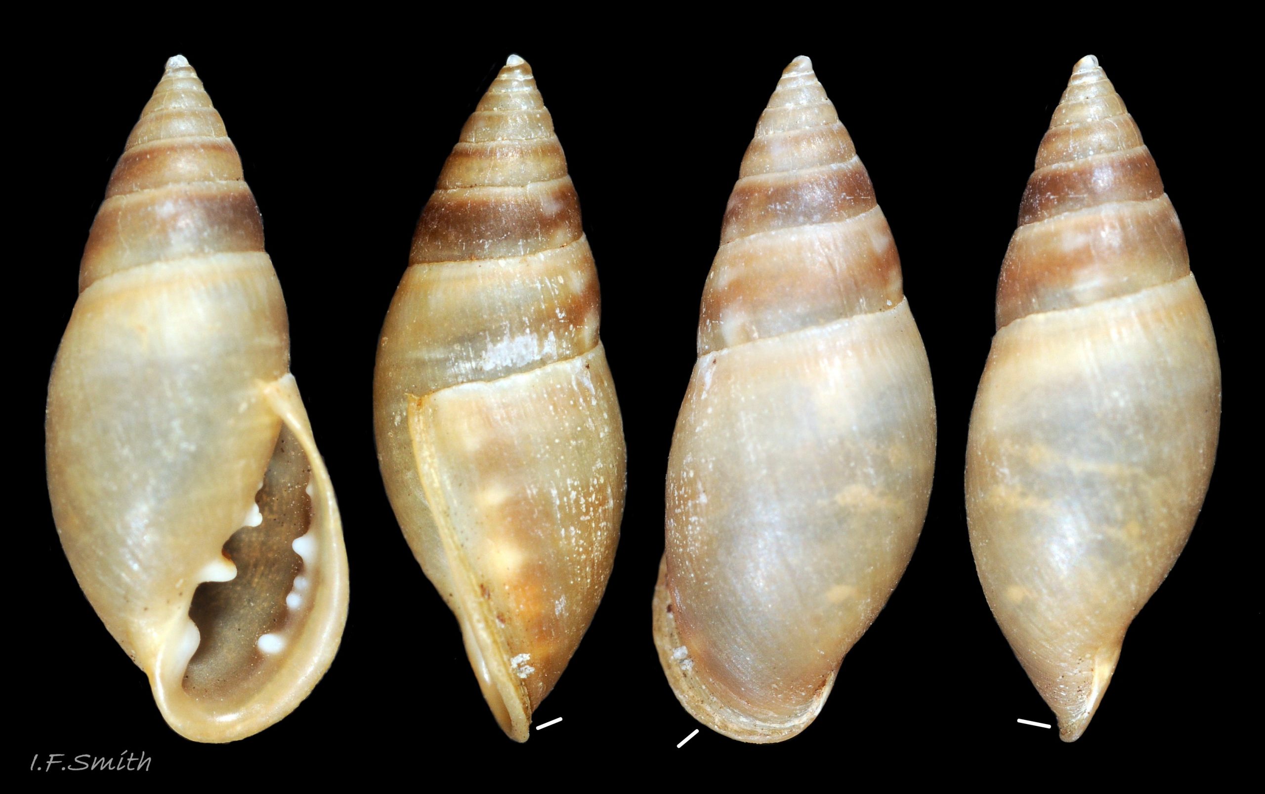
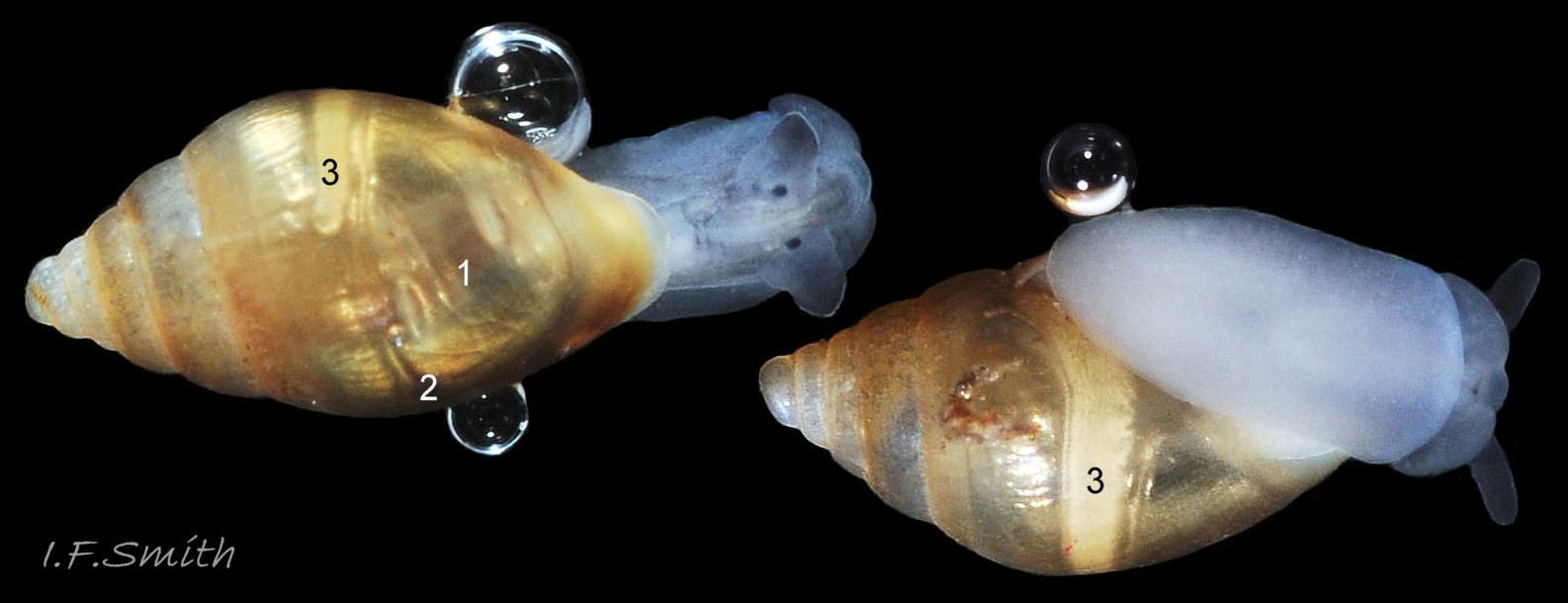
01 Myosotella myosotis form denticulata.
02 Myosotella myosotis form denticulata.
03 Myosotella myosotis form denticulata.
04 Myosotella myosotis form denticulata.
05 Myosotella myosotis form denticulata.
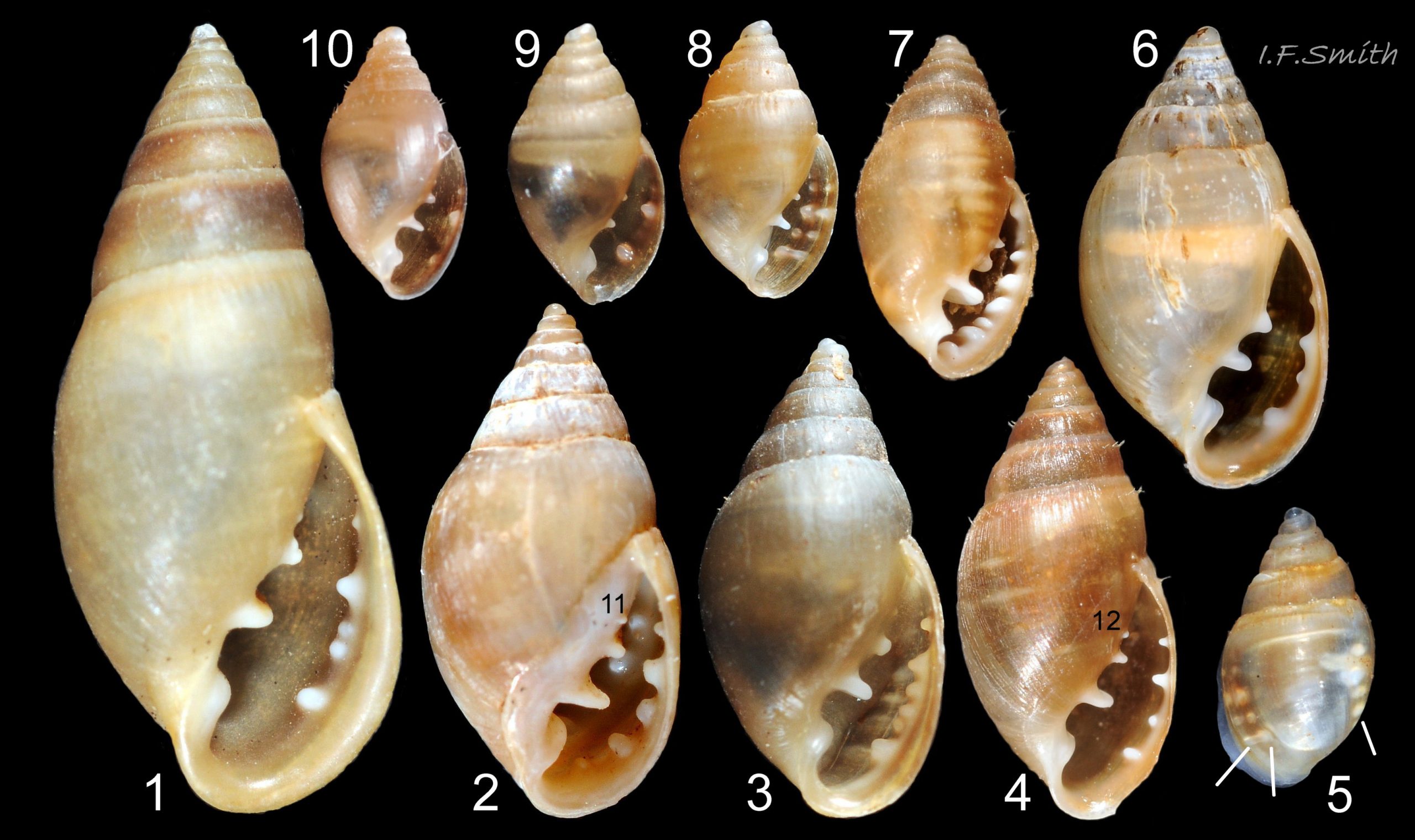
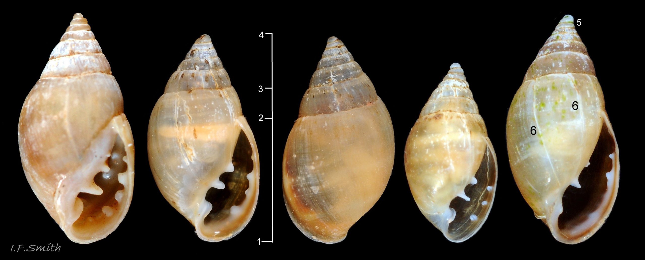
06 Myosotella myosotis form denticulata.
07 Myosotella myosotis form denticulata.
08 Myosotella myosotis form denticulata.
09 Myosotella myosotis form denticulata.
10 Myosotella myosotis form denticulata.
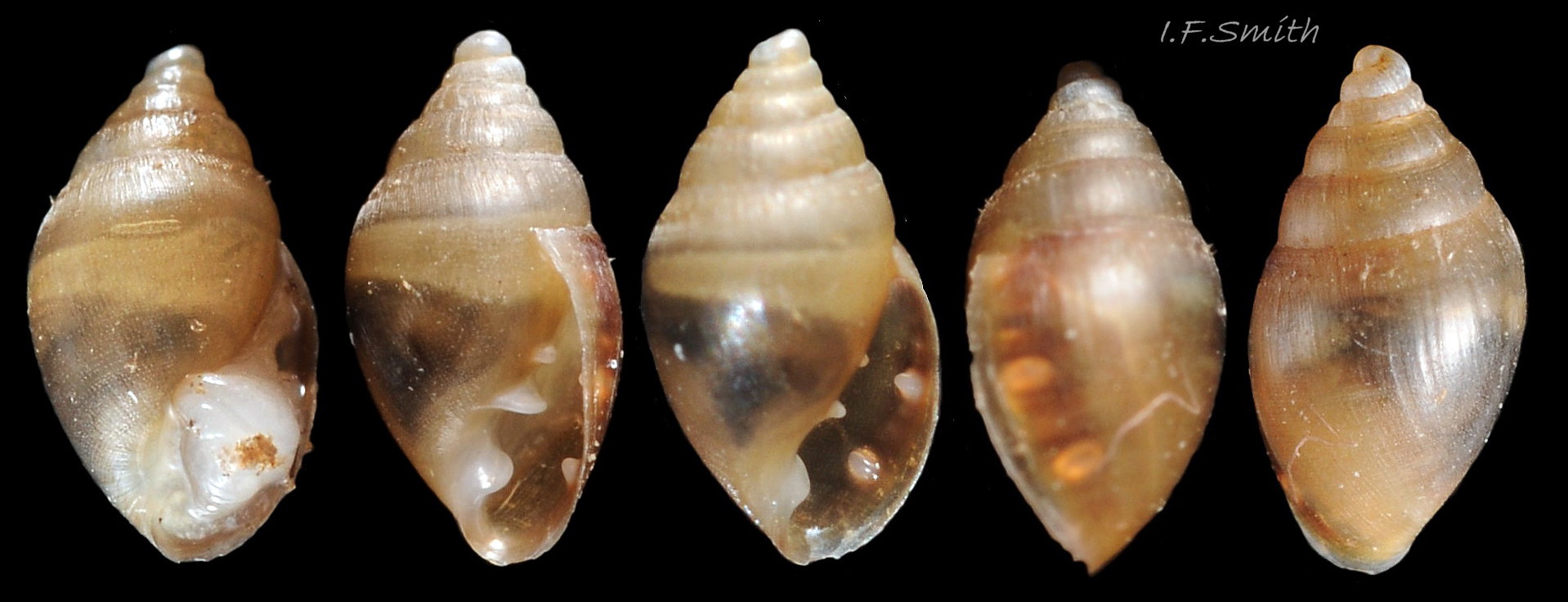
11 Myosotella myosotis form denticulata.
12 Myosotella myosotis form denticulata.
13 Myosotella myosotis form denticulata.
14 Myosotella myosotis form denticulata.
15 Myosotella myosotis form denticulata.
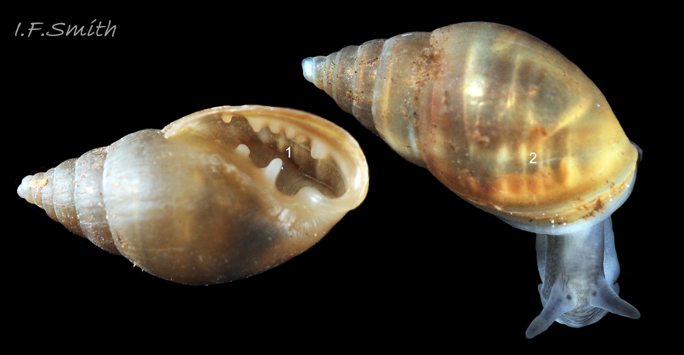
16 Myosotella myosotis form denticulata.
17 Myosotella myosotis form denticulata.
18 Myosotella myosotis form denticulata.

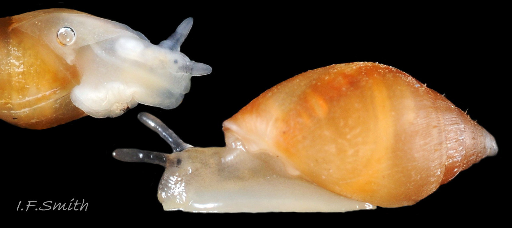
19 Myosotella myosotis form denticulata.
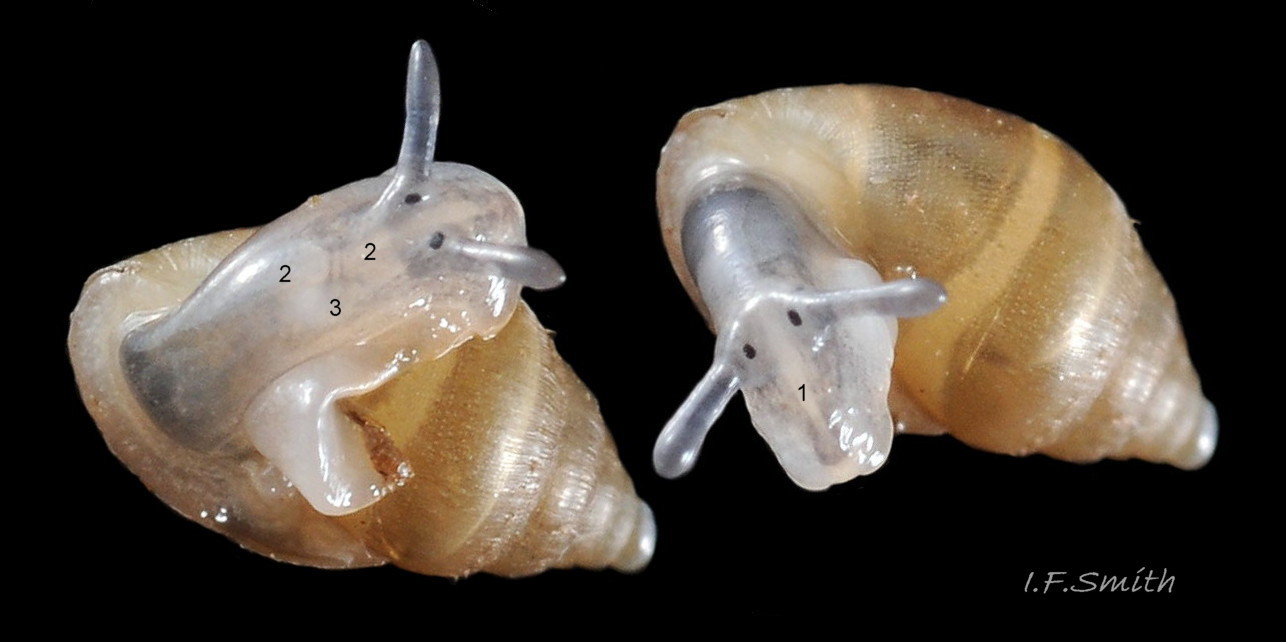
20 Myosotella myosotis form denticulata.
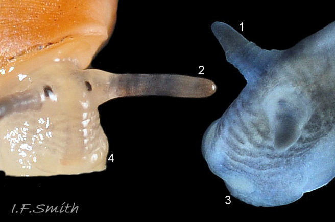
21 Myosotella myosotis form denticulata.
22 Myosotella myosotis form denticulata.
23 Myosotella myosotis form denticulata.
24 Myosotella myosotis form denticulata.
25 Myosotella myosotis form denticulata.
26 Myosotella myosotis form denticulata.
27 Myosotella myosotis form denticulata.
28 Myosotella myosotis form denticulata COMPARE Myosotella myosotis.
29 Myosotella myosotis form denticulata COMPARE Myosotella myosotis.
30 Myosotella myosotis form denticulata COMPARE Myosotella myosotis.
31 Myosotella myosotis form denticulata COMPARE Myosotella myosotis.
32 Myosotella myosotis form denticulata COMPARE Myosotella myosotis.
33 Myosotella denticulata. COMPARE Leucophytia bidentata.
Myosotella myosotis form denticulata (Montagu, 1803)
Preface
Specimens illustrated in this account which were supplied to Amgueddfa Cymru (the Natural History Museum, Wales) were sequenced by Ben Rowson who found no difference in the DNA of M. myosotis and M. denticulata and concluded that they were a single species; Myosotella myosotis (Draparnaud, 1801). This has been accepted by WoRMS; see www.marinespecies.org/aphia.php?p=taxdetails&id=139672 ]
A possibility, raised by Martins (2013), is that the true M. myosotis (Draparnaud, 1801) occurs in the Mediterranean and that both British shell forms are ecotypes of M. denticulata (Montagu, 1803). This account, written before molecular sequencing united them, describes the form previously regarded as M. denticulata.
Because of its special habitat intermediate between terrestrial and marine, this species, and its Leucophytia relative in the family Ellobiidae, are omitted from some identification guides, while variously appearing in others devoted solely to either terrestrial, marine or even freshwater mollusca.
Synonyms: Voluta denticulata Montagu, 1803; Voluta ringens W. Turton, 1819; Ovatella denticulata (Montagu, 1803); Alexia ringicula Locard, 1893; Conovulus denticulatus in Forbes & Hanley (1853); Melampus myosotis (part of) in Jeffreys (1869);
Vernacular Probably applied at times to both M. denticulata and M. myosotis: Mouse-eared Alexia, Mouse ear(ed) snail (English); Clust llygoden (Welsh); Evesnegl (Danish); Muizenoortje (Dutch); Ovatelle naine des vases (French); Stranddvärgsnäcka (Swedish); Mäuseöhrchen (German);
Applied to just this form: Many-toothed mouse-ear (English); Gewoon muizenoortje (Dutch);
Description
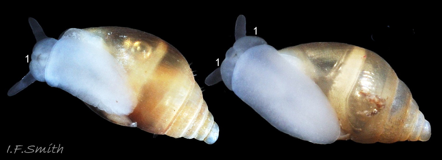
When in water, shell is more translucent, lighter and brighter in colour, and less reflective, than when in air 01 Myosotella myosotis form denticulata. . The following shell description is of specimens in air.
Shell
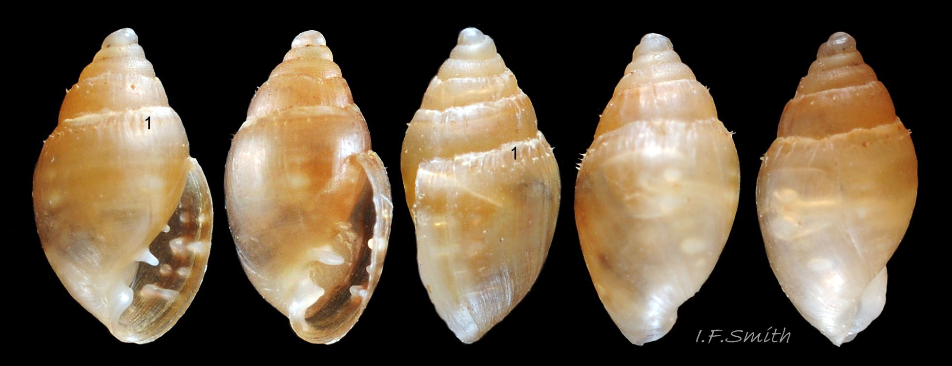
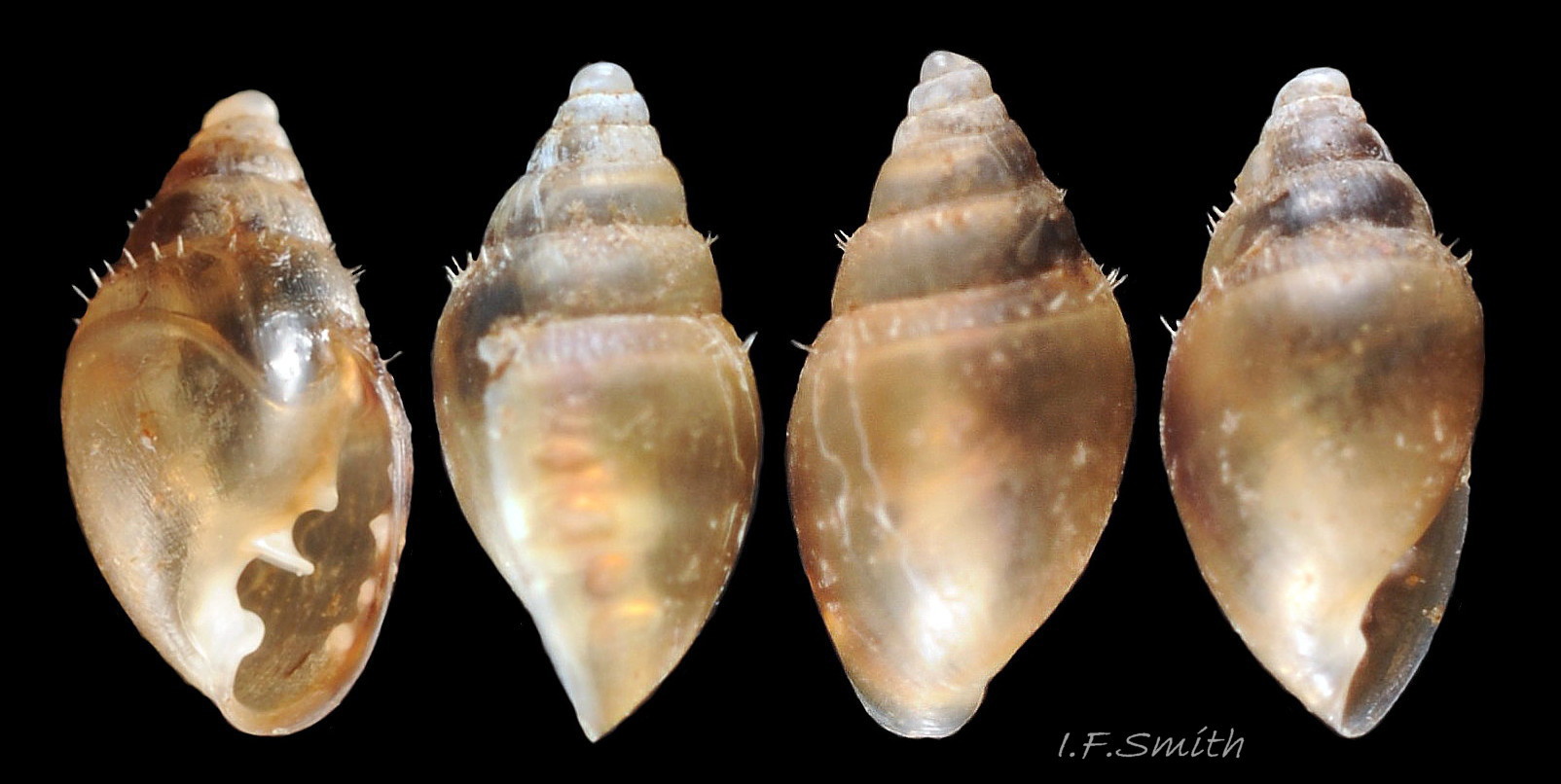
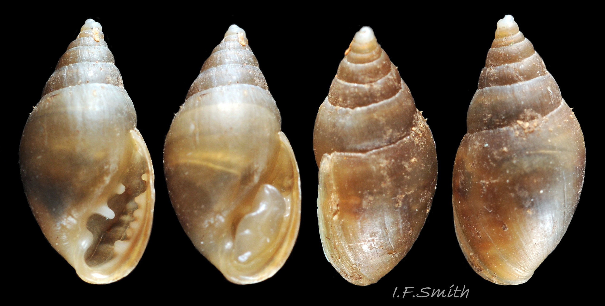
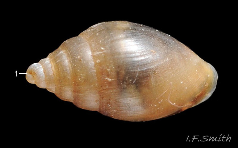
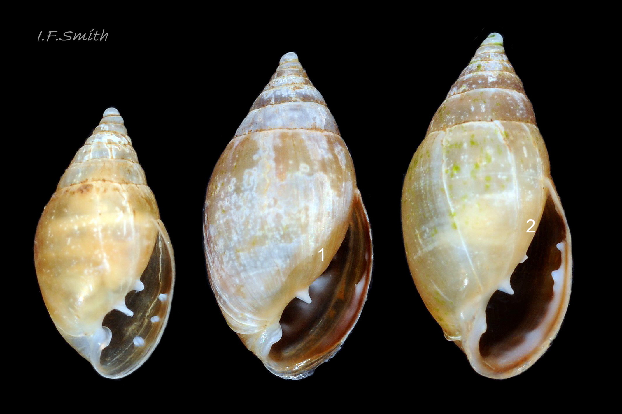
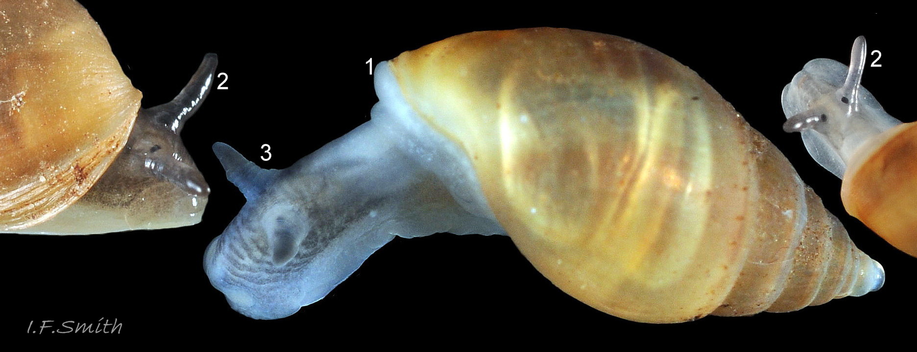
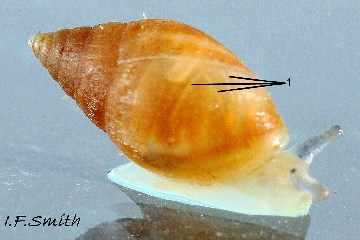
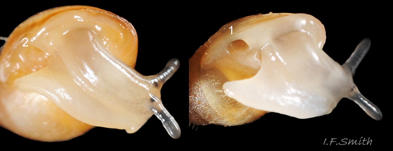
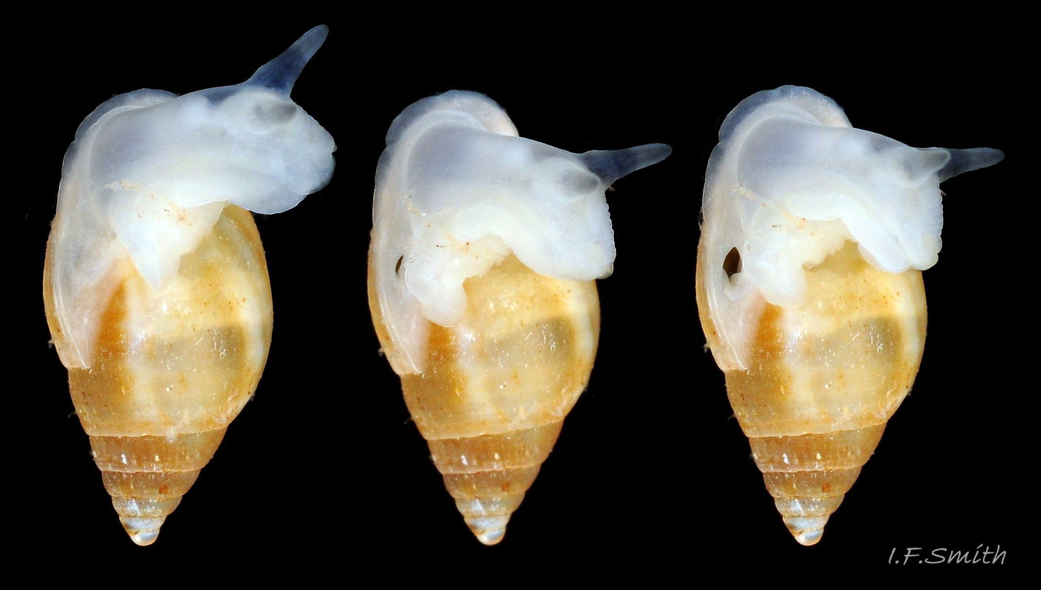
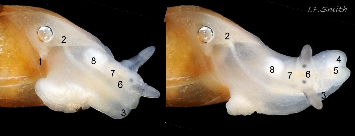
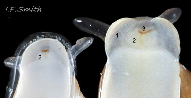
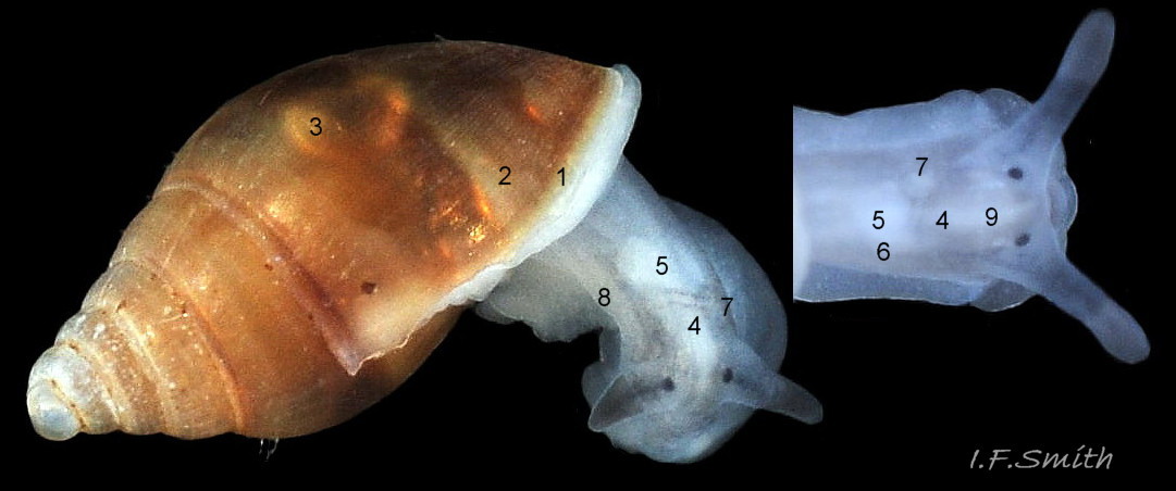
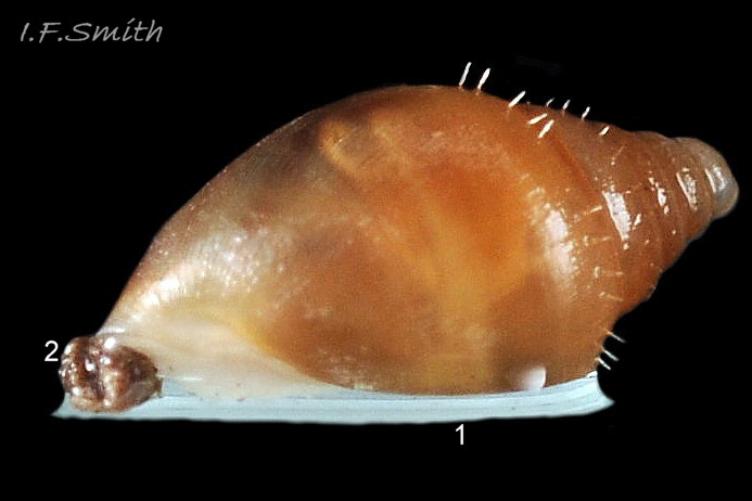
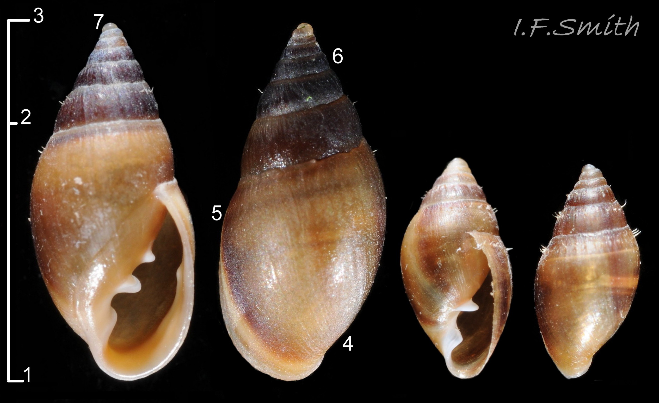
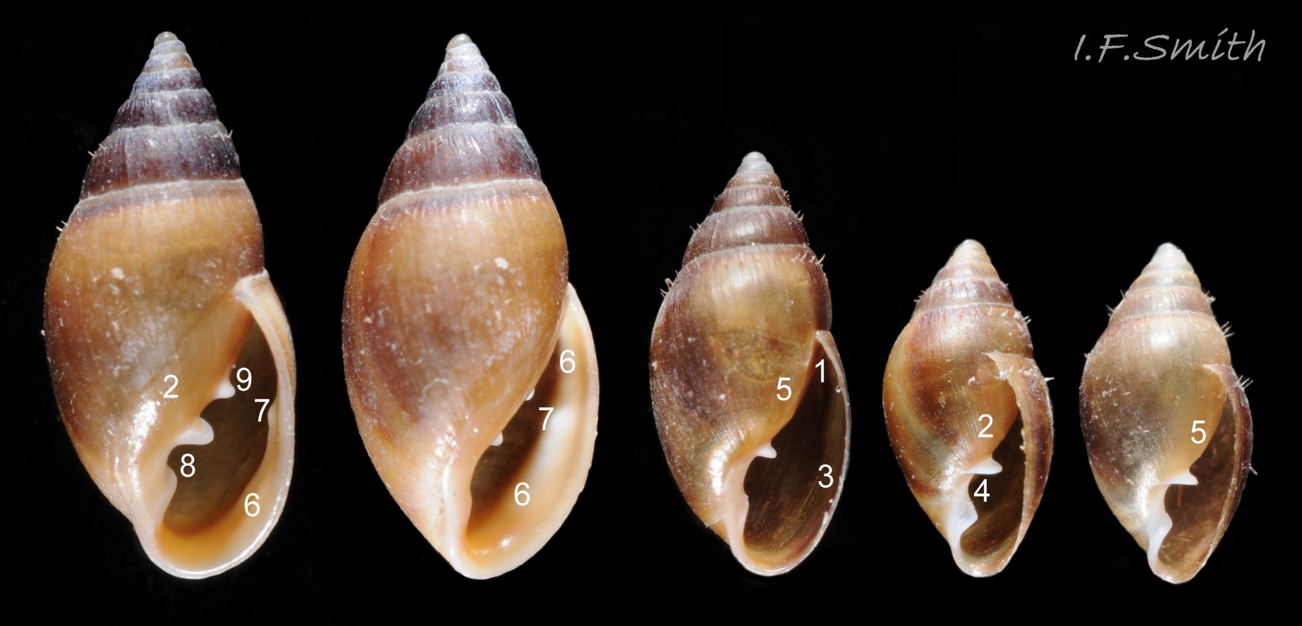
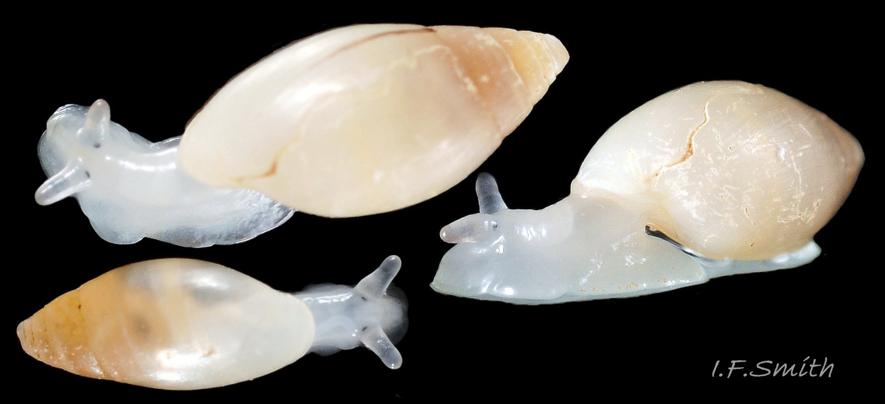
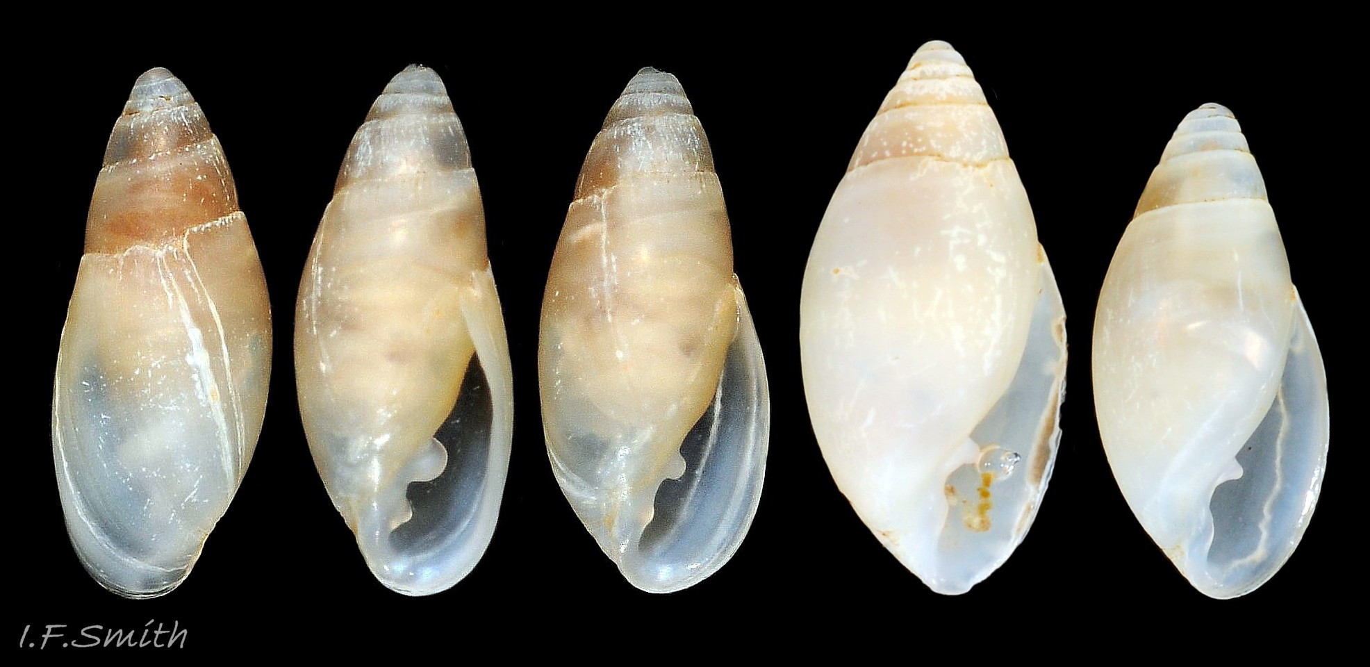
Juvenile shell usually less than 6 mm high. Adult shells often less than 6mm , usual maximum 7.5 mm, exceptionally 10 mm 02 Myosotella myosotis form denticulata. . Fusiform shell, width c.45% to 55% of height 03 Myosotella myosotis form denticulata. . Small spire with sharp apex; body whorl c. 73% of shell height, usually a little less on small specimens. Apex slightly twisted 03 Myosotella myosotis form denticulata. due to change from sinistral protoconch to dextral teleoconch. Shell-wall thin, opaque or slightly translucent, with a silky sheen when clean 04 Myosotella myosotis form denticulata. . Up to 8 moderately convex whorls separated by distinct shallow sutures. On juveniles, the periostracum is drawn into a row of bristles below the sutures 05 Myosotella myosotis form denticulata. , but they are worn off over time. Earliest juveniles with three or fewer whorls lack periostracum and bristles; their shells are white-translucent with punctate spiral lines which may persist for a time as the shell grows ; occasionally visible through periostracum on later whorls 06 Myosotella myosotis form denticulata. . Very fine, closely spaced, costal lines sometimes visible on adults, especially on spire whorls 06 Myosotella myosotis form denticulata. , often most clearly developed on subsutural ramp 04 Myosotella myosotis form denticulata. . Adults have growth lines; most easily seen when periostracum is worn 03 Myosotella myosotis form denticulata. , less so when periostracum is intact 07 Myosotella myosotis form denticulata. . Usually no umbilicus except for an umbilicus-like slit in the apex caused by the change from the sinistral larval shell (protoconch) to a dextral shell 08 Myosotella myosotis form denticulata. . Within the shell, when it reaches 2½ whorls, the columella and septa between the spire whorls are resorbed by the mantle, leaving an open space except for the columella and septum of the body whorl. Aperture 50% to 60% of shell height 03 Myosotella myosotis form denticulata. , juveniles usually nearer the higher limit; shaped like a narrow ear with a rounded base and a sharp adapical angle 02 Myosotella myosotis form denticulata. . Thin palatal (outer) lip on specimens over 3mm high has two to seven (or more) protrusions (folds/teeth/denticles) 02 Myosotella myosotis form denticulata. which may be set into a pale calcareous ridge within the aperture near the palatal rim. Further sets of protrusions are often present further back in the aperture, marking previous positions of palatal lip 02 Myosotella myosotis form denticulata. . The palatal lip is sometimes weakly reflected on large adults 09 Myosotella myosotis form denticulata. . The columellar-parietal lip (inner lip of aperture) has three or four protrusions . The parietal lip consists of a wide glazed area on the body whorl, but is often difficult to discern 02 Myosotella myosotis form denticulata. & 10 Myosotella myosotis form denticulata. . Juveniles less than 3mm high may not have developed protrusions sufficiently to be distinguished from M. myosotis. For a clear view of the features within the aperture, including far-back rows of teeth, the animal may need a prod with a small brush to make it withdraw, and the shell requires tilting at different angles 11 Myosotella myosotis form denticulata. . There is no operculum . Exterior colour varies from yellowish brown to brown 02 Myosotella myosotis form denticulata. . The protoconch and juvenile shell up to about 1.4mm height are white, and are retained as a white apex on the adult 03 Myosotella myosotis form denticulata. & 14 Myosotella myosotis form denticulata. . On dead stranded shells the periostracum often peels off and the colour bleaches to whitish 10 Myosotella myosotis form denticulata. .
Body
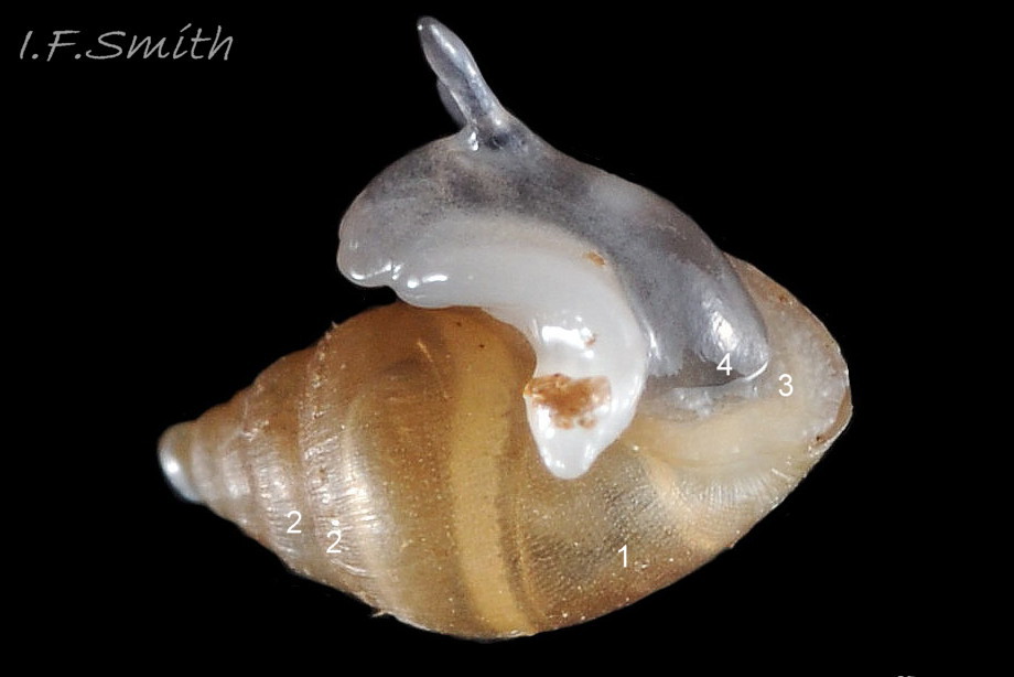
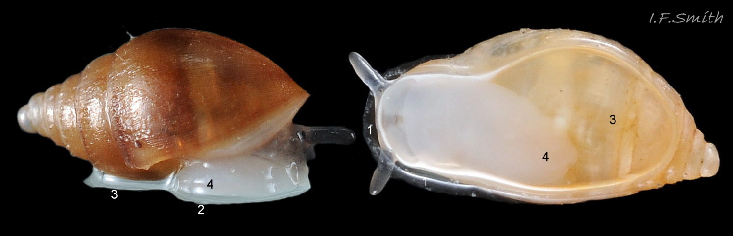
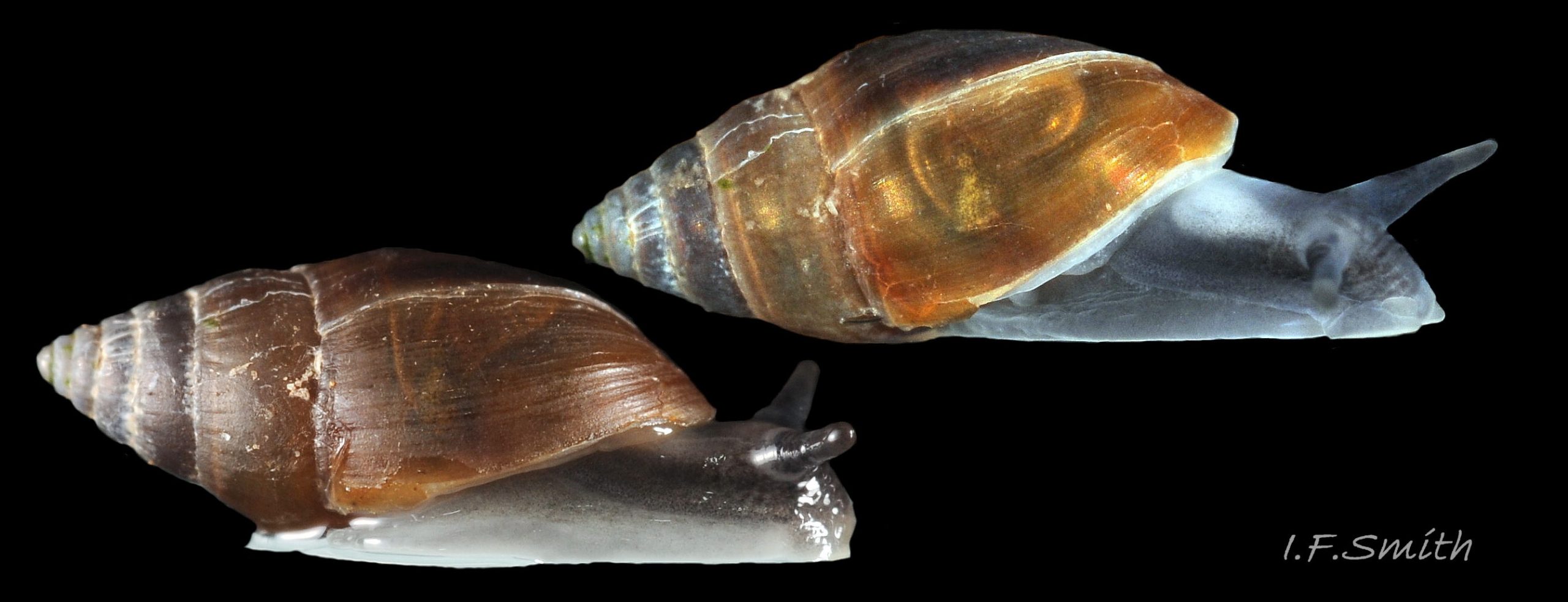
Specimens from non-salting conditions have white or very pale grey flesh; colour on an individual varies with degree of extension and whether in air or water 12 Myosotella myosotis form denticulata. . The colour of the occasional ones from under stones on saltings is similar to that of M. myosotis with darker grey arranged in transverse bands across the dorsum, and colour intensity usually increases with size/age 13 Myosotella myosotis form denticulata. . The tentacles on all forms are usually grey or greyish. Sides of foot are paler than the dorsum of grey specimens 12 Myosotella myosotis form denticulata. & 13 Myosotella myosotis form denticulata. . The mantle sometimes projects a short way beyond the aperture rim of the palatal lip, but is not reflected onto it 13 Myosotella myosotis form denticulata. . The parietal lip on the body whorl is a glaze formed by the mantle extending onto it 02 Myosotella myosotis form denticulata. & 10 Myosotella myosotis form denticulata. . The mantle cavity, the roof of which contains a network of haemolymph vessels 14 Myosotella myosotis form denticulata. , functions as a lung for respiration. It is sealed off from the exterior by a thick, white or brownish-white, mantle-collar which fits closely round the body as it extends or retracts 06 Myosotella myosotis form denticulata. & 24 Myosotella myosotis form denticulata. . The collar has a pneumostome which, when in air, can be opened and closed 15 Myosotella myosotis form denticulata. for respiration and humidity control but, when immersed, does not effectively retain air or exclude water 16 Myosotella myosotis form denticulata. & 17 Myosotella myosotis form denticulata. . The rectum and part of the intestine, visible through translucent shells in water 17 Myosotella myosotis form denticulata. , runs along the rear edge of the roof of the mantle cavity to the anus 18 Myosotella myosotis form denticulata. which opens to the exterior in a folded part of the mantle-collar in the adapical angle of the aperture close to the pneumostome . The head has two cephalic tentacles; nearly linear with a bluntly pointed tip (subulate) when dry, and conical and paler when swollen with water 13 Myosotella myosotis form denticulata. . When not fully extended, they are contracted, becoming annulated in the basal half 19 Myosotella myosotis form denticulata. , not retracted by inversion into the body. The tentacles widely diverge from their bases near the midline of the head 20 Myosotella myosotis form denticulata. . The distal half of the tentacles, sometimes slightly bulbous, is opaque grey, sometimes with a brownish tint 21 Myosotella myosotis form denticulata. , and contains sensory chemoreceptor cells (Wondrak, 1984). There is an internal black eye within the posteromesial base of each tentacle 19 Myosotella myosotis form denticulata. . The head in front of the tentacles forms a broad, slightly bilobed “muzzle” (Forbes & Hanley,1853) 22 Myosotella myosotis form denticulata. which can be variably configured, but not cylindrically to form a snout like that of many marine gastropods. Near the anterior edge of the muzzle are two button-like, pads (“fungiform bodies” of Wondrak, 1984) which contain sensory cells 21 Myosotella myosotis form denticulata. , but they are inconspicuous on animals with white flesh. Ventrally, the mouth is protected by white outer-lip lobes. When feeding, the ventrally translucent-white muzzle is spread out flat on the substrate and the outer lips moved aside to expose the mouth edged anteriorly by the rim of the red-brown jaw 23 Myosotella myosotis form denticulata. , and to allow the extension of the anterior of the radula covered in thousands of white teeth. When translucent, the muzzle may reveal dorsally the oral tube leading from the mouth to the buccal mass, and the oesophagus passing from it towards the stomach 20 Myosotella myosotis form denticulata. . On weakly pigmented, translucent specimens the dumbbell-shaped, dorsal part of the nerve ring with two cerebral ganglia may be visible 20 Myosotella myosotis form denticulata. . The ring encircles the oesophagus. It and its ganglia that innervate organs on the head are the nearest approximation in gastropods to a centralised brain, but other ganglia distributed on nerve cords around the body innervate other organs. The anterior edge of the translucent white sole is broad and gently curved or almost straight, sometimes with a slightly indented middle, and tapers to a rounded posterior 22 Myosotella myosotis form denticulata. . M. denticulata is a protandrous hermaphrodite. The common genital aperture is hidden beneath the mantle on the right of the animal. The female opening is covered by a thin lip of integument which continues forwards as a narrow fold enclosing the vas deferens 18 Myosotella myosotis form denticulata. to the male aperture on the right of the head from which penis with vas deferens can be everted for mating by hydrostatic pressure of haemolymph.
When immersed in water, the body absorbs water, swells, and it and the shell become paler and more translucent, sometimes, revealing internal organs 24 Myosotella myosotis form denticulata. , 17 Myosotella myosotis form denticulata. ,18 Myosotella myosotis form denticulata. & 20 Myosotella myosotis form denticulata. . A dissection was not made for this species/ecotype. Most published anatomy accounts are of M. Myosotis sensu lato which includes this species. Dissections can be seen of M. myosotis in its account at Myosotella myosotis images 32 to 37.
Key identification features
Features 1 to 4, below, accord with Forbes & Hanley (1853) and Gittenberger (2004). The former aggregated M. myosotis sensu stricto with M. denticulata but “scrupulously kept apart their description.” Many currently used identification guides aggregate them and their features under M. myosotis sensu lato. Consequently, distribution maps on GBIF and NBN include many M. denticulata occurrences under “M. myosotis”, and the M. denticulata maps have under-representation of its occurrence.
To observe aperture sculpture the animal must be well withdrawn, and the shell tilted at different angles. Sometimes the outer (palatal) lip sculpture of an earlier growth stage is visible deep into the aperture and should be used if the sculpture on new growth has not yet developed. It is advisable to examine several specimens of different sizes from a site; sometimes both are present..
Myosotella denticulata(Montagu, 1803).
1. Live shell brown (beachworn shells may be dull whitish). Usual adult height 3.5 mm to 7.5 mm, exceptionally 10 mm 02 Myosotella myosotis form denticulata. .
2. Inner (columellar/parietal) lip has 3 or 4 apertural protrusions 02 Myosotella myosotis form denticulata. .
3.Outer (palatal) lip has 2 to 7 (or more) apertural protrusions 02 Myosotella myosotis form denticulata. sometimes set into a pale ridge which occasionally submerges them. [If no protrusions, check further back in aperture for protrusions on earlier lip position; may be visible from exterior through translucent shell, with or without connecting streaks.]
4. In its typical non-salting habitat, the flesh colour of normally extended dorsal body is white or very pale whitish grey, with darker grey tentacles 12 Myosotella myosotis form denticulata. . But when it occurs in muddier conditions, it may be as dark as M. myosotis 13 Myosotella myosotis form denticulata. .
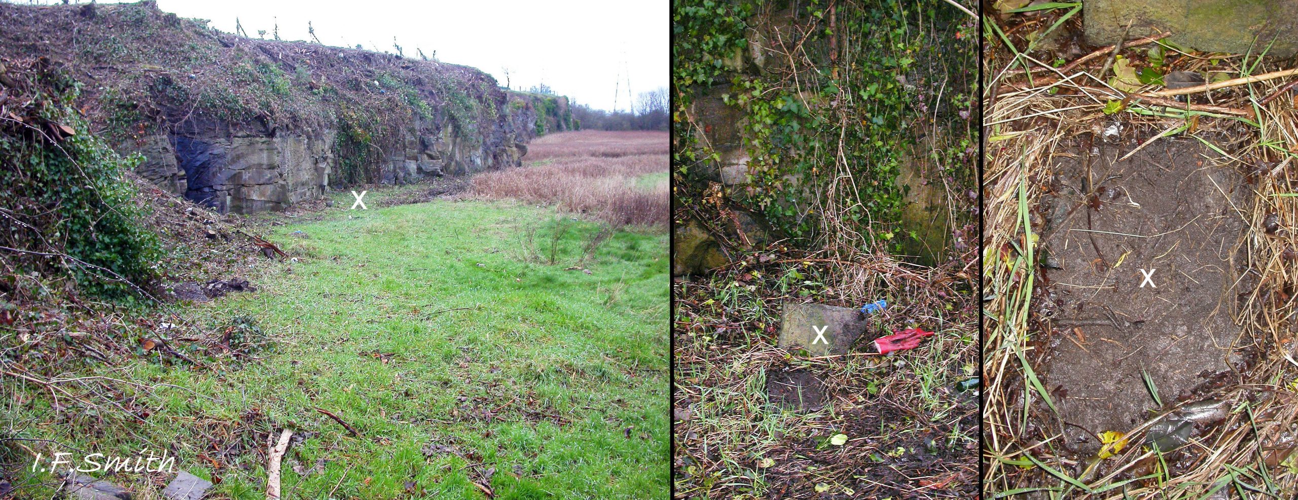
5. Habitat: typically under slightly embedded stones at Extreme High Water Spring level and above (supralittoral) on sheltered coast without salting vegetation at fully marine salinity. Occasionally under stones on landward edge of Saltmarsh-grass sward by tidal rivers with low salinity 25 Myosotella myosotis form denticulata. .
Similar species/ecotype
Myosotella myosotis
(Full account Myosotella myosotis)
1. Live shell brown 28 Myosotella myosotis form denticulata (beachworn shells may be dull whitish 29 Myosotella myosotis form denticulata). Usual adult height 6.5mm to 8mm, exceptionally 10mm .
2. Inner (columellar/parietal) lip has only 2 or 3 apertural protrusions 30 Myosotella myosotis form denticulata .
3. Outer (palatal) lip has a single apertural denticle or none 30 Myosotella myosotis form denticulata Some have a pale apertural ridge running close to the lip.
4. Flesh colour of normally extended dorsal body is grey 31 Myosotella myosotis form denticulata Shade and intensity varies with age, extension and whether in air or water, but not pure white when adult.
5. Habitat: among vegetation, often under driftwood, on low salinity estuarine saltings and Saltmarsh-grass sward by tidal rivers a little above and below EHWS. Locally abundant. (May occur with M. denticulata under stones on/near saltings 25 Myosotella myosotis form denticulata. .)
Leucophytia bidentata (Montagu, 1808).
(Full account Leucophytia bidentata)
1.Live shell slightly-translucent ivory-white; yellow viscera may show through spire 32 Myosotella myosotis form denticulata. Usual adult height to 5 mm, occasionally to 7 mm. Sutures shallower and whorls less rounded than on M. myosotis 33 Myosotella denticulata.
2. Inner (columellar/parietal) lip has 2 protrusions within the aperture; not more 33 Myosotella denticulata.
3. Outer (palatal) lip has no protrusions or rib (sometimes in a photo, a strong growth line might be mistaken for a rib 33 Myosotella denticulata.
4. Flesh colour of normally extended dorsal body is almost pure white 32 Myosotella myosotis form denticulata, but when contracted into body-whorl colour saturation gives it a cream appearance.
5.Lives in deep, silty, rock crevices between High Water Neap level and Low Water Spring level. Also under stones embedded into soil-like substrate at Extreme High Water Spring level and a little above on sheltered coast where it is often with M. denticulata.
Habits and ecology
M. denticulata lives typically under slightly embedded stones at Extreme High Water Spring level and slightly above (supralittoral) on sheltered coast without salting vegetation at fully marine salinity; often in company with Leucophytia bidentata and some terrestrial invertebrates. Occasionally, it also occurs under stones on the landward edge of Saltmarsh-grass sward by tidal rivers with low salinity 25 Myosotella myosotis form denticulata. , often with more numerous M. myosotis, Assiminea grayana and some terrestrial invertebrates. It does not live in permanently submerged in pools, but can survive and be active for short periods of immersion. As there is no operculum to reduce dessication, the species is an obligatory hygrophile. Its spindle shaped shell is well adapted for moving through small gaps under stones. When moving, the foot and shell are cushioned on a layer of watery mucus which is sometimes mistaken for the foot 26 Myosotella myosotis form denticulata. & 27 Myosotella myosotis form denticulata. which usually underlies little more than the aperture . M. denticulata senses its surroundings with its tentacles and the two button-like pads (“fungiform bodies” of Wondrak, 1984) 21 Myosotella myosotis form denticulata. near the anterior edge of the muzzle. In its usually dark habitat, its eyes probably function as little more than light detectors to trigger negative phototaxic motion when exposed to light.
It is a euryhaline species capable of surviving immersion in water from 0 p.p.t to full marine salinity or more, but individuals require time to adapt to changes in salinity and may become inactive/moribund when abruptly immersed in water they are unaccustomed to.
Respiration is of atmospheric air in the mantle cavity which is sealed by a collar of thickened mantle 06 Myosotella myosotis form denticulata. that firmly embraces the body but allows it to extend-from/retract-into the shell 24 Myosotella myosotis form denticulata. . A pneumostome (respiratory pore) in the collar 15 Myosotella myosotis form denticulata. can be opened for inhalation/exhalation of air or closed to seal the cavity against dehydration. The roof of the mantle cavity contains a network of haemolymph vessels 14 Myosotella myosotis form denticulata. and is very thin, enabling oxygen from inhaled air to diffuse into the vessels and for carbon dioxide to leave with the exhaled air. When immersed, air escapes 17 Myosotella myosotis form denticulata. from the mantle cavity and water enters as the pneumostome is not tightly closed 16 Myosotella myosotis form denticulata. .
When feeding, the muzzle is spread out on the substrate and the radula is extended 23 Myosotella myosotis form denticulata. to gather, with the red jaw as a backstop, decaying vegetation, diatoms (Wiese & Richling, 2008) and sediment rich in organic material which are bound into food boli with mucus from the supra pedal gland brought to the mouth along a median groove. Unlike marine prosobranch gastropods, which defecate into a mantle cavity that is cleared by water currents, M. denticulata has a rectum that opens to the exterior through an anus in the mantle collar 18 Myosotella myosotis form denticulata. , near to, but separate from, the pneumostome so that faeces are expelled without fouling the respiratory mantle-cavity. The soft faeces are wet and loosely bound with mucus when fresh 27 Myosotella myosotis form denticulata. . There is no operculum 11 Myosotella myosotis form denticulata. to provide protection against intrusion by predators, but the numerous protrusions narrow the aperture to impede attack. The aperture protrusions of M. denticulata may have developed in response to the different (more threatening?) predators present in its habitat, which is more terrestrial than that of M. myosotis.
Reproduction: (Details assumed from published accounts of M. myosotis sensu lato.) M. denticulata is a protandrous hermaphrodite which changes its sexual function in the wild when 1½ to 2 years old, so younger, 1 to 1½ years, fully mature males mate with older, over 1½ years, females (Schultes, 2014) using the stout, conical penis everted from the side near the posterior of the right tentacle. Female deposits 15 to 80 egg capsules in a small, yellow or white, frog-spawn-like mass (Morton, 1954 and Gittenberger, 2004). Each ovoid capsule contains a single ovum. The cases are attached to each other in a loosely convoluted chain by a filament (chalaziform process) at each end. The closely packed cases with intervening clear fluid are contained in a tough binding membrane which is attached to stones. There is a larval veliger stage, with sinistral shell, which is passed entirely within the ovum (Morton, 1954).
Distribution and status
Europe from England to Mediterranean and Azores. GBIF map, www.gbif.org/species/4359191
Locally common in suitable habitat with rocks in Britain but records from vegetated saltings are likely to be the species/ecotype M. myosotis sensu stricto. NBN map
species.nbnatlas.org/species/NHMSYS0001702111
Irish distribution, National biodiversity data centre, in Mollusc Ireland: www.habitas.org.uk/molluscireland/species.asp?ID=121
Acknowledgements
I gratefully thank Ben Rowson of the National Museum Wales for his help with the account, but any errors or omissions are mine.
Links and references
Anderson, R. MolluscIreland, accessed January 2019. www.habitas.org.uk/molluscireland/species.asp?ID=121
Forbes, E. & Hanley S. 1849-53. A history of the British mollusca and their shells. vol. 4 (1853), 190 – 197 & plate CXXV. London, van Voorst. (AsConovulus denticulatus var. myosotis.)
Free pdf at archive.org/details/historyofbritish04forbe/page/190
plate at archive.org/details/historyofbritish04forbe/page/n565
Fretter, V. & Peake, J. 1975. Pulmonates functional anatomy and physiology. Vol.1. London. Academic Press.
Gittenberger, E. et al. 2004. De Nederlandse zoetwatermollusken. Leiden, Netherlands, Nationaal Natuurhistorisch Museum Naturalis.
Heller J. 2015. Marine Ancestors of most Land Snails: Pulmonates. In: Sea Snails. Springer, Cham. link.springer.com/chapter/10.1007%2F978-3-319-15452-7_10
Jeffreys, J.G. 1862-69. British conchology. vol. 5 (1869). London, van Voorst. (As Melampus myosotis (including var. ringens = Myosotella denticulata); Free pdf at archive.org/stream/britishconcholog05jeffr#page/106/mode/2up . Use slide at base of page to select pp.106-109.)
Martins, A.M. de F. 1996. Anatomy and systematics of the western Atlantic Ellobidae (Gastropoda: Pulmonata). Malacologia 37(2): 163 – 332.
www.biodiversitylibrary.org/page/13113594#page/179/mode/1up
Martins, A.M. de F. & Mendes, A.R.M. 2013. Do cosmopolitans speciate? Anatomical diversity of Myosotella in Azores. Centro de Investigação em Biodiversidade e Recursos Genéticos. Ponta Delgada, Açores, Portugal. Poster for World Congress of Malacology 2013 in pdf: www.researchgate.net/publication/264339925_Do_cosmopolita… .
Montagu, G. 1808. Supplement to: 1803 Testacea Britannica, or, Natural history of British shells, marine, land, and fresh-water, including the most minute : systematically arranged and embellished with figures. London, J. White.
Description of Leucophtia bidentata as Voluta bidentata pp. 100-101.
www.biodiversitylibrary.org/page/24430722#page/806/mode/1up
Plate 30, :
www.biodiversitylibrary.org/page/24430722#page/917/mode/1up
Morton, J. E. 1955. The functional morphology of the British Ellobiidae (Gastropoda Pulmonata) with special reference to the digestive and reproductive systems. Phil. Trans. R. Soc. Ser. B .
239, No. 661: 89-160 www.jstor.org/stable/92507
Schultes, F.W. 2014. Species summary for Ovatella myosotis (Draparnoud, 1801). AnimalBase. SUB Göttingen. www.animalbase.uni-goettingen.de/zooweb/servlet/AnimalBas… Accessed January 2019.
Watson, H. I943. Notes on a list of the British non-marine Mollusca. J. Conch. 22: 13 – 22.
Wiese, V. & Richling, I. 2008. Das Mäuseöhrchen Myosotella myosotis (Draparnaud 1801). Arbeitskreis Mollusken NRW.
www.mollusken-nrw.de/weichtier_des_jahres/weichtier2008.htm
Wondrak, G. 1984. Ultrastructure of the sensory epithelia of oral tube, fungiform sensory bodies, and terminal knobs of tentacles of Ovatella
myosotis. Draparnaud (Archaeopulmonata, Gastropoda) J. Morphol. 181: 333-347 .
onlinelibrary.wiley.com/doi/pdf/10.1002/jmor.1051810307
Current taxonomy:
www.marinespecies.org/aphia.php?p=taxdetails&id=139673
Glossary
adapical angle = angle at which outer lip meets body-whorl.
boli = (sing. bolus) small rounded masses, especially of triturated food material.
cerebral = to do with integration of sensory and neural functions to initiate and coordinate body activity.
chalaziform = resembling the two spiral bands (chalazae) in a bird’s egg that attach the yolk to opposite ends of the lining membrane.
columella = solid or hollow axial “little column” around which gastropod shell spirals; hidden inside shell, except on final whorl next to lower part of inner lip of aperture where hollow ones may end in an umbilicus or siphonal canal.
columellar = (adj.) of or near central axis of coiled gastropod.
columellar lip = lower (abapical) part of inner lip of aperture.
costa (pl. costae) = rib running across a whorl of a gastropod shell at approximately right-angles to direction of coiling and any spiral striae.
costal (adj.) = of, or arranged like, costae.
dextral = (of gastropod shell) in apertural view with spire uppermost, the aperture is on the right. Most gastropod species adults have dextral shells.
distal = away from centre of body or from point of attachment.
diverticula = (for digestion) blind ended tubules in the digestive gland that receive nutrients for digestion.
EHWS = extreme high water spring tide.
euryhaline = able to tolerate a wide variation in salinty.
fusiform = slender, spindle-shaped, tapering almost equally towards both ends.
ganglia = (sing. ganglion) knots on a nerve cord containing sensory cell bodies that conduct impulses to (innervate) organs of the body.
haemolymph = circulating fluid in molluscs that carries nutrients, waste and hormones. Analagous to vertebrate blood, but most molluscs have copper-based haemocyanin in it instead of red haemoglobin to carry oxygen. It may be tinged blue when oxygenated; colourless when depleted of oxygen.
halophyte = plant tolerant of saline soil and periodic tidal immersion, usually on saltmarshes, estuarine shores and sides of tidal rivers.
hygrophile = living in moist, humid, but not submerged, conditions.
(obligatory hygrophile = only able to live in such conditions.)
mantle = sheet of tissue that secretes the shell, covers the viscera and forms a cavity in gastropods. In terrestrial gastropods (‘pulmonates’) the cavity roof contains a network of haemolymph (‘blood’) vessels enabling the cavity to act like a lung.
mesial = on or facing towards the midline of the body.
operculum = plate of horny conchiolin, rarely calcareous, used to close shell aperture of prosobranch gastropods.
palatal lip = outer lip of gastropod aperture.
parietal lip ( or parietal wall) = upper part of inner side of gastropod aperture, often lacking clear lip structure with just a glaze on side of whorl adapically of columellar lip.
periostracum = thin horny layer of proteinaceous material often coating shells.
posteromesial = at the rear facing towards the midline of the body.
prosobranch = member of Prosobranchia, one of three subclasses into which the class Gastropoda (slugs and snails) was divided during the 20th Century (other two were Pulmonata and Opisthobranchia). This classification is no longer used by scientists, but prosobranch is a useful informal term to signify (mainly marine) snails breathing with a ctenidium (comblike gill inside mantle cavity), an operculum, and a shell which can accommodate the whole body.
protandrous hermaphrodite = each individual starts mature life as a functioning male, later changing to female function.
protoconch = apical whorls produced during embryonic and larval stages of gastropod; often different in form from other whorls (teleoconch).
protrusions = teeth, denticles, folds, lamellae or cogs (terms used by various authors).
punctate = with pinprick-like depressions.
resorb = absorb what was previously secreted; break it down into component materials and disperse into the circulation.
resorption = the process of absorbing what was previously secreted by breaking it down into component materials and dispersal into the circulation.
salting = area of salt tolerant vascular plants rooted in sediment between mean high water mark (MHW) and extreme high water of spring tides (EHWS). [Preferred synonym for “saltmarsh” as much of salting not marshy.]
septa = plural of septum; internal partition separating two chambers/ shell-whorls of a gastropod.
septum = internal partition separating two chambers/ shell-whorls of a gastropod.
sinistral = (of gastropod shell) in apertural view with spire uppermost, the aperture is on the left. Most gastropod species adults have dextral shells.
subsutural = close below the suture when shell positioned with apex uppermost.
subulate = slender and tapering to a point like onion leaf or awl.
suture = groove or line where whorls of gastropod shell adjoin.
teleoconch = entire gastropod shell other than the apical, embryonic & larval stage protoconch.
triturate = reduce to small particles.
vascular plants = plants that have vascular tissues to transport water and nutrients through the plant. Include all seed-bearing plants, ferns and horsetails. Usually terrestrial or in freshwater or brackish water; a few, such as Zostera, live in fully marine salinity water.