Click image to enlarge with full caption. Main text below slider.
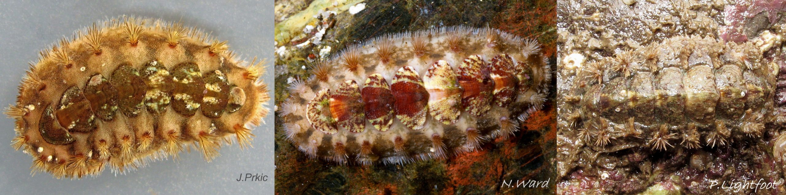
01 Acanthochitona crinita
02 Acanthochitona crinita
03 Acanthochitona crinita
04 Acanthochitona crinita
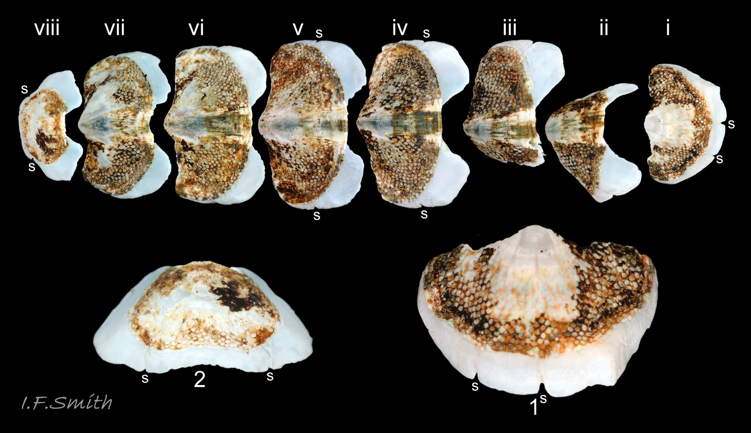
05 Acanthochitona crinita
06 Acanthochitona crinita
07 Acanthochitona crinita
08 Acanthochitona crinita
09 Acanthochitona crinita
10 Acanthochitona crinita
11 Acanthochitona crinita
12 Acanthochitona crinita
13 Acanthochitona crinita
14 Acanthochitona crinita
15 Acanthochitona crinita
16 Acanthochitona crinita
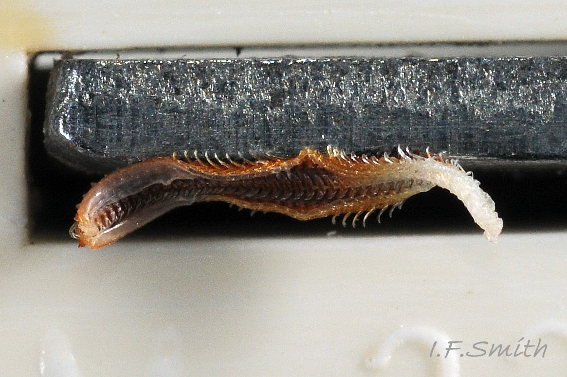
17 Acanthochitona crinita
18 Acanthochitona crinita
19 Acanthochitona crinita
20 Acanthochitona crinita
21 Acanthochitona crinita
22 Acanthochitona crinita
23 Acanthochitona crinita
24 Acanthochitona crinita
25 Acanthochitona crinita
26 Acanthochitona crinita
27 Acanthochitona crinita
28 Acanthochitona crinita
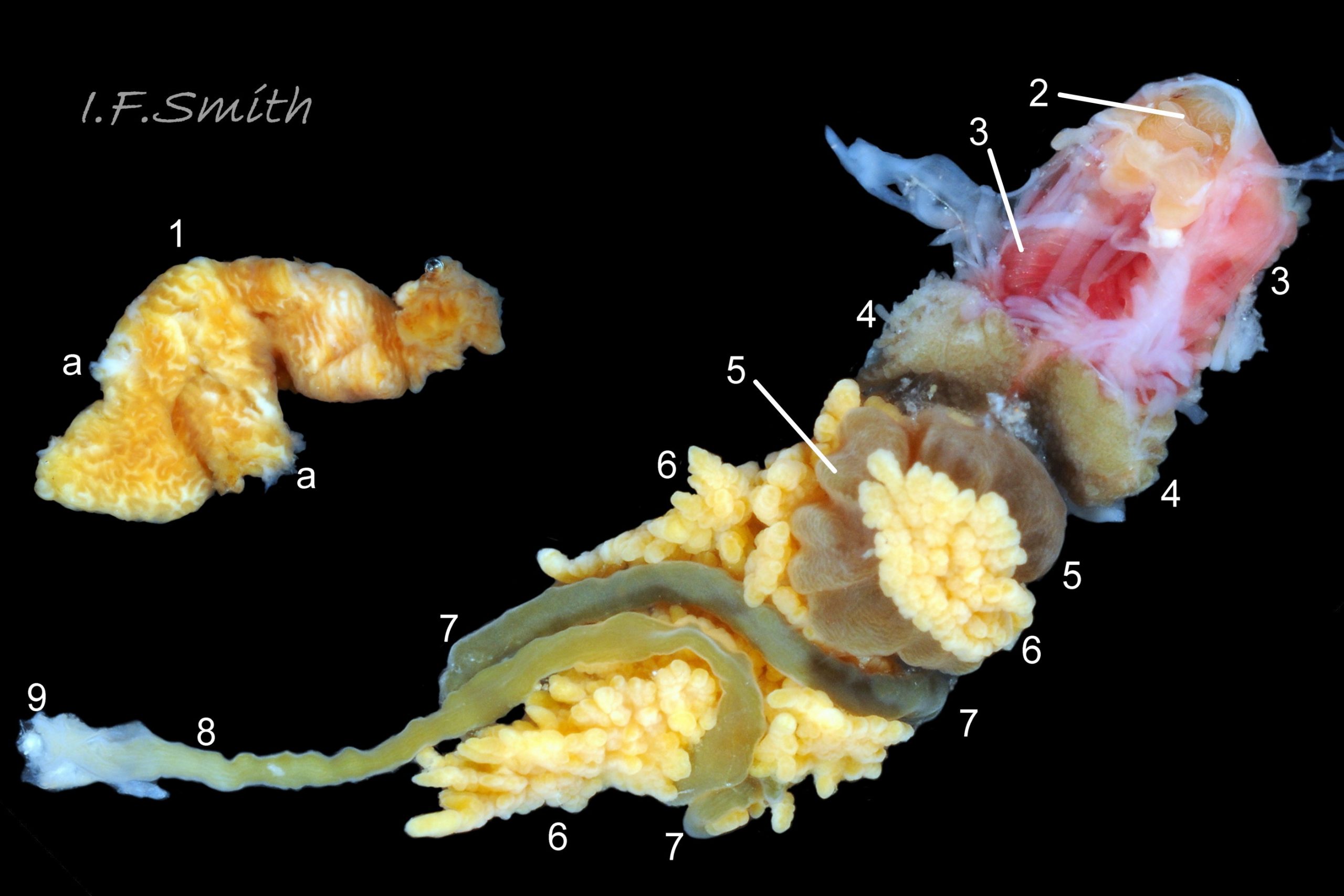
29 Acanthochitona crinita
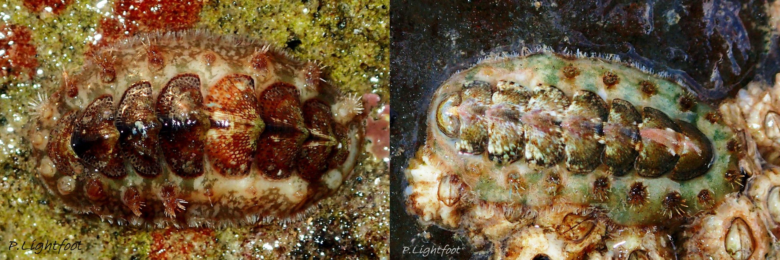
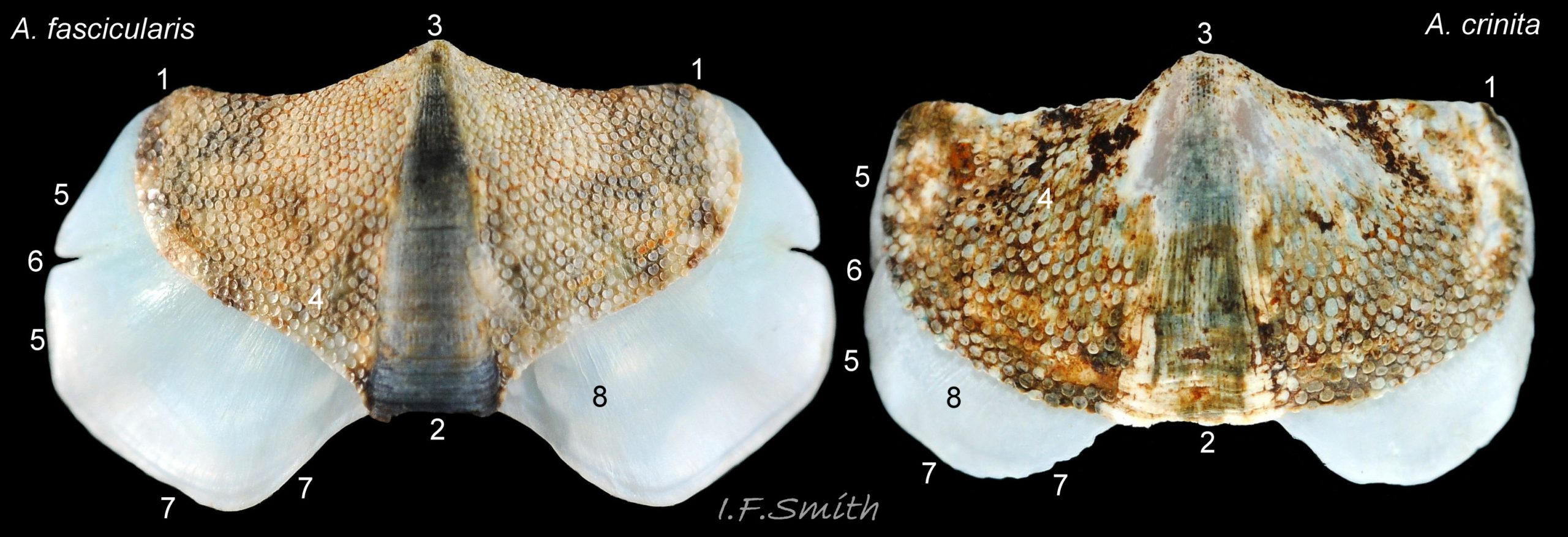
30 Acanthochitona crinita COMPARISON Acanthochitona fascicularis
31 Acanthochitona crinita
32 Acanthochitona crinita
33 Acanthochitona crinita
Acanthochitona crinita (Pennant, 1777)
Current taxonomy: World Register of Marine Species (WoRMS)
www.marinespecies.org/aphia.php?p=taxdetails&id=138675
A pdf of this and other species accounts can be downloaded from www.researchgate.net/profile/Ian_Smith19/research
Synonyms: Chiton crinitus Pennant, 1777; Chiton fascicularis var. attenuata Jeffreys, 1865; regarded as Chiton fascicularis Linneaeus, 1767 in Forbes & Hanley; Acanthochitona crinitus (Pennant, 1777) spelling variation. [There is uncertainty about which species Linnaeus called Chiton fascicularis. His specimen was from Algeria. Care is needed when using old publications.]
Vernacular: Bristled chiton (English); Lleuen wrychog y graig (Welsh); petit chiton épineux (French); børsteleddsnegel (Norwegian); Quitón espinoso (Spanish);
GLOSSARY BELOW This account uses the standardised terminology for chitons proposed by Schwabe (2010). Some of Jones & Baxter (1987) alternatives are indicated in the glossary as a.k.a..
The images in this account are mainly of a single large specimen. Users are advised to view these Flickr links to see the range of intraspecies variation:
Paula Lightfoot flic.kr/p/o4iVD1
Siôn Roberts flic.kr/p/4iTtK2
Nicholas Berkley flic.kr/p/zVYVey
Nature22 website, chiton page, has good selection of A. crinita varieties. nature22.com/estran22/mollusques/polyplacodentale/chitons…
Shell Description
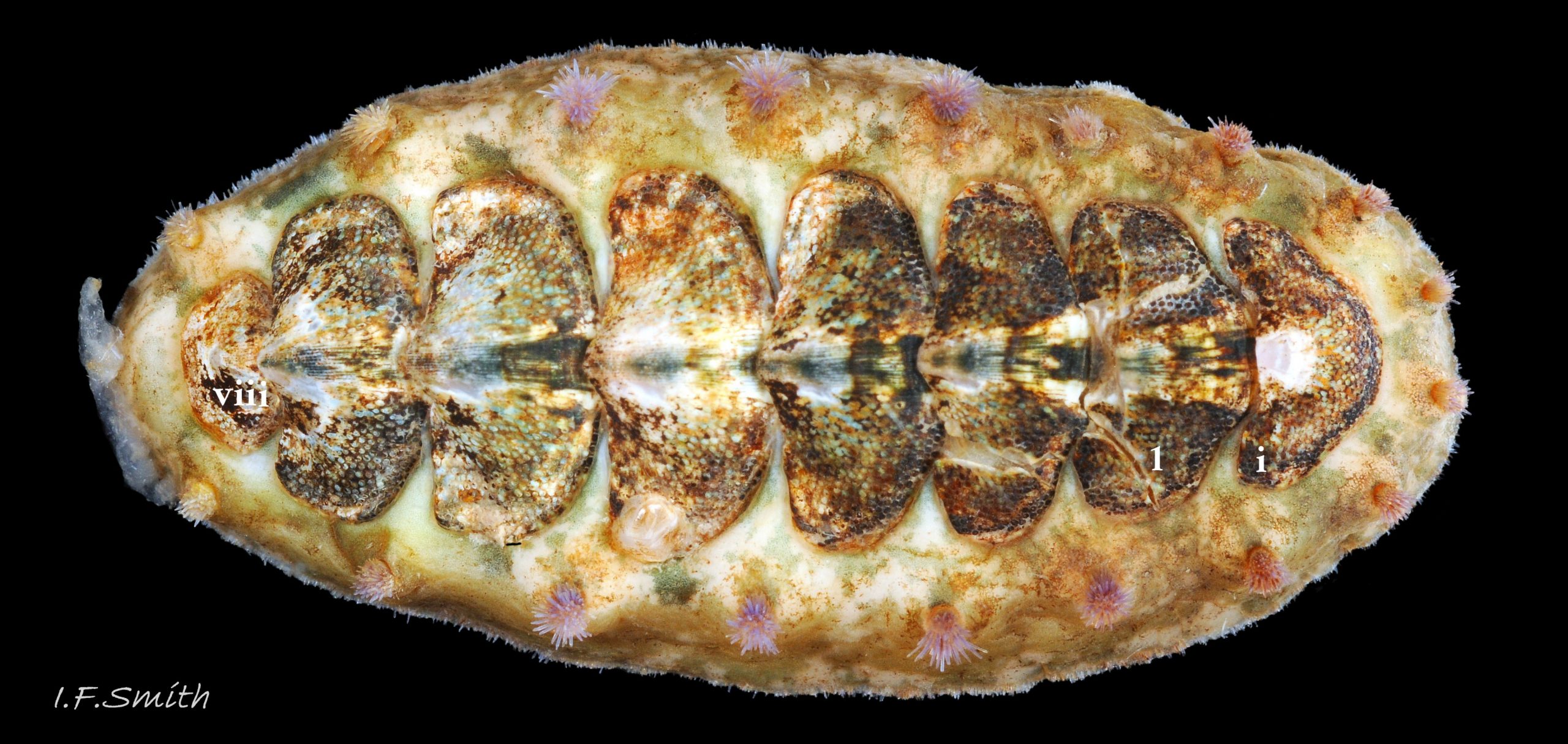
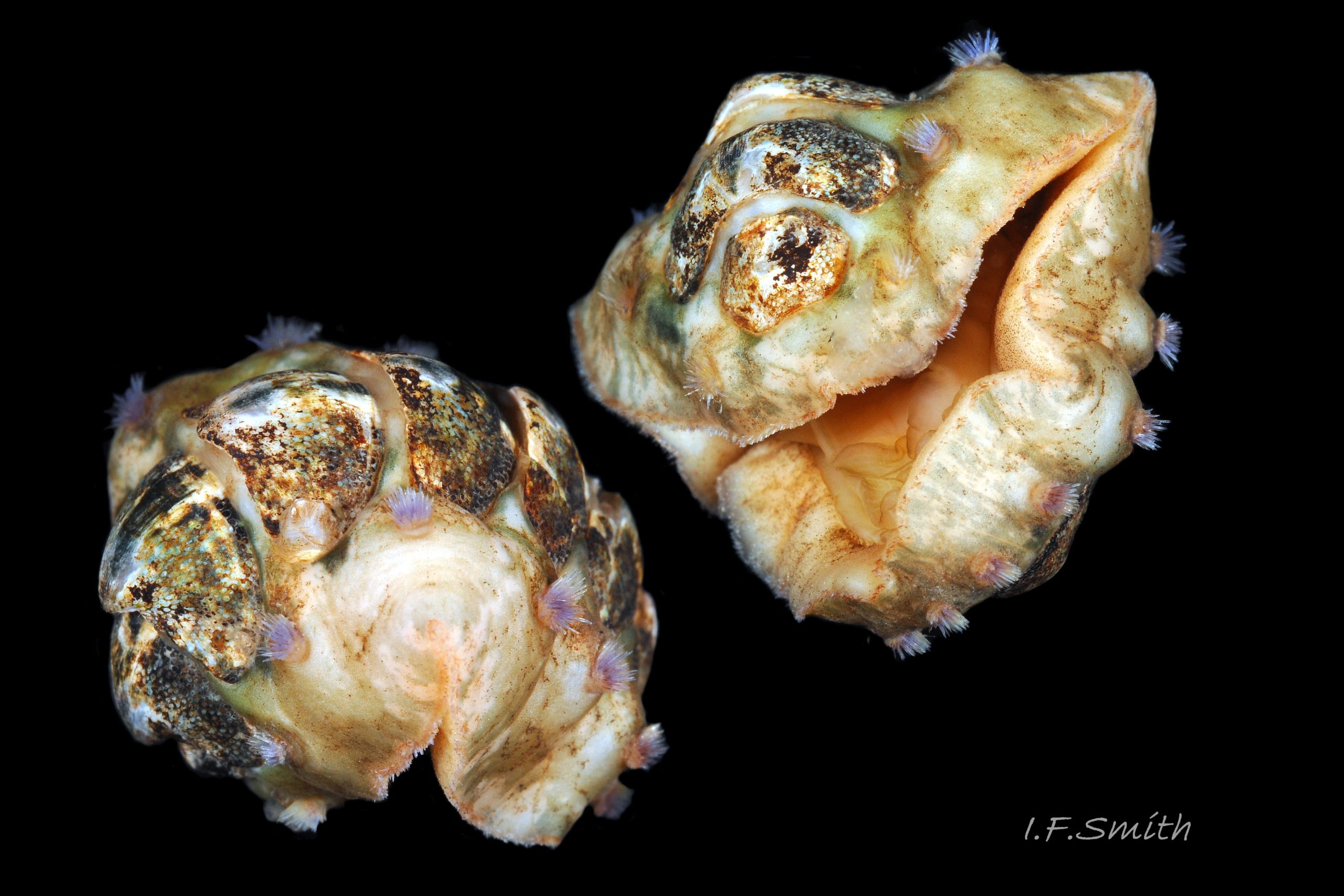
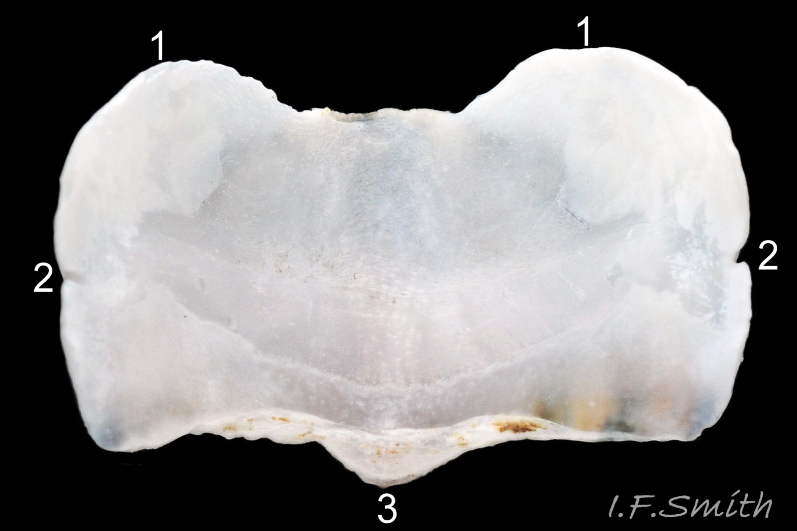
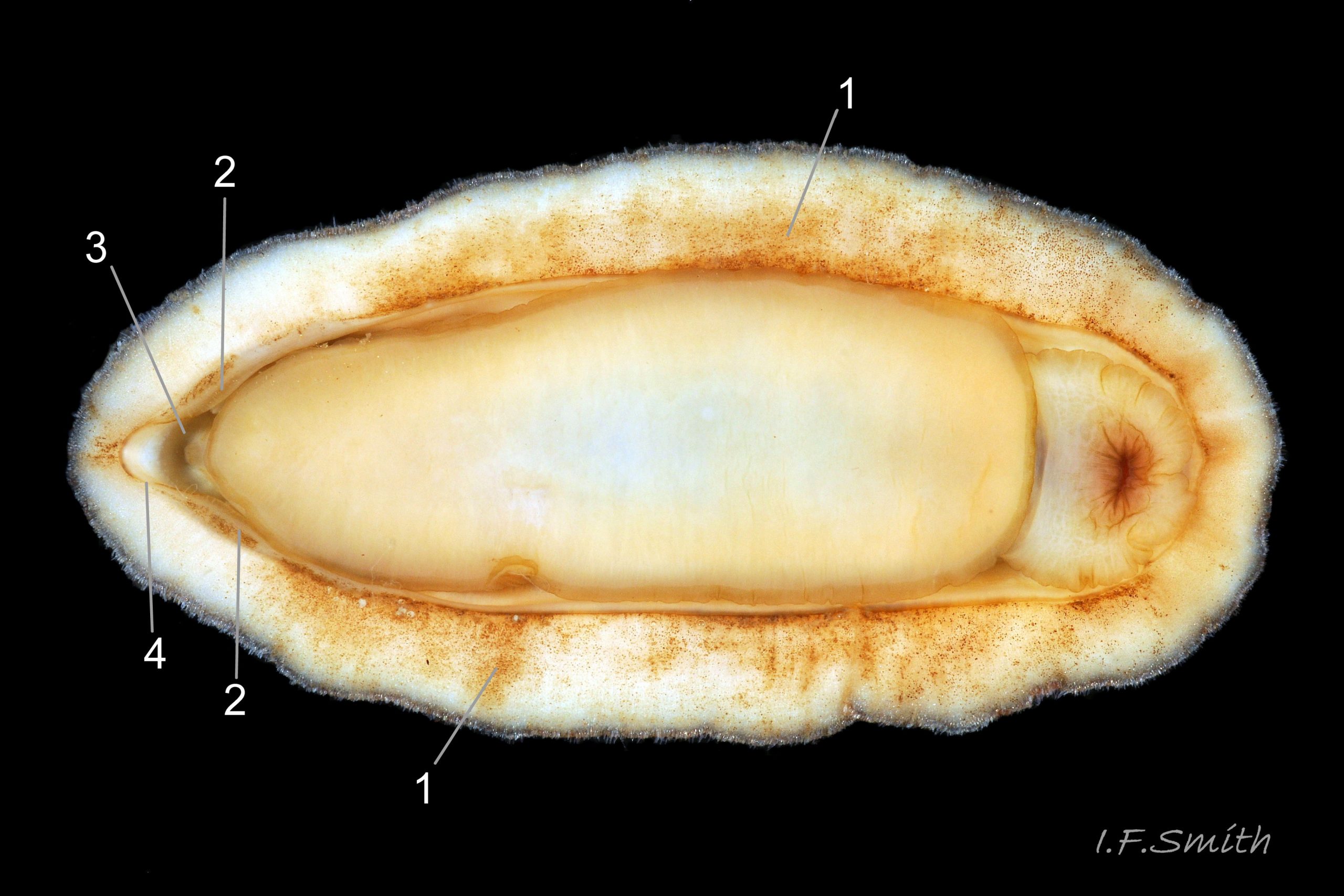
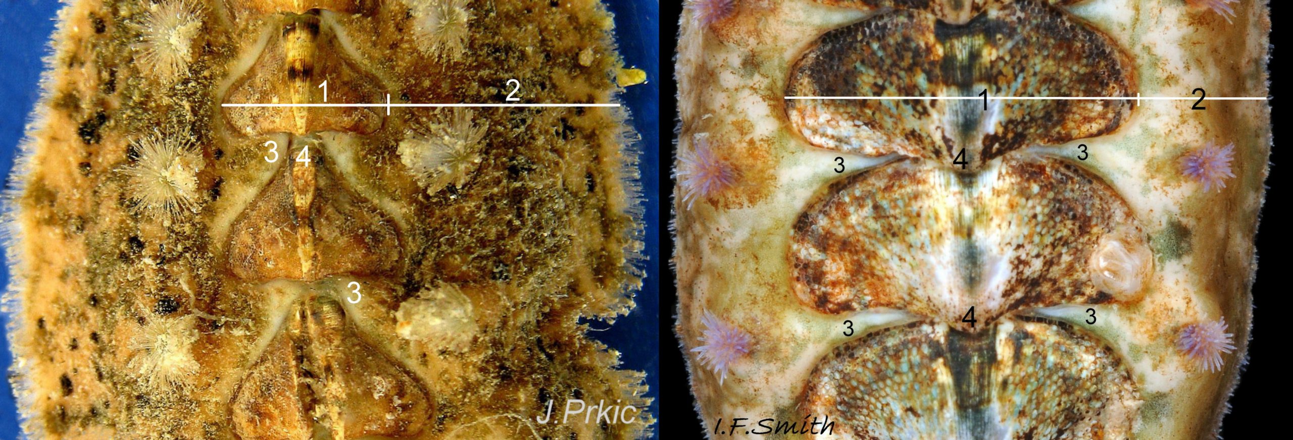
Usually to 13mm long and 6mm wide, occasionally 21mm, or exceptionally 34mm, long. Dorso-ventrally flattened, bilaterally symmetrical. In dorsal view, outline elliptical; width about 50% of length when extended 01 Acanthochitona crinita , slightly wider when contracted 02 Acanthochitona crinita . Intermediate shell valves (ii to vii) occupy 50 to 60% of chiton’s width; girdle is wide, but valves occupy more of the chiton’s width than the girdle on either side 03 Acanthochitona crinita . Slender girdle extension from each side passes between valves but, when animal adhering to substrate, extensions do not meet and are separated by parts of valves measuring about 20% of animal’s width 03 Acanthochitona crinita ; however, girdle can often be seen for 100% of width of chiton when it curls 04 Acanthochitona crinita .
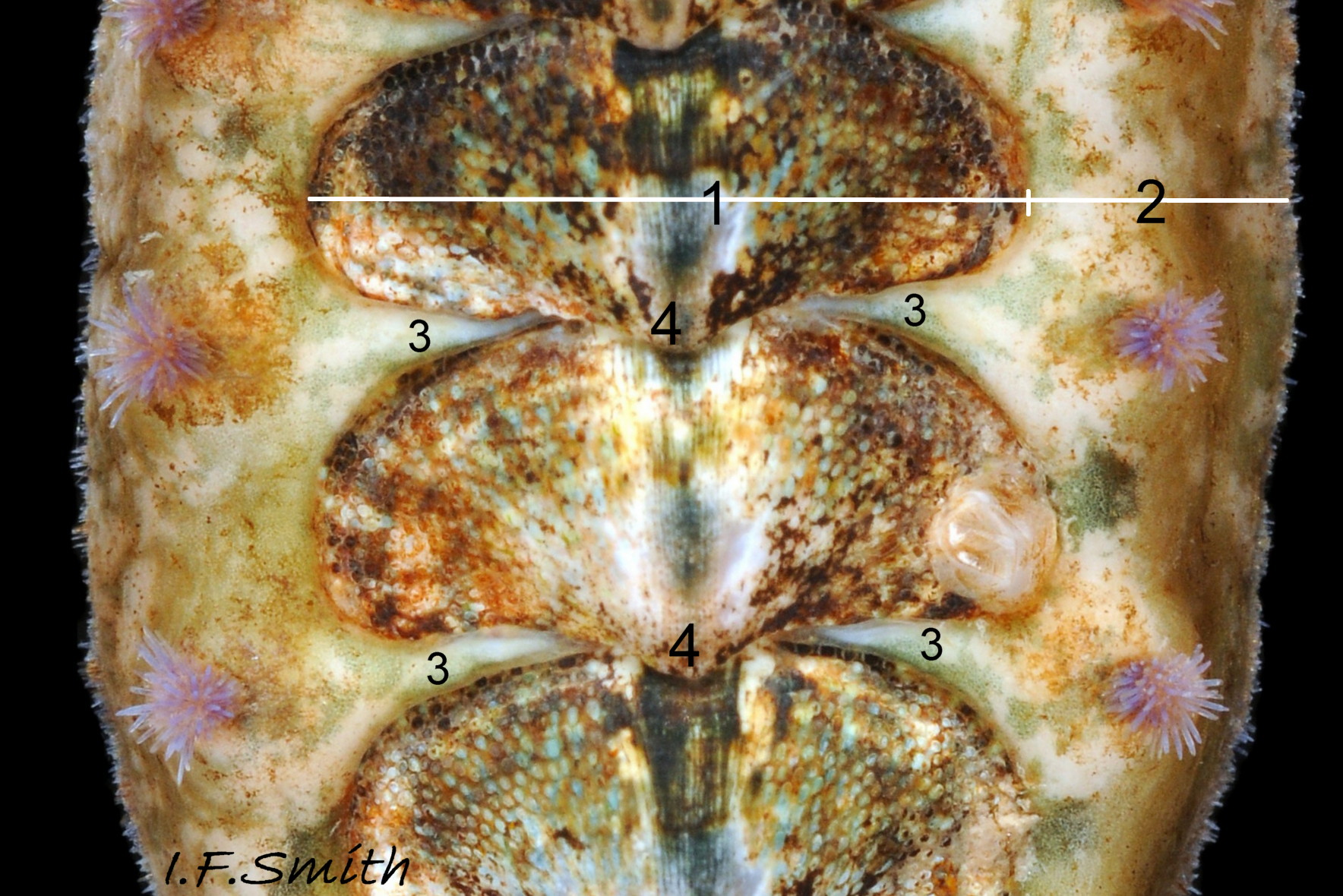
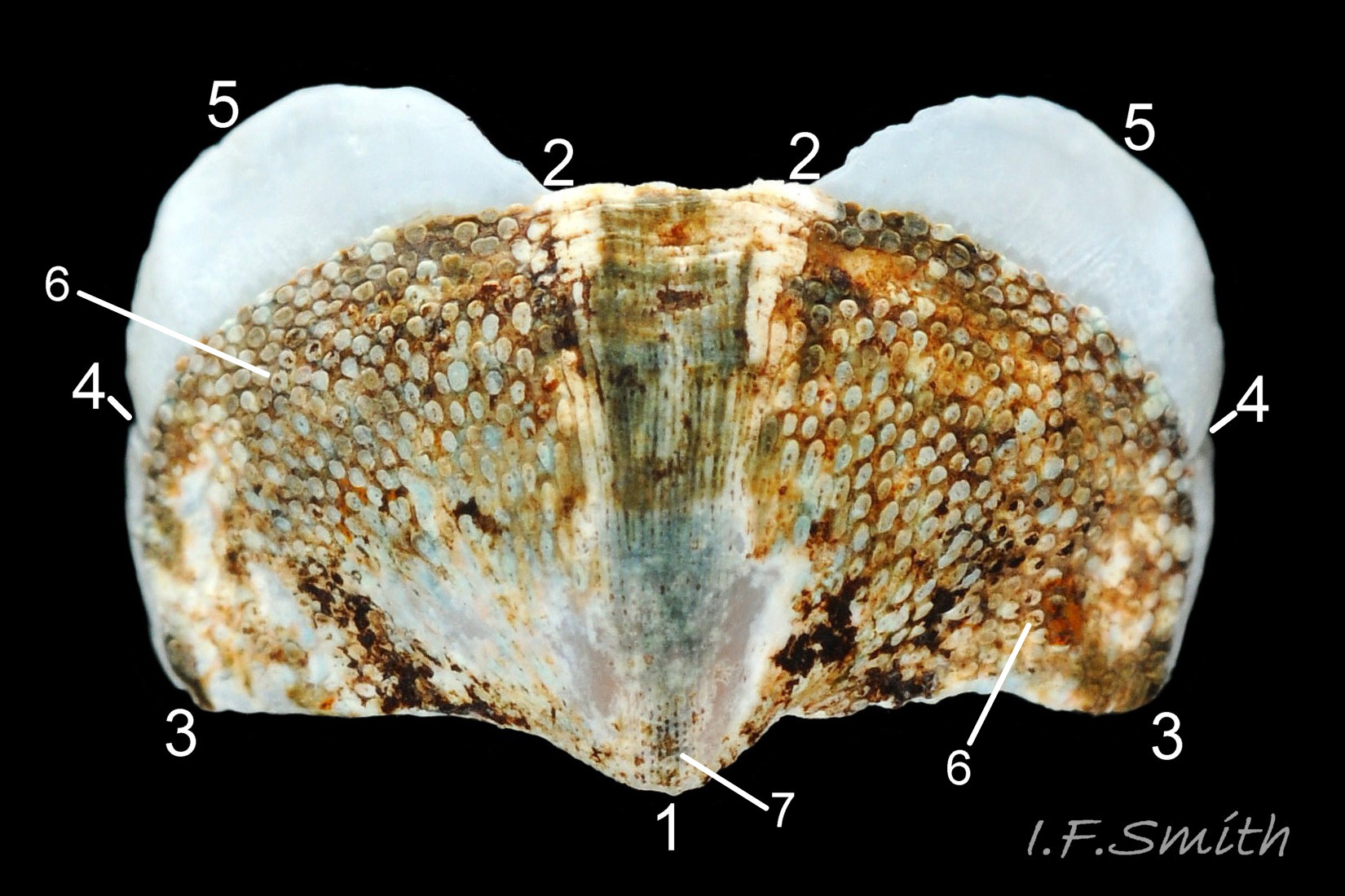
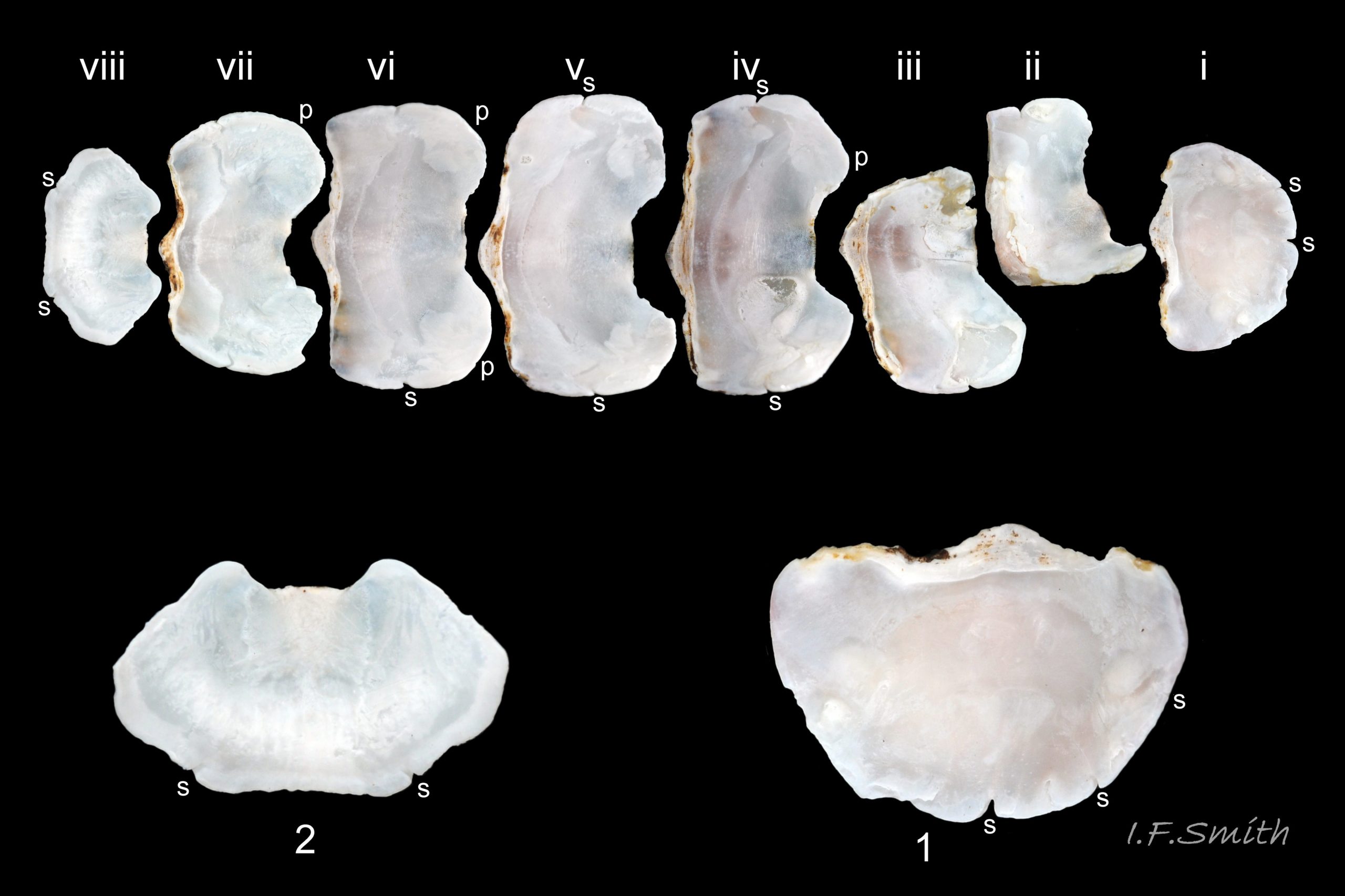
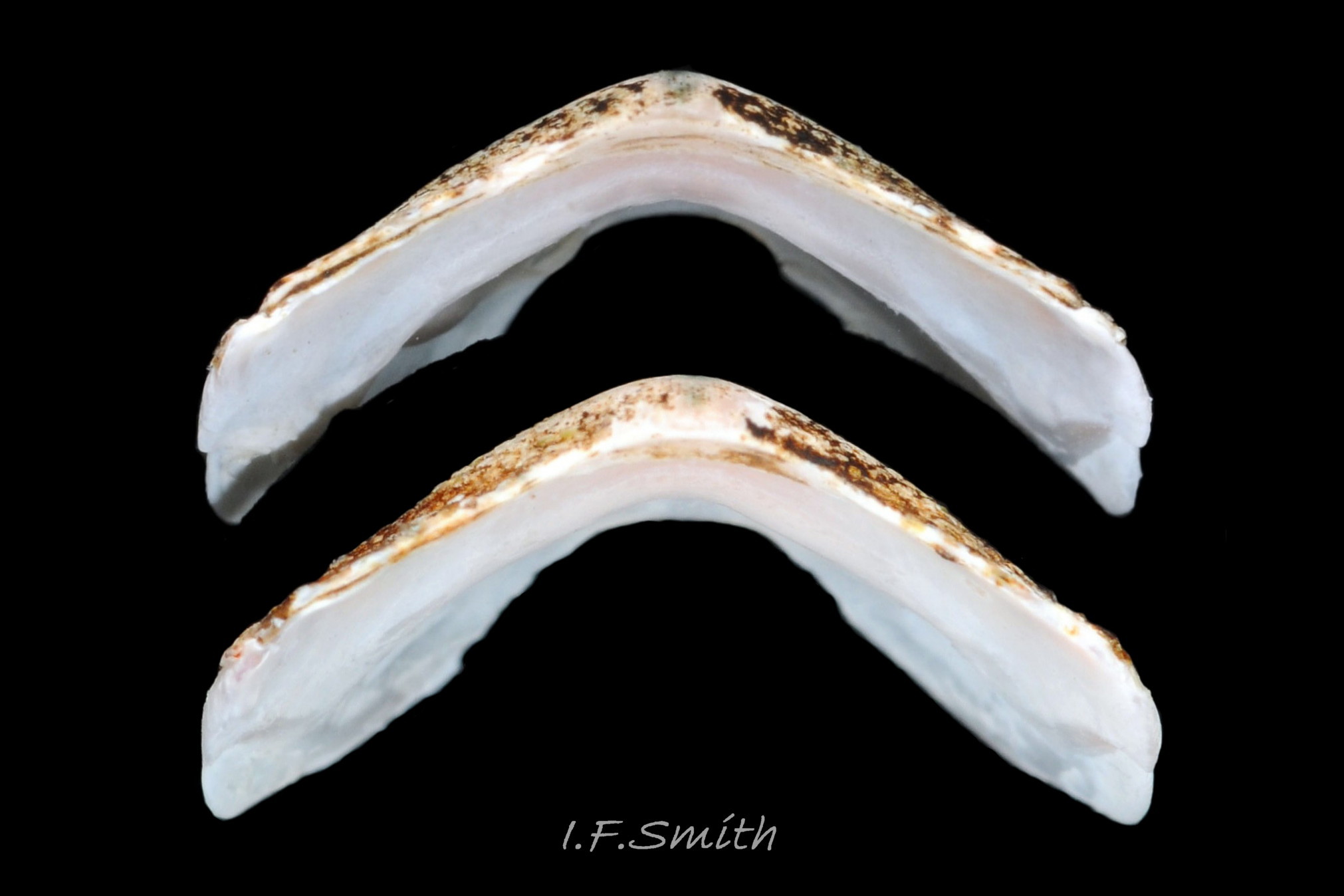
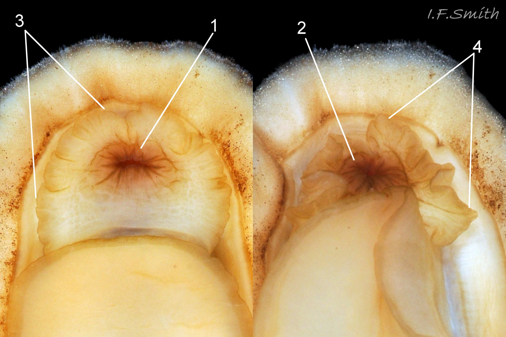
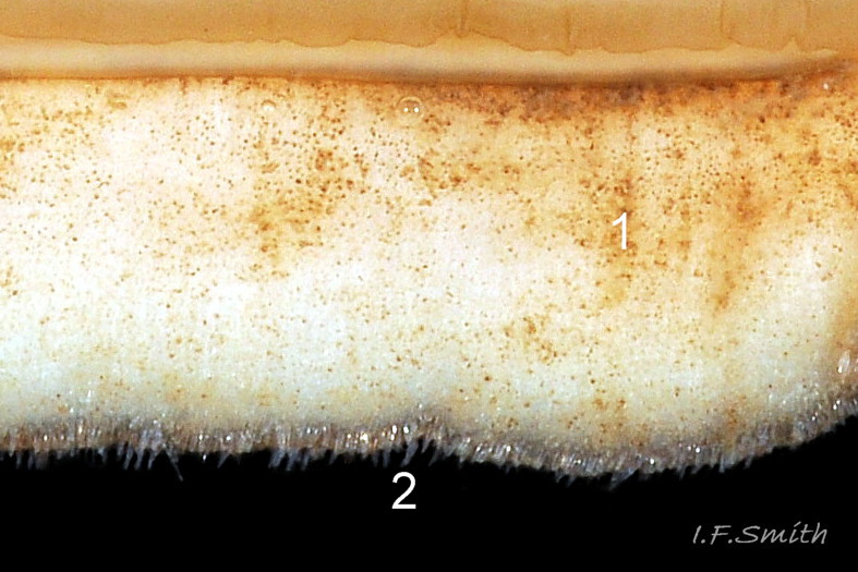
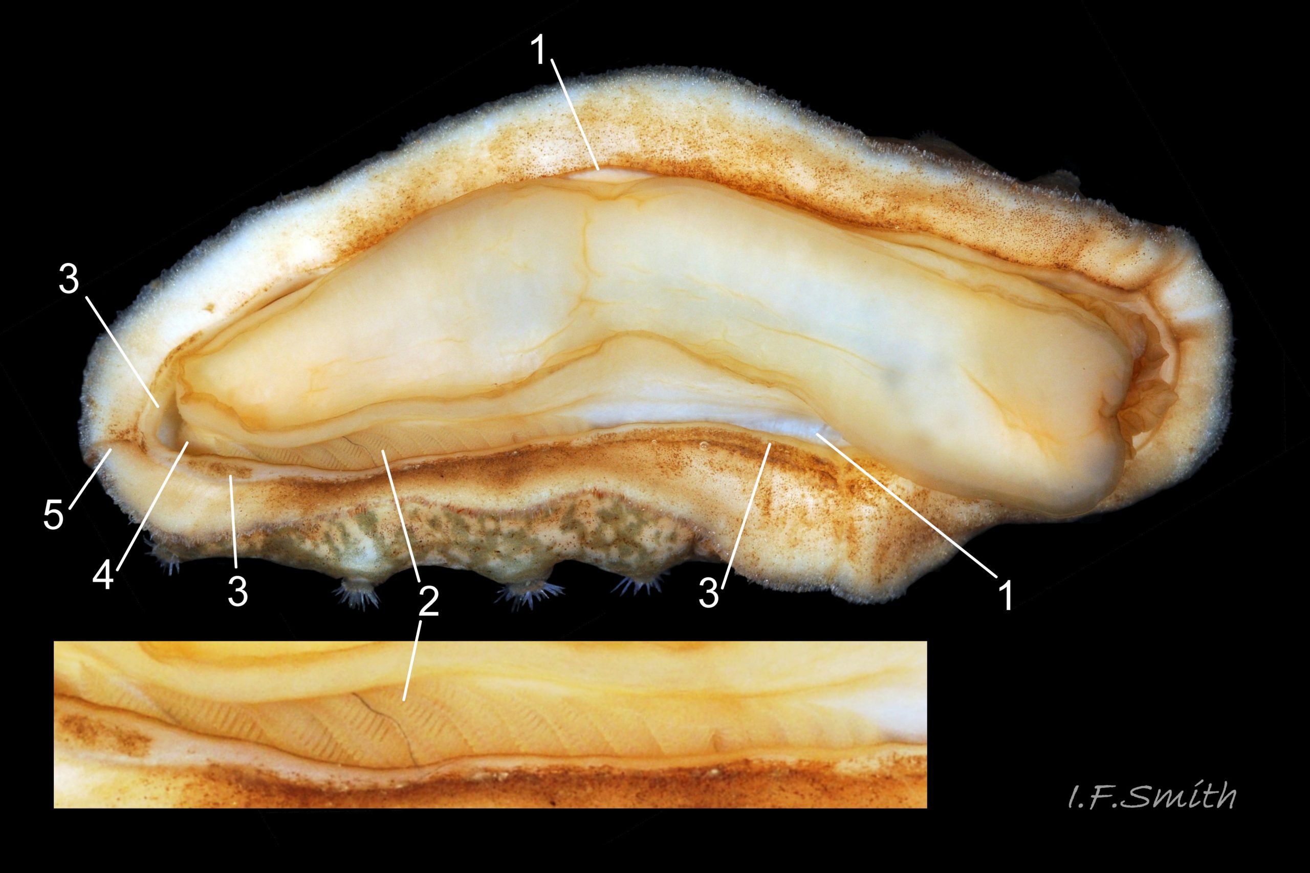
Eight overlapping valves 05 Acanthochitona crinita principally made of 1) tegmentum: dorsal coloured layer of aragonite 06 Acanthochitona crinita, 11 Acanthochitona crinita permeated and weakened by canals 07 Acanthochitona crinita & 08 Acanthochitona crinita , and 2) articulamentum: thick ventral layer of white aragonite 09 Acanthochitona crinita, 10 Acanthochitona, 11 Acanthochitona crinita , also 3) properiostracum: outermost proteinaceous layer containing colour and 4) myostracum: thin discontinuous innermost layer (only visible in section examined under microscope). Valves quite thick, but aragonite permeated by canals is brittle so fractures frequent on living specimens, though parts are usually kept in place by the girdle and strong shell musculature 02 Acanthochitona crinita . Head valve (i) on live animal shaped like Napoleonic bicorn hat 01 Acanthochitona crinita . From above, coloured tegmentum of intermediate valves (ii to vii) resembles a bow with the prominent posterior beak forming the hand drawing the string 06 Acanthochitona crinita . Intermediate valves slightly keeled, with flat side slopes when viewed from posterior 11 Acanthochitona crinita . Tail valve (viii) very small relative to other valves, inequilateral hexagon with curved corners 01 Acanthochitona crinita & 05 Acanthochitona crinita . Distinct beak on each of intermediate valves (ii to vii) visible in lateral view 12 Acanthochitona crinita .
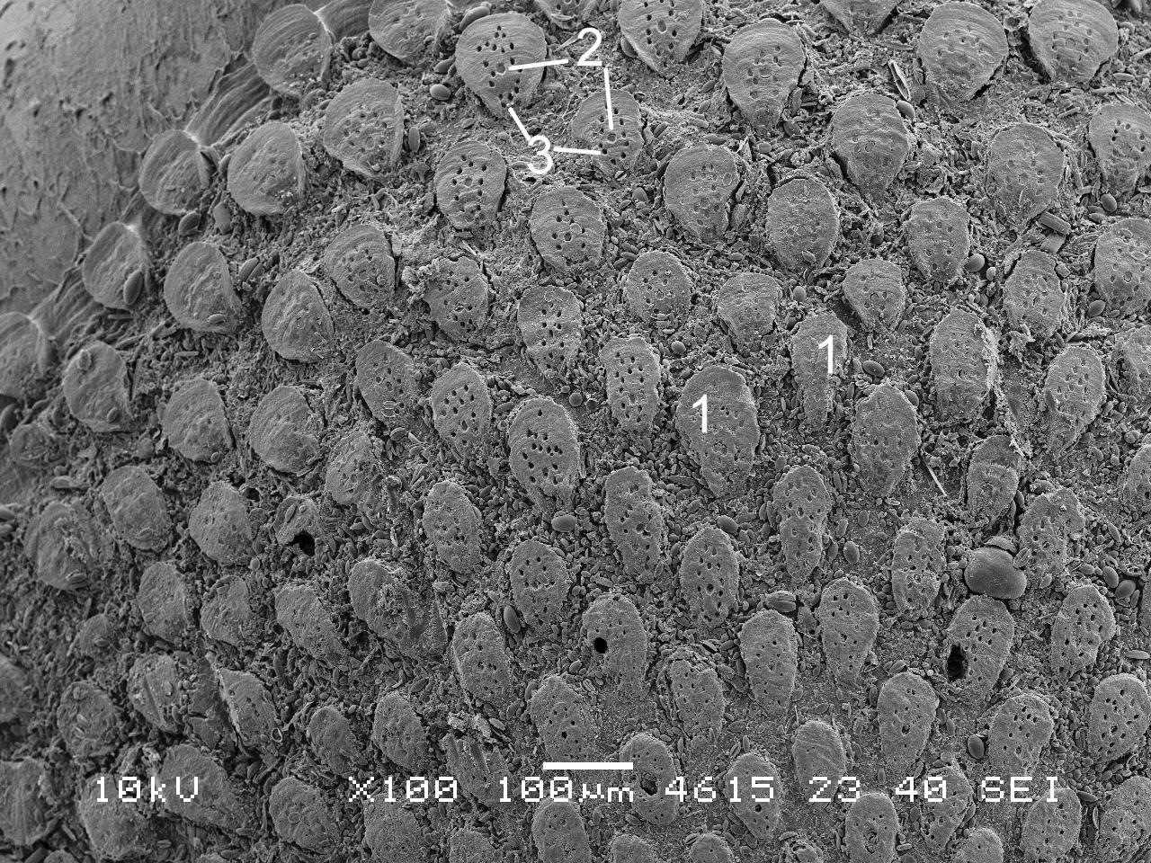
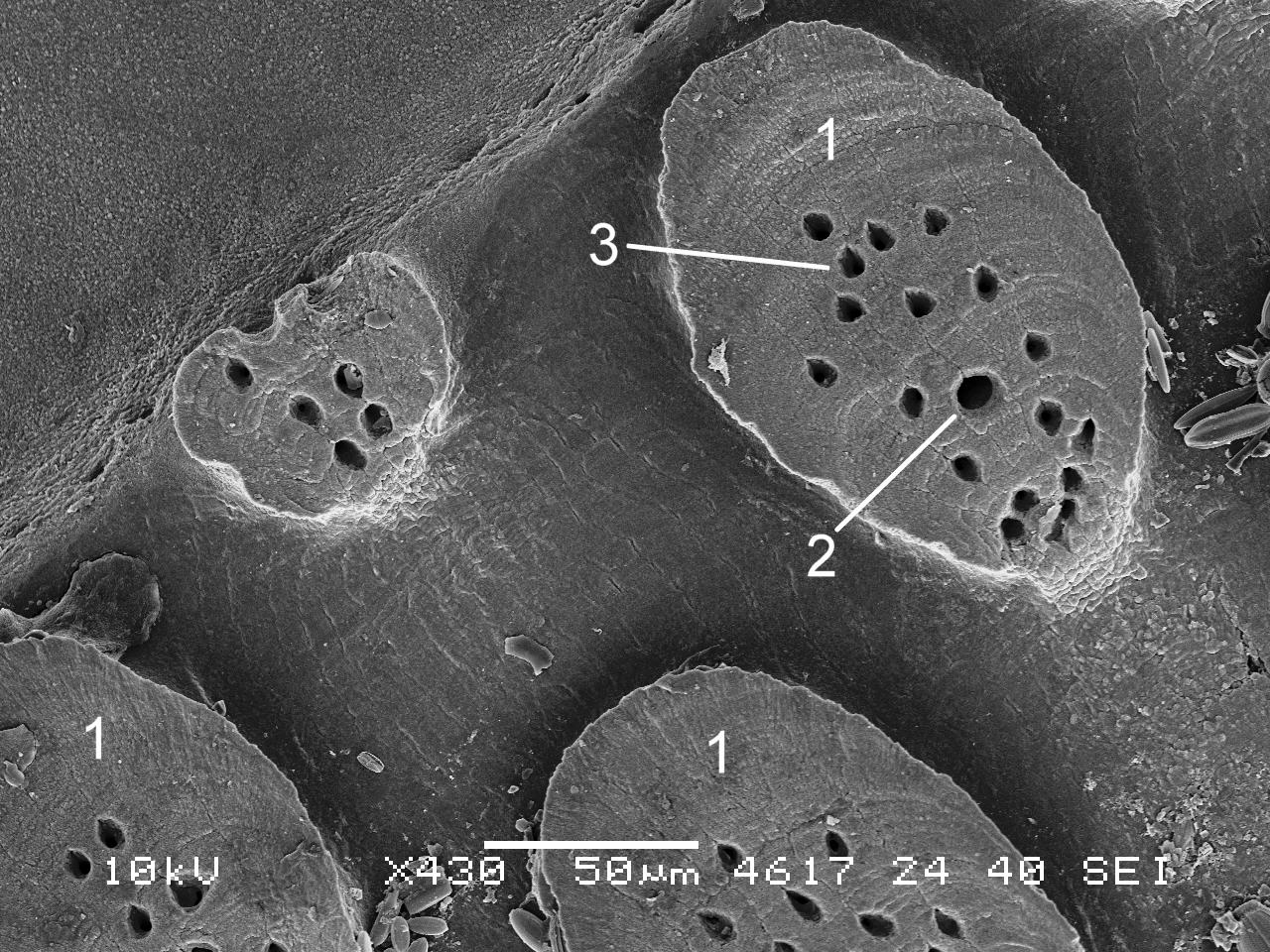
Canals permeate tegmentum and terminate on its dorsal surface in flat-topped granules that are widest and tilted up at anterior 07 Acanthochitona crinita . Each granule has a single megalaesthete (large pore with underlying canal) surrounded by 12 to 25 smaller micraesthetes 08 Acanthochitona crinita. On a single specimen, granules can vary from large (easily seen at X3, 06 Acanthochitona crinita ) to small, oval to tear shaped and from wide random arrangement to close and even.
Two insertion plates, separated by single slit, embedded in girdle on each side of valves ii to vii 06 Acanthochitona crinita ; head valve (i) typically has five slits (but may be fused) and tail valve (viii) has two slits that may be indistinct 05 Acanthochitona crinita . Typical slit formula 5/1/2, but can vary. Valves ii to viii each have two very large rounded apophyses on anterior edge. Apophyses extend under next valve forwards.
Dorsal surface of valves coarsely granular apart from jugal area of valves ii to vii and central area of valve viii which have small granules fused into slight longitudinal ridges bearing small aesthete pores 06 Acanthochitona crinita . Lateral and pleural areas not differentiated as separating diagonal ridge undeveloped. Growth lines obscured by granules. Principal dorsal colours of shell and girdle usually dull grey/green (image S.Roberts) flic.kr/p/4iTtK2 or brown 01 Acanthochitona crinita, sometimes with some orange 13 Acanthochitona crinita , yellow or white 13.1Ac ; jugal area usually paler, often with some dark streaks running in same direction as jugal ribs 06 Acanthochitona crinita . Often coated with detritus or epibiota which further dull appearance (P.Lightfoot) flic.kr/p/o4iVD1 .
Body Description
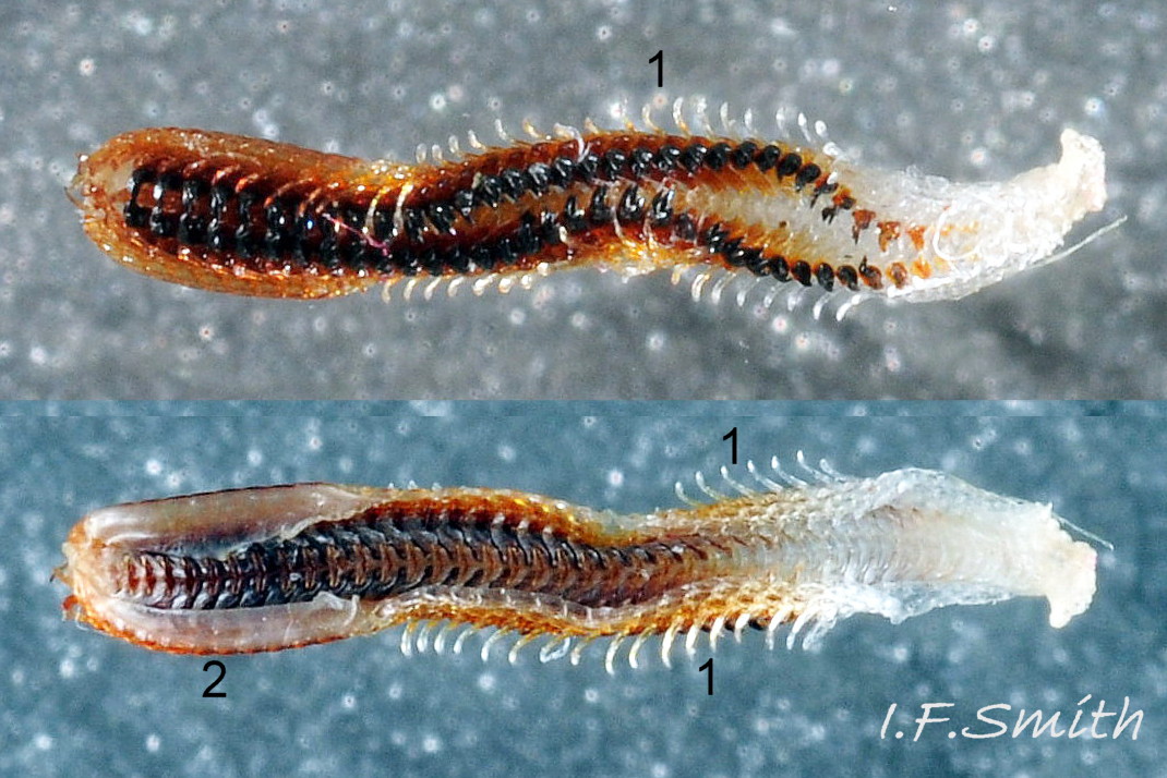
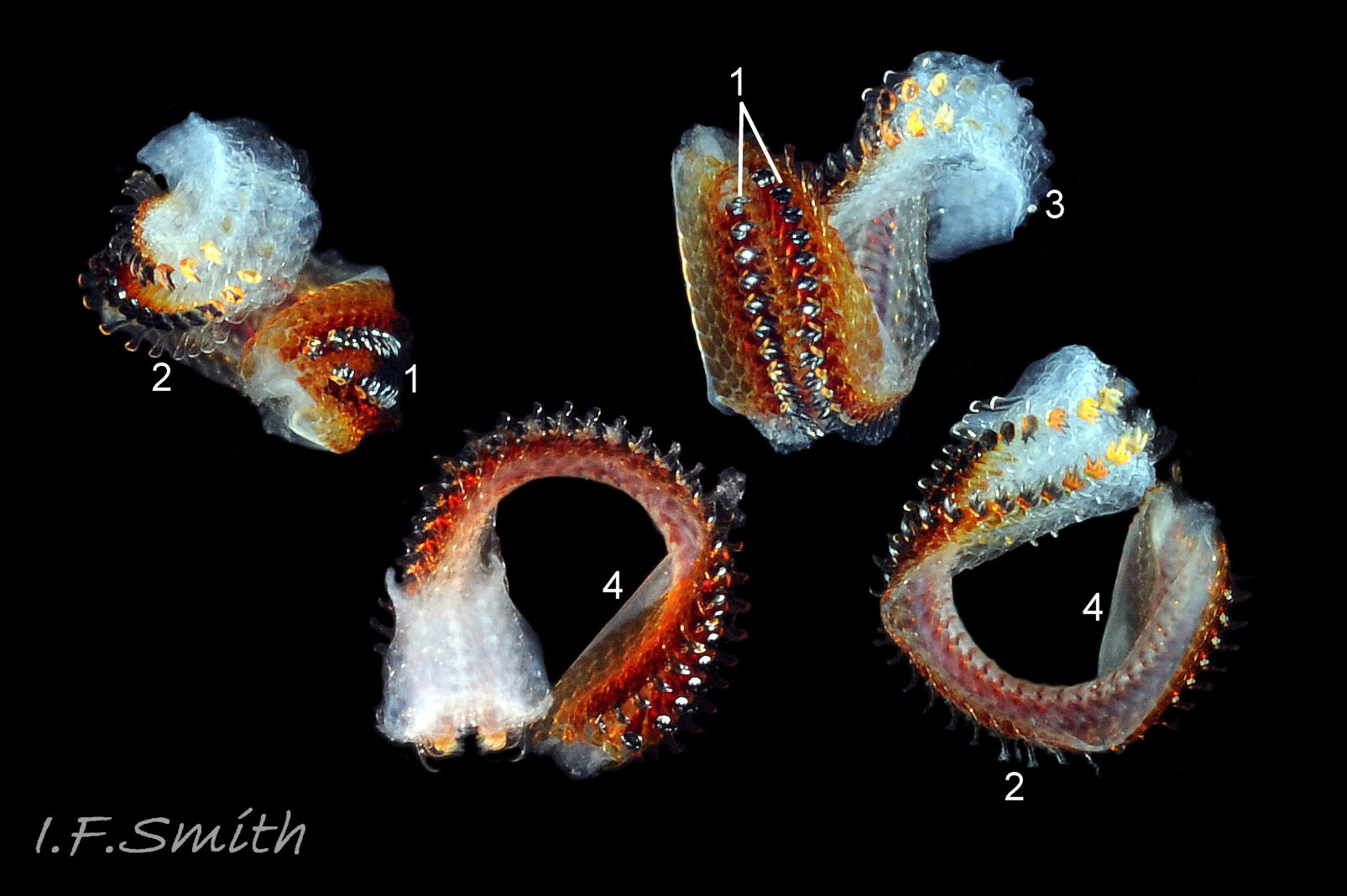
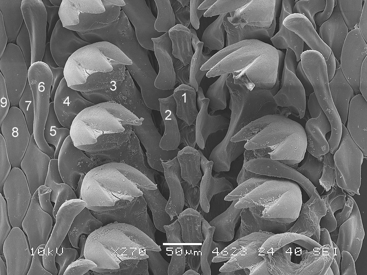
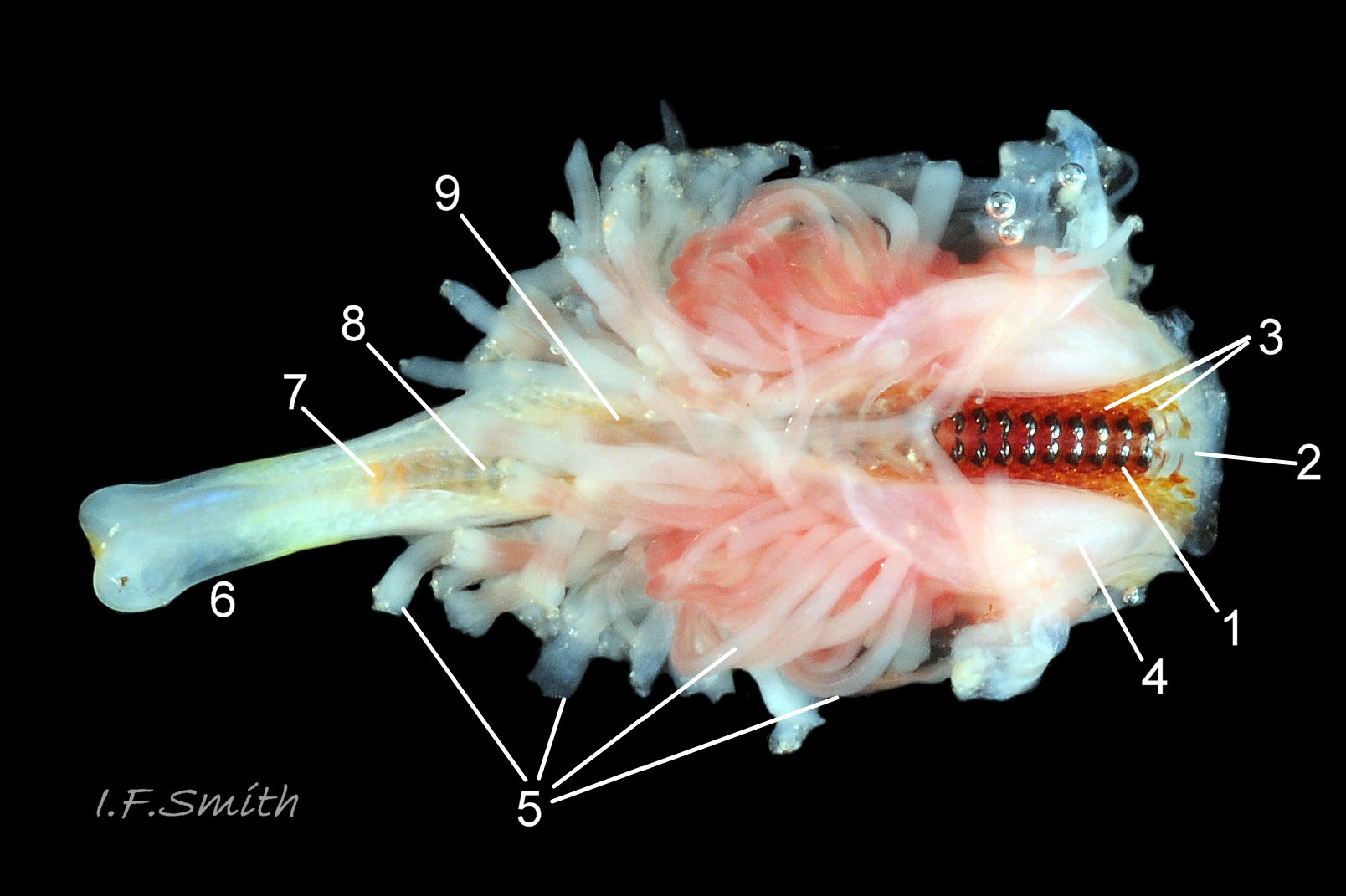
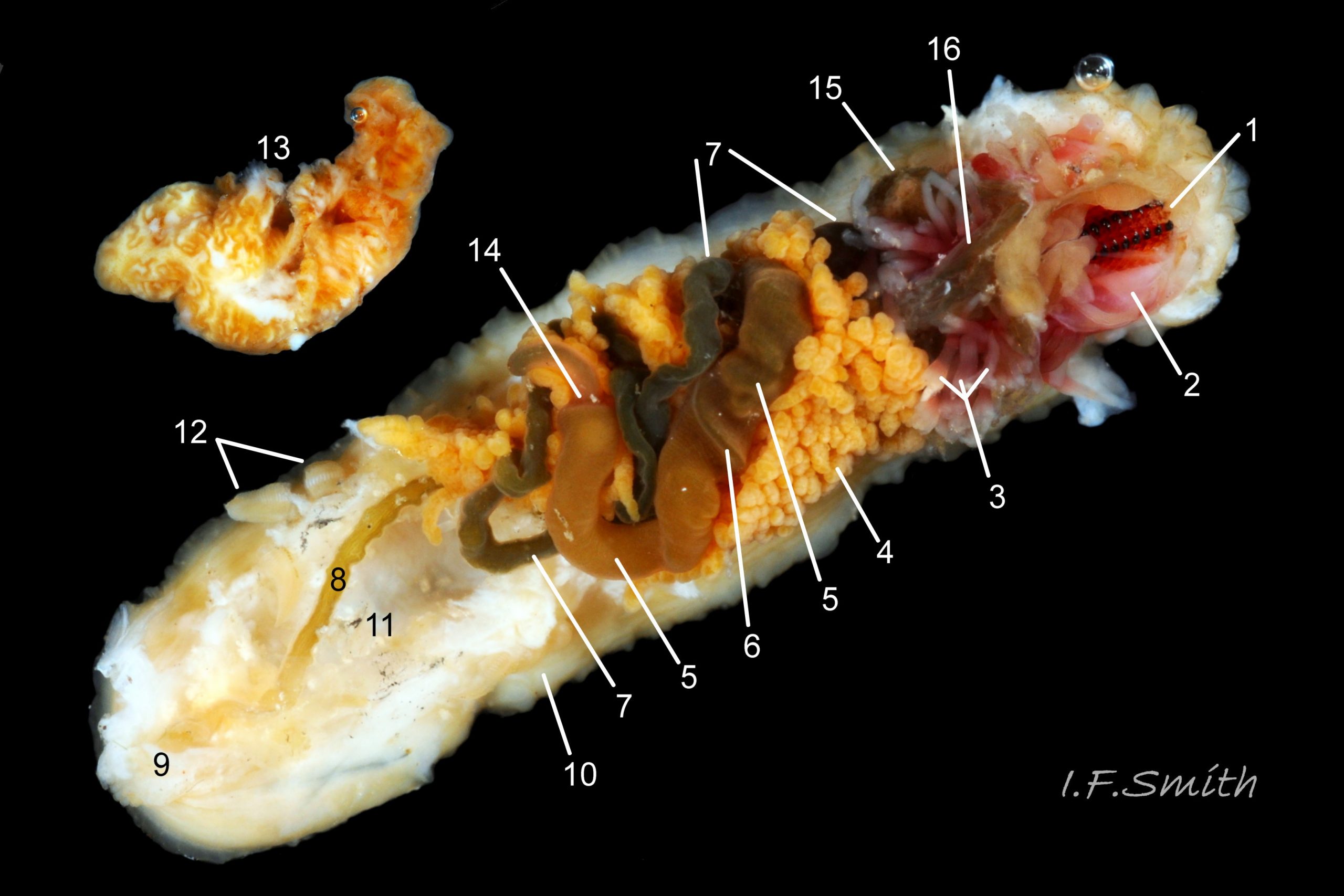
Head and foot never protrude into view naturally on live animal, can only be seen if animal removed from substrate, or placed on glass. Most of head occupied by large transverse slit-mouth with wrinkled lips, surrounded by large 14 Acanthochitona crinita , no eyes or sensory tentacles. As on chitons generally, broad radula 15 Acanthochitona crinita with extremely strong hard teeth of chitin mineralized with magnetite 16 Acanthochitona crinita (so iron rich that adheres to magnet 17 Acanthochitona crinita ) in rows of seventeen 18 Acanthochitona crinita . Teeth pale when formed at rear of radular sac 19 Acanthochitona crinita , darken and harden with magnetite as moved forwards 16 Acanthochitona crinita . In each row a pair of major lateral teeth, the principal scrapers, are the largest and darkest. Major (third) uncinal tooth sweeps up loosened food particles 18 Acanthochitona crinita . Anterior of radula flanked by hyaline shield and supported by reddish myaglobin rich odontophore when grazing rock surface 19 Acanthochitona crinita .
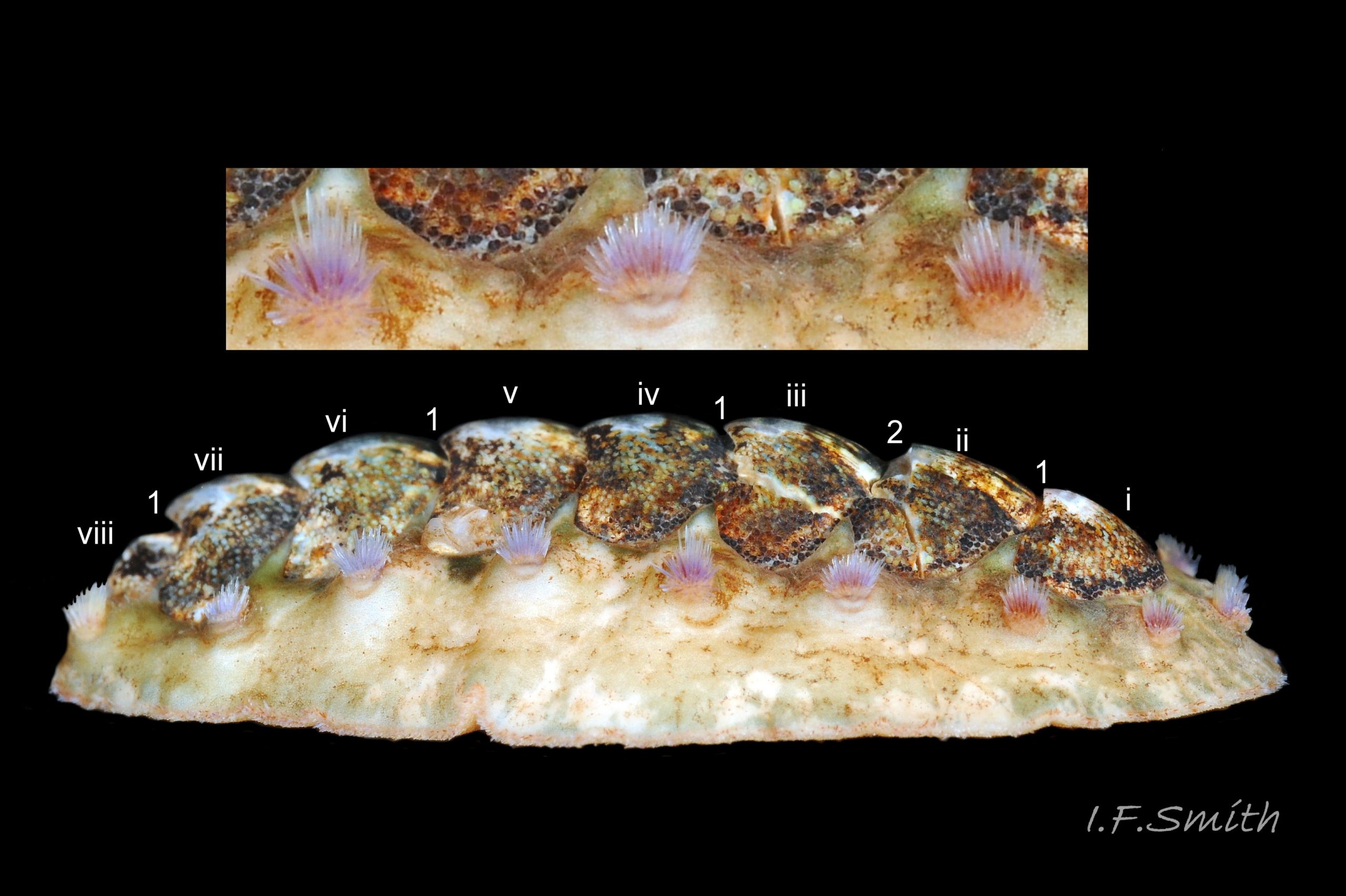
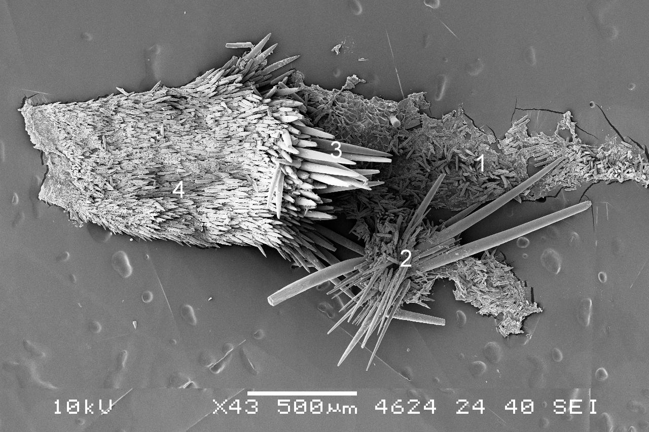
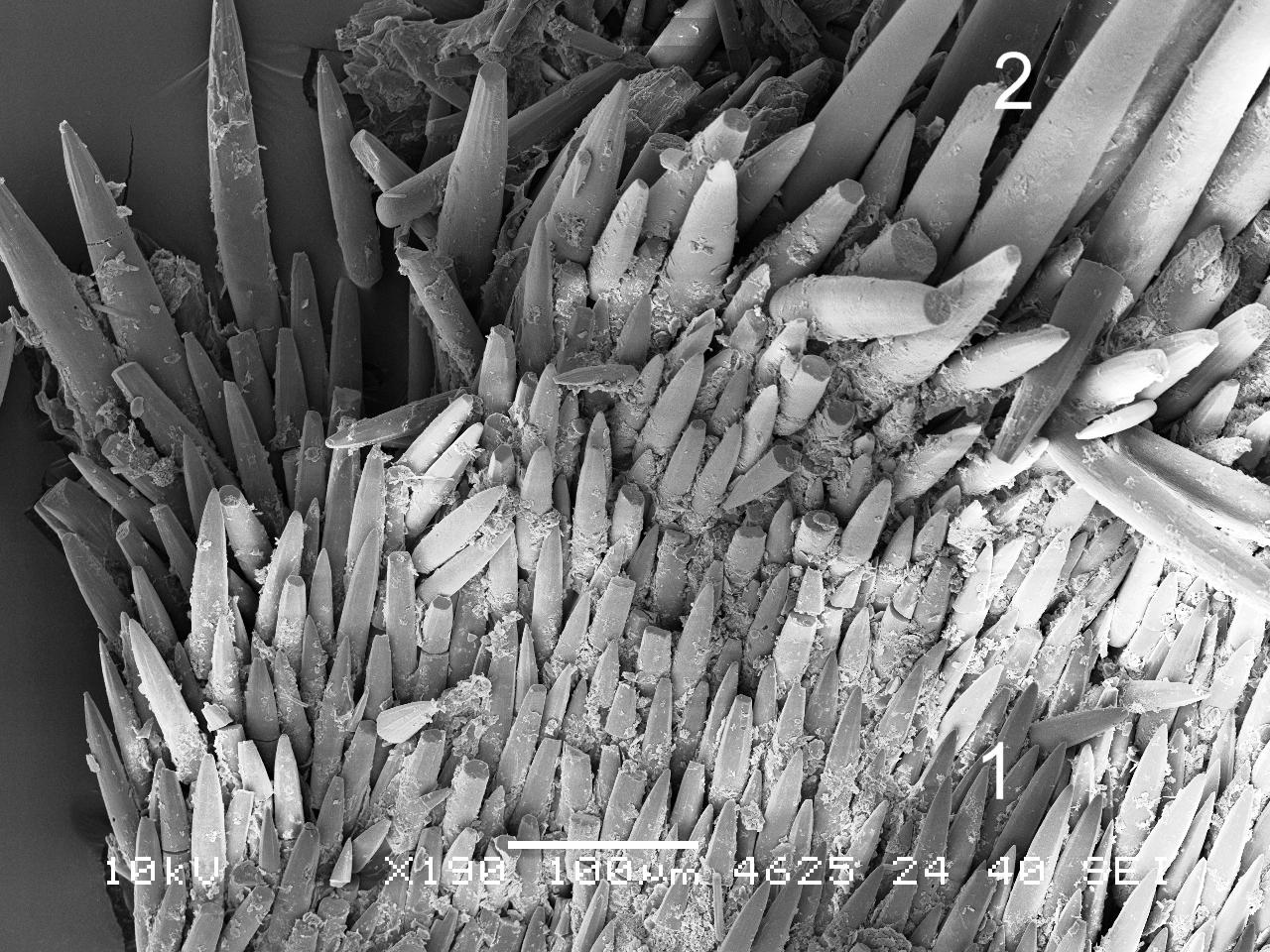
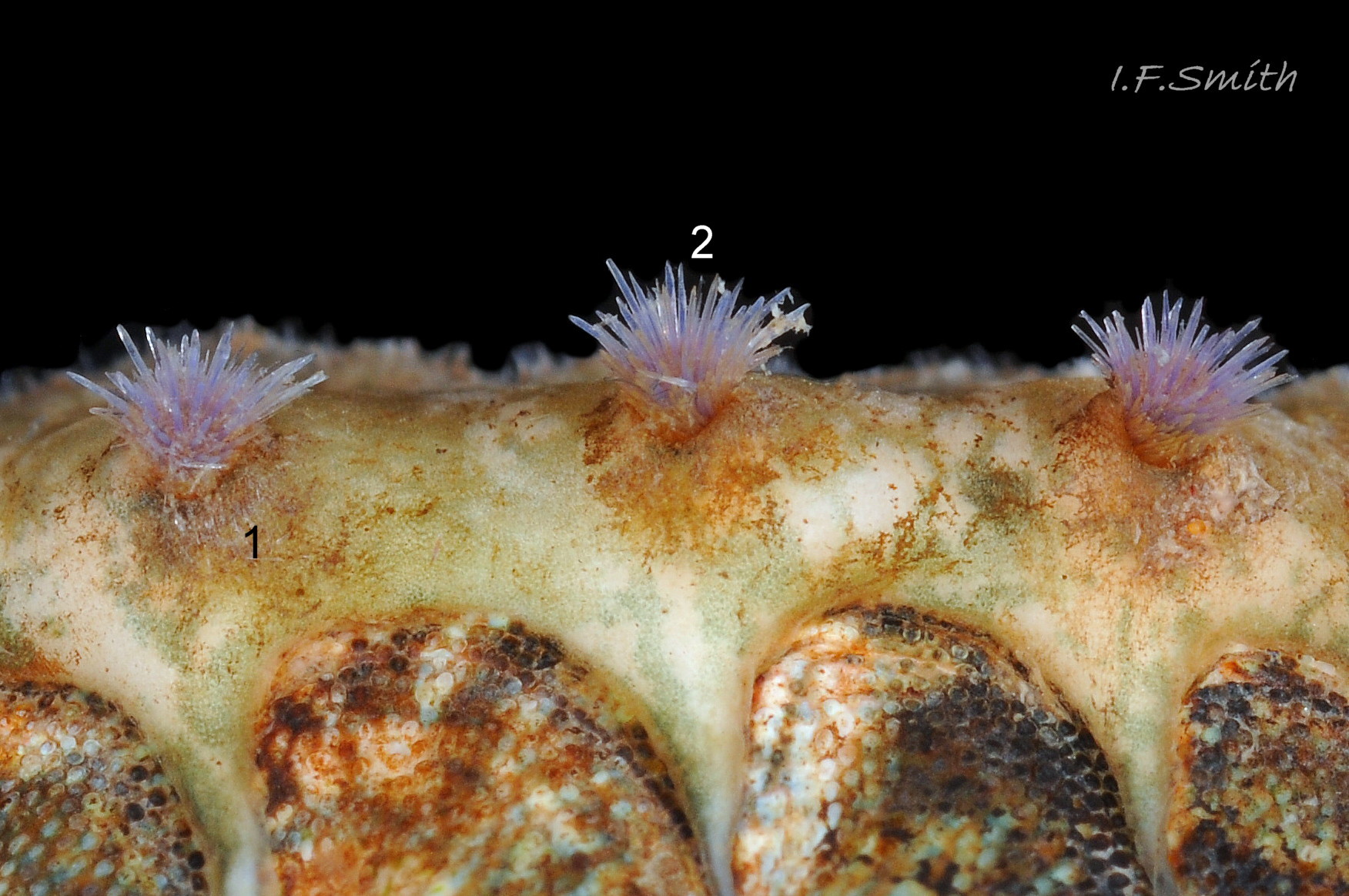
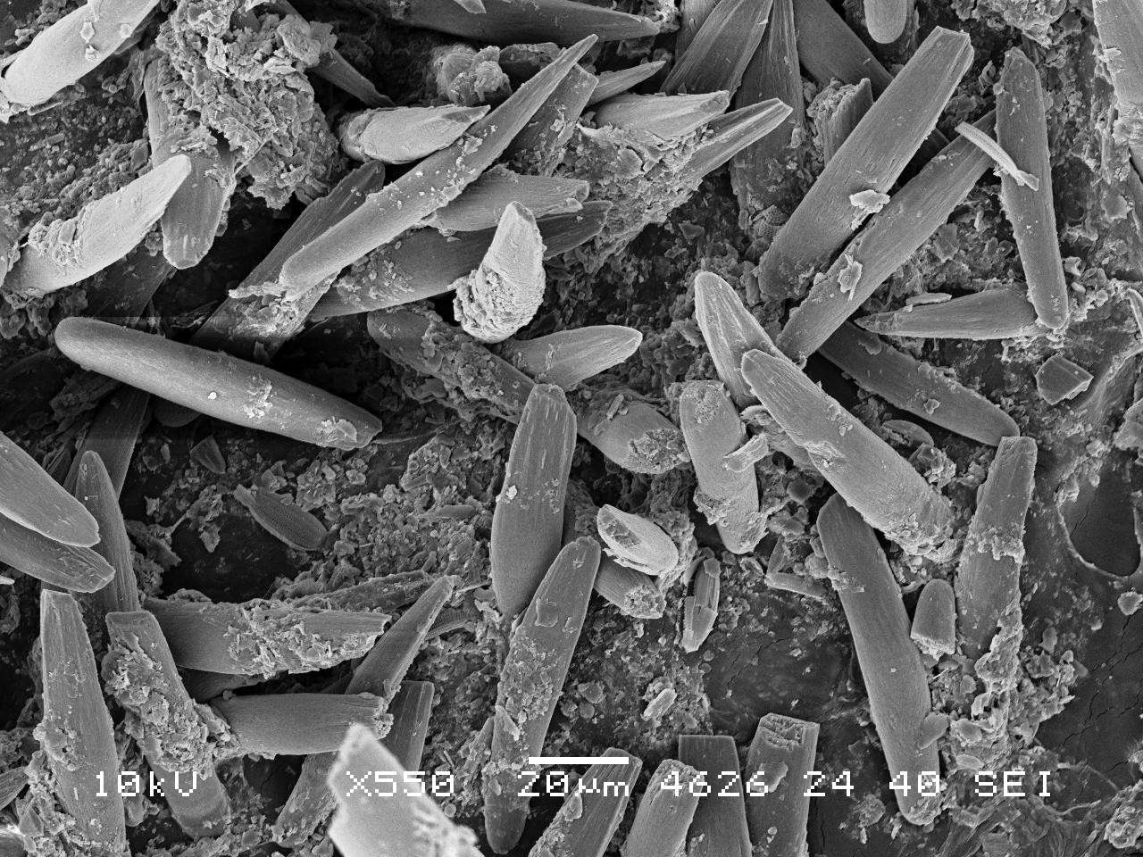
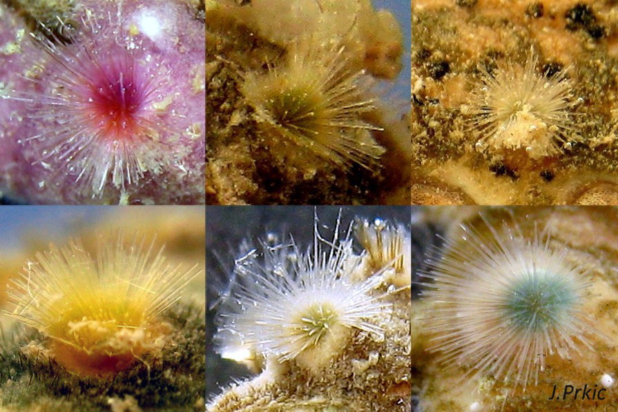
Aesthetes (sensory tissue) fill canals that permeate the tegmentum and parts of articulamentum; terminate in flat topped granules as sense organs on dorsal surface of valves 06 Acanthochitona crinita, 07 Acanthochitona crinita & 08 Acanthochitona crinita. Immediately below the valves, mantle is tough thin translucent epidermis, but greatly thickened where reflected around periphery to form a fleshy girdle into which ends of valves deeply embedded 04 Acanthochitona crinita . On this species, girdle extends across the chiton between the valves, but only fully exposed when animal curls into a ball. Ventral surface of girdle is covered with conical, outwardly directed scale-like spicules, often some stained brownish 20 Acanthochitona crinita, 21 Acanthochitona crinita , 22 Acanthochitona crinita . Dorsal surface of girdle has unaligned, short, straight spines that are spatulate and slightly ribbed distally 21 Acanthochitona crinita 23 Acanthochitona crinita, 24 Acanthochitona crinita and 18 tufts of long (0.6 to 1.0mm) bristles which may be colourless or tinted orange, red or purple; arranged one tuft at each valve intersection and four bordering head valve i 01 Acanthochitona crinita, 12 Acanthochitona crinita 23 Acanthochitona crinita . Peripheral margin of girdle has dense, long spines, though not as long as bristles in tufts 21 Acanthochitona crinita 25 Acanthochitona crinita . Bristles and spines appear not to grow as fast as general body size as they often look relatively small on large specimens, and much longer relatively on small young specimens. Hyponotum can be flexed outwards to form channel at posterior for release of faeces, ova or sperm 26 Acanthochitona crinita .
Open narrow mantle-cavity runs around whole animal; in large specimen contains about fifteen ctenidia on each side in the posterior half only (merobranch arrangement) 27 Acanthochitona crinita . Number of ctenidia varies with age. Between mantle-cavity and hyponotum the mantle-fold is unobtrusive 27 Acanthochitona crinita but can cover ctenidia when in place 26 Acanthochitona crinita . Anus opens into mantle-cavity at posterior near channel formed by deflection/ depression of hyponotum for expulsion of faeces 26 Acanthochitona crinita . Nephridiopores and gonopores open laterally into posterior quarter of cavity. No penis as external fertilization. Foot, elongate ellipse, sole whitish centrally, becoming yellowish towards periphery, no medial dividing line 26 Acanthochitona crinita . Foot spreads widely, concealing pallial cavity with help of mantle fold when gripping substrate.
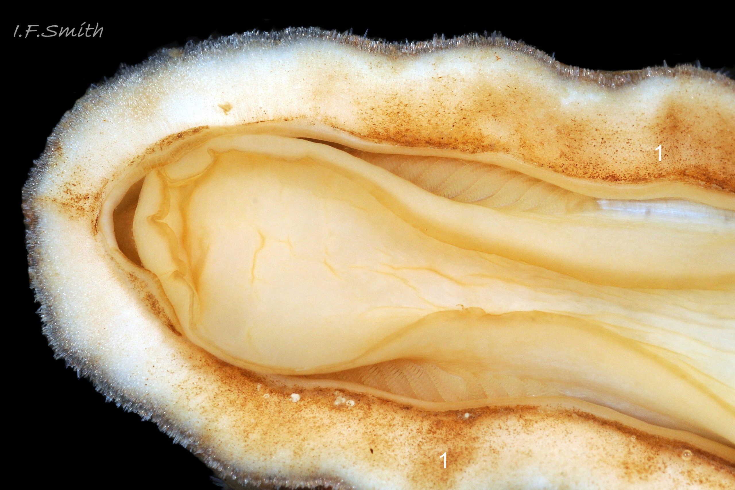
Basic dissection by removal of shell with girdle and shell musculature 28 Acanthochitona crinita , and removal of foot 29 Acanthochitona crinita , reveals the following: radula, hyaline shield, radula retractor muscles, nephridia, stomach, intestine, rectum, anus, dorsal surface of foot, gonads, salivary glands and pharynx (more detail in captions of images 28 Acanthochitona crinita and 29 Acanthochitona crinita ).
Key identification features
Acanthochitona crinita
1: Eighteen conspicuous colourless or colourful tufts of bristles on girdle 01 Acanthochitona crinita & 12 Acanthochitona crinita . Occasional slight variation in number.
2: Slender girdle extension from each side passes between valves but, when animal adhering to substrate, extensions do not meet and are separated by part of valve measuring about 20% of animal’s width 03 Acanthochitona crinita .
3: Girdle is wide, but valves occupy much more of the chiton’s width than the girdle on either side 01 Acanthochitona crinita
4: Dorsal surface of valves has large (and small), often tear shape, flat topped granules, easily visible at X3 if not covered by detritus or epibiota 06 Acanthochitona crinita .
5: From above, coloured tegmentum of intermediate valves (ii to vii) resembles a bow with the prominent posterior beak forming the hand drawing the string 05 Acanthochitona crinita .
Similar species
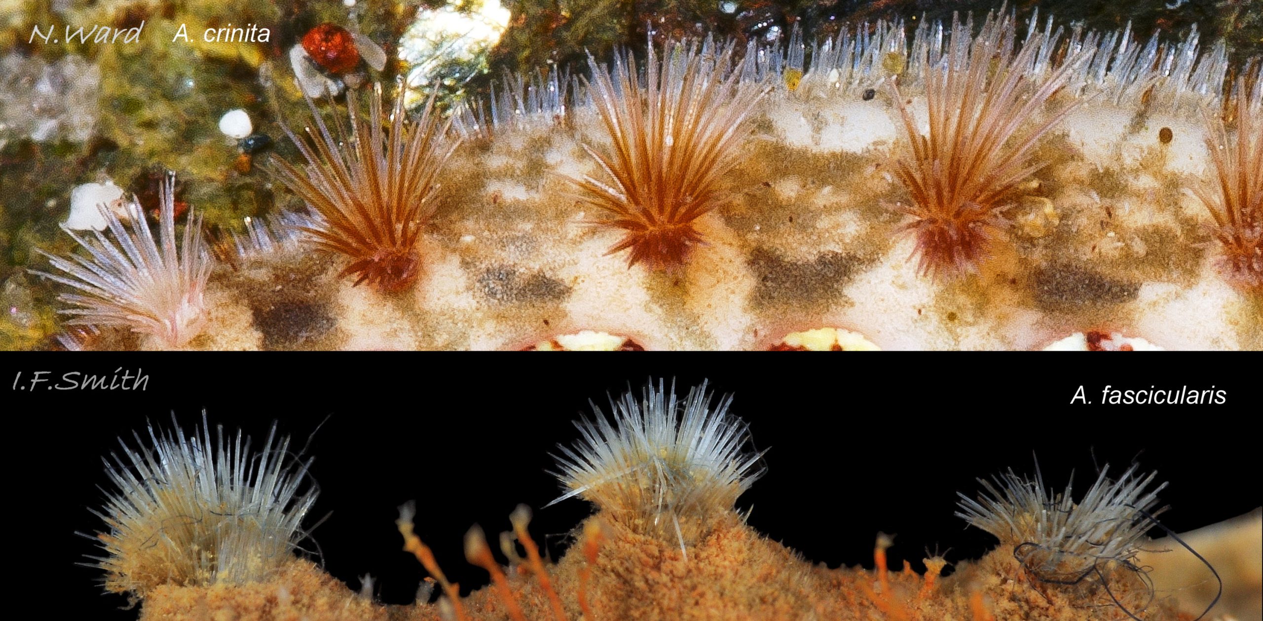
Acanthochitona fascicularis (Linnaeus, 1767).
1: Eighteen conspicuous colourless or colourful tufts of bristles on girdle 30 Acanthochitona crinita COMPARISON Acanthochitona fascicularis , occasional slight variation in number.
2: Broad extension of girdle from each side passes between valves. When animal adhering to substrate, girdle extensions meet or are only separated by very small parts of valves measuring less than 10% of animal’s width 31 Acanthochitona crinita, 32 Acanthochitona crinita .
3: Width of valve about same width as, or narrower than, girdle to side of it 31 Acanthochitona crinita & 32 Acanthochitona crinita .
4: Dorsal surface of valves, apart from ribbed jugal area, has small, evenly sized, rounded, flat topped granules arranged in fibonacci like curves. Not easily seen unless magnified 33 Acanthochitona crinita
5: From above, coloured tegmentum of intermediate valves (ii to vii) varies in shape. 31 Acanthochitona crinita & 32 Acanthochitona crinita
Acanthochitona discrepans (Brown, 1827).
A. discrepans also has eighteen tufts of bristles, and its other features intergrade with A. crinita. Jones & Baxter and others regard them as inseparable. However, A. discrepans is an accepted species on WoRMS; their image is at www.marinespecies.org/aphia.php?p=image&pic=70144 .
Mediterranean Acanthochitona
There are other tufted species in the Mediterranean, such as A. pilosa, Schmidt-Petersen, Schwabe & Haszprunar, 2015 www.researchgate.net/publication/281628035_Acanthochitona… .
Specimens from the Adriatic of A. crinita 13 Acanthochitona crinita appear similar to some specimens from southern England (N.Berkley) flic.kr/p/zVYVey .
Habits and ecology
Lives in rock crevices and on and under stones that are lightly embedded in sand or gravel, on lower shore and Laminaria zone at LWS, also sublittorally to about 50 metres. Shell articulates to conform closely to uneven rock surface; large foot and flat hyponotum grip surface firmly 26 Acanthochitona crinita . When alarmed, can increase grip suctorially by raising part of hyponotum to form partial vacuum, and if displaced from substrate, can roll into a ball 04 Acanthochitona crinita . Respiration: cilia on ctenidia and mantle create inhalent water-current entering pallial cavity wherever girdle is raised at anterior. Adjacent ctenidia have interlocking cilia so all work as a unit (as in many bivalves) 27 Acanthochitona crinita . Water current passes through ctenidia and then along cavity as exhalent current to exit at posterior through channel formed by deflection of hyponotum of girdle 26 Acanthochitona crinita .
In absence of eyes or of tactile or chemoreceptor tentacles on head, senses environment through aesthetes exposed on surface of shell 08 Acanthochitona crinita. Precise functions of aesthetes uncertain, but focusing lens and receptive retina found in aesthetes of some chiton species, but not all. Other proposed aesthete functions include chemoreception, mechanoreception, replenishment of properiostracum materials, and secretion of protective substances. Also sensory organs on girdle.
Feeds by scraping micro algae and associated organisms from the rock surface using its hard radula of chitin mineralized with magnetite 19 Acanthochitona crinita & 28 Acanthochitona crinita . Though primitive in several ways, has elaborate digestive system with no trace of primitive rotating style in stomach. Long coiled intestine compacts faeces into pellets. Water current in pallial cavity carries excreta from lateral nephridiopores to posterior, where faecal pellets from anus 26 Acanthochitona crinita join the flow; all expelled at posterior through channel in hyponotum of girdle.
Travels by monotaxic retrograde compression waves on sole of foot.
Breeding: dioecious. Water current in pallial cavity carries sperm or ova from lateral gonopores 29 Acanthochitona crinita to posterior and out through channel in deflected girdle 26 Acanthochitona crinita . As fertilization is external, synchronised emission of sperm and ova needed to ensure success; trigger in many chiton species is moon-phase/ state of tides. Planktonic trochophore larvae hatch and metamorphose into small adult-form young without intervening veliger stage.
Distribution and status
Widespread, usually in small numbers, occasionally locally common.
Norway to Portugal and western Mediterranean. Not inner Baltic. GBIF map www.gbif.org/species/2305904 . All around Britain and Ireland on suitable substrate, except north east Irish Sea and Flamborough to Kent, NBN UK map at species.nbnatlas.org/species/NBNSYS0000173552
Acknowledgements
I am most grateful to the Naturalis Biodiversity Center, Netherlands, for permission to feature SEM images of its specimens, and to Jakov Prkic for use of his images and helpful discussion. Links to the Flickr images of Paula Lightfoot, Siôn Roberts and Nicholas Berkley are a valued addition to the account. I thank Enrico Schwabe and Jeroen Goud for help and cooperation. I am especially indebted to Dr Julia Sigwart, Director of the Marine Laboratory, Queens University, Belfast, for detailed advice with the text and interpretation of the images; any errors or omissions are my (IFS) responsibility.
Links and references
Botelho, A. 2013. Zoologger: mollusc grows hardest teeth in the world New Scientist 3rd October 2013 www.newscientist.com/article/dn24329-zoologger-mollusc-gr…
Fernandez, C.Z., Vendrasco, M.J. & Runnegar,B. 2007. Aesthete canal morphology in twelve species of chiton (Polyplacophora). The Veliger 49(2) 51 – 69 www.researchgate.net/publication/246548141_Aesthete_Canal…
Forbes, E. & Hanley S. 1849-53. A history of the British mollusca and their shells. vol. 2 (1853) London, van Voorst. (As Chiton fascicularis Linnaeus); Free pdf at archive.org/stream/historyofbritish02forbe#page/392/mode/2up Use slide at base of page to select pp.393-396.)
Fox, R. 2007. Invertebrate Anatomy On Line. Katharina tunicata lanwebs.lander.edu/faculty/rsfox/invertebrates/katharina….
Hayward, P.J. & Ryland, J.S. 1995. Handbook of the marine fauna of north-west Europe. Oxford, Oxford University Press.
Jardim, J.A. & Simone, L.R.L. 2010. Redescription of Hanleya brachyplax (Polyplacophora, Hanleyidae) from the south-southeastern Brazilian coast. Pap. Avulsos Zool. (São Paulo) 50 no.40
www.scielo.br/scielo.php?pid=S0031-10492010004000001&…
Jeffreys, J.G. 1862-69. British conchology. vol. 3 (1865). London, van Voorst. (As Chiton fascicularis var. attenuata); Free pdf at archive.org/stream/britishconcholog03jeffr#page/210/mode/2up . Use slide at base of page to select pp.211-213.)
Jones, A.M. & Baxter, J.M. 1987. Molluscs: Caudofoveata, Solenogastres, Polyplacophora and Scaphopoda London, Linnean Society, and Estuarine and Brackish-water Sciences Association.
Kaas, P. & Van Belle, R.A. 1985. Monograph of living chitons Vol 1. Viderup, Denmark. Brill. Preview at
books.google.co.uk/books?id=CKautf2EUPkC&pg=PA7&s…
Matthews, G. 1967. The identification of British chitons. Papers for Students No.9. London, Conchological Society of Great Britain and Ireland.
Nature22 website, chiton page. nature22.com/estran22/mollusques/polyplacodentale/chitons…
Naturalis Biodiversity Center www.naturalis.nl/en/
Schwabe, E. 2010. Illustrated summary of chiton terminology. Spixiana 33(2):171-194.
Free pdf at verlag-pfeil.de/04biol/pdf/spix33_2_02.pdf
Glossary
μm – 0.001 mm
aesthete (in chitons) = one of complex of canals filled with sensory tissue that permeate tegmentum and parts of articulamentum. Occur in bundles of a large megalaesthete surrounded by several smaller radiating micraesthetes that open as sensory macropores and micropores on dorsal surface of valves. Some are photoreceptors; other function(s) uncertain, may include chemoreception, mechanoreception, properiostracum replenishment and/or secretion of protective substances.
a.k.a. = also known as.
antemucronal area = area situated to anterior of mucro.
apophysis (pl. apophyses) = natural protruberance within a shell for attachment of muscles. On chitons, anterior extension of articulamentum which underlies preceding valve; on all valves except head valve.
aragonite = orthorhombic crystalline mineral-form of calcium carbonate www.minerals.net/mineral/aragonite.aspx . Less common on land than calcite, but, currently, the more frequent mineral-form in oceans and living mollusc shells.
articulamentum = ventral shell-layer of chiton valves, usually hard, white, porcelaneous aragonite and often differently coloured in central part. (Partially overlain ventrally by inconspicuous myostracum layer.)
buccal mass = anterior of digestive system including an odontophore that supports anterior of radula, and a complex of muscles to operate them and other mouthparts. Often red or pink with myoglobin.
caecum = a blind pouch (cul de sac)
calcite = trigonal crystalline mineral-form of calcium carbonate www.minerals.net/mineral/calcite.aspx . More common on land than aragonite, but, currently, the less frequent mineral-form in oceans and living mollusc shells. More resistant than aragonite to acid rain corrosion; forms outer shell layer of shore-dwelling Littorina species in cool climates. (Corrosion of calcium carbonate faster at cold temperatures).
chemoreception = sensing of chemicals; “smell / taste”.
cilia = (pl.) microscopic linear extensions of membrane that move in rhythmic waves to create locomotion, or move particles and liquids e.g. inhalent water currents. (“cilium” singular). (scanning electron microscope image at 18 Lepidochitona cinerea )
ciliary = (adj.) relating to or involving cilia.
ciliated = (adj.) coated with cilia.
chitin = semitransparent flexible horny protein.
chitinous = (adj.) made of chitin.
coll. = in the collection of (named person or institution) (compare with legit).
ctenidium = comb-like molluscan gill; usually an axis with a row of filaments either side.
ctenidia (pl.) = comb-like molluscan gills; usually an axis with a row of filaments either side.
dioecious = having separate male and female individuals, not hermaphrodite.
direct = (of locomotion waves on foot) waves travel from posterior to anterior.
distal = away from centre of body or point of attachment.
epibiota = organisms living on surface of another organism.
ELWS = extreme low water spring tide (usually near March and September equinoxes).
girdle = (on chiton) peripheral band of thickened, reflexed mantle that encloses ends of valves.
gonopore = opening through which eggs or sperm are released.
haemoglobin = oxygen-carrying substance in blood; scarlet when oxygenated.
holobranch (of chitons) = arrangement of gills in pallial groove that extends full length of foot.
hyponotum = ventral surface of chiton’s girdle.
in litt. (abbreviation of Latin “in litteris”) = in upublished correspondence.
insertion plate (on most chitons) = extension of articulamentum on lateral margin of intermediate valves, anterior margin of head valve and posterior margin of tail valve. Inserts into, and anchors valve to, the girdle muscle block.
intermediate valve = (of chiton) any valve (ii to vii), except head valve (i) and tail valve (viii).
jugal area = triangular middle section of central area of intermediate valves, with apex pointing to posterior; discernible when defined by differences of colour and/or sculpture (dorsal surface).
jugal tract = triangular middle section of central area of intermediate valves, with apex pointing to posterior; discernible when defined by densely arranged aesthete pores (ventral surface).
jugum = triangular middle section of central area of intermediate valves. (See jugal area and jugal tract.)
lateral area (on intermediate valve of chiton) = triangular area with its base along lateral edge of valve and its apex near the centre of the posterior edge. a.k.a. lateral triangle.
legit (abbreviation; leg.) = collected/ found by (compare with coll.)
LWS = low water spring tide, two periods of a few days each month when tide falls lowest.
magnetite = mineral of iron oxide, hardest material made by any living organism (Botelho, 2013).
major lateral teeth (o chiton) = largest two teeth in each row of seventeen on chiton radulae; principal scraping blades. Often darkened more than other teeth by high magnetite mineralization.
mantle = sheet of tissue covering visceral mass of molluscs. Secretes shell of shelled species, and forms part or all of dorsal body surface (notum) of those without shells. On chitons, forms pallial cavity and is toughened to form the girdle surrounding the shell valves. (See mantle skirt.)
megalaesthete = (see aesthete).
merobranch = (of chitons) gills in pallial groove only in posterior two-thirds of animal.
micraesthete = (see aesthete).
monotaxic = (of locomotion waves on foot) single series of waves across complete width of foot.
motile = (adj.) able to move spontaneously; applicable to whole animals or to parts of them such as cells, gametes or cilia.
mucro = (on chiton) projection on tail valve (viii) demarking posterior from rest of valve. Varies in prominence and position.
mucro = a short sharp point at the end of a feature or organ.
mucronate = bluntly rounded end with small terminal point. eflora.library.usyd.edu.au/glossary/mucronate
myoglobin = red oxygen-binding protein in muscle tissue; often in buccal-mass muscles of gastropods. Similar to red haemoglobin in vertebrate blood, but green haemocyanin is usual oxygen-carrier in mollusc blood. See www.researchgate.net/publication/251227038_Radular_myoglo…
myostracum = microscopically thin discontinuous innermost layer of chiton valve.
nephridium (pl. nephridia) = cilia-lined excretory/osmoregulatory tubule (kidney).
nephridiopore = opening of nephridium for excretion. a.k.a. nephropore, or renal pore.
odontophore = firm, approximately ellipsoid, structure of cartilage supporting radula. Protruded like a tongue to operate radula. Often reddish from myoglobin, and medially grooved.
penes (pl. of penis) = male copulatory organs.
perinotum = dorsal surface of chiton’s girdle.
phototactic (adj.) = of species that moves towards light (positively phototactic) or away from it (negatively phototactic).
plankton = animals and plants that drift in pelagic zone (main body of water). cf. “nekton”.
pleural area (on intermediate valve of chiton) = triangular area with its base along anterior edge of valve and its apex near the centre of the posterior edge. a.k.a. median triangle.
porcelaneous = resembling vitreous glazed ceramic material.
postmucronal = situated to posterior of mucro on tail valve.
properiostracum (on chitons) = proteinaceous material covering the shell. Different composition from periostracum of most other molluscs.
radula = ribbon of chitin bearing chitinous teeth that is extruded on a tongue-like odontophore of cartilage to rasp food. On chitons and limpets, teeth are usually impregnated with magnetite, a hard magnetic mineral of iron.
en.wikipedia.org/wiki/Radula
radular sac = tube, ending in a caecum, where radula is created and stored.
rec. = recorded by (person who submitted record, may be different from leg. and coll. persons/institution).
retrograde (of locomotion waves on foot) = waves travel from anterior to posterior.
scar = mark on shell made by attachment point of muscle or other body part.
side slope (on chiton) = shape in profile view (from posterior or anterior) of lateral areas of intermediate valves; may be straight, convex, concave or a combination of these.
sublittoral = below level of low water spring tide
tegmentum = outer shell-layer of chiton valves, usually porous and relatively soft. (Covered by properiostracum when live.)
trochophore = spherical or pear-shaped larvae that move with aid of girdle of cilia. Stage preceding veliger, passed within gastropod egg in most spp. but free in plankton for limpets, Trochidae, Tricolia pullus and (with no veligers) chitons.
veliger = shelled larva of marine gastropod or bivalve mollusc which swims by beating cilia of a velum (bilobed flap). Stage may be passed in plankton or within liquid-filled egg-capsule.