“Head scar” confusion
Many authors use the “head scar” on the shell interior as a feature for distinguishing British species of limpet. This term is anatomically inaccurate, and is vague and confusing in respect to location and extent.
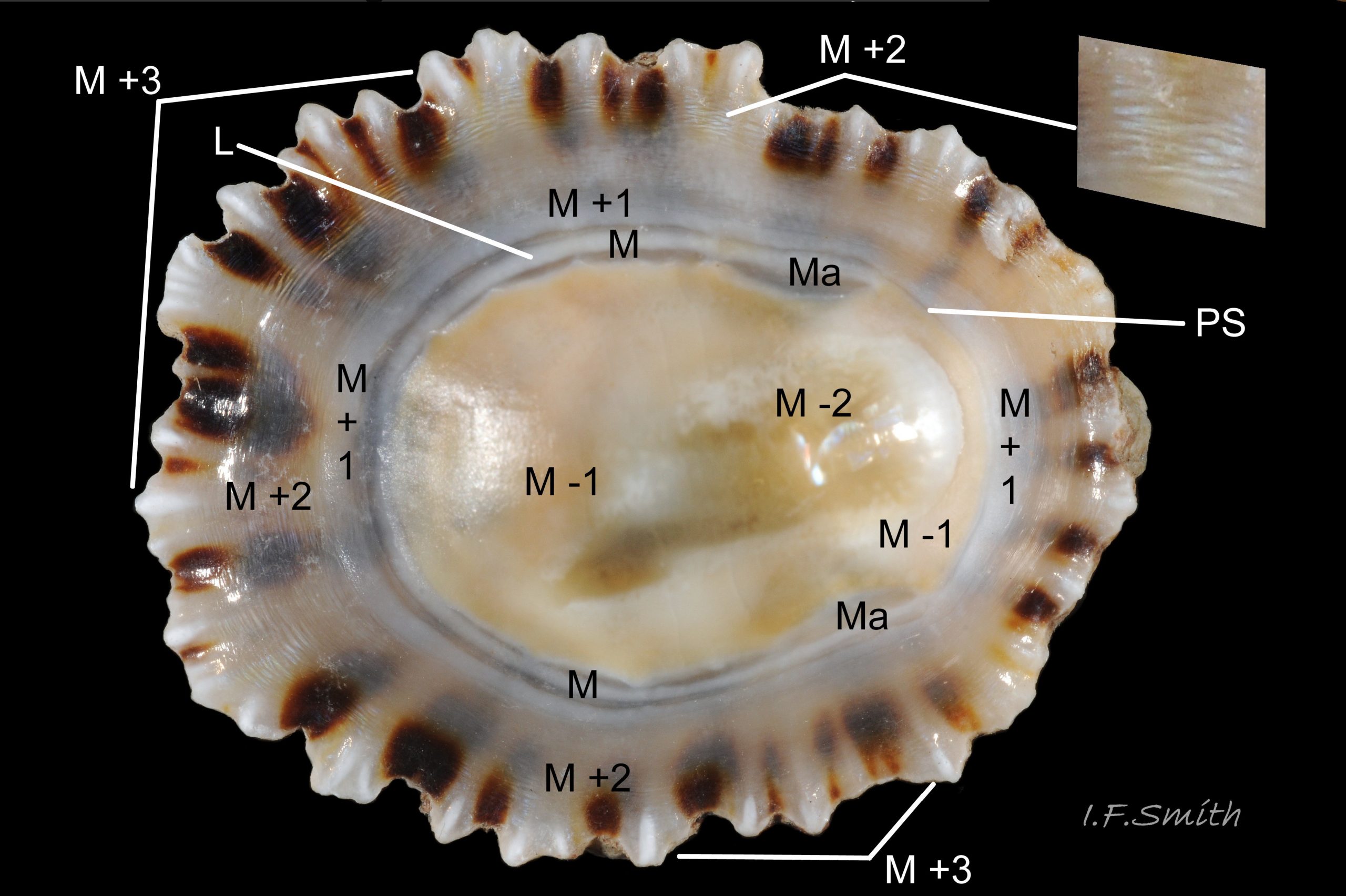
Scars on the interior of shells mark the attachment points of muscles and other body parts. A limpet’s head is contained within the nuchal cavity; it is not attached to the shell or to the mantle forming the cavity-roof so cannot form a scar 6P 06 Patella shell. Underside of live Patella vulgata. .
The main scar inside a limpet shell is a horseshoe-shaped mark made by the shell-muscle (M). The open end of the horseshoe is closed by a thin pallial scar (PS), and the enclosed area resembles a wide-necked amphora (M -1). It contains a thin layer of porcelaneous material that lacks pigment when deposited, but often subsequently acquires a coloured stain, probably from the digestive gland.
The "head scar" is variously regarded as :
1) the whole of the amphora-shape,
2) just the amphora-neck which marks the position of the nuchal cavity,
3) a patch of material within the amphora near the vertex of the shell that is differentiated by greater reflectivity or iridescence and, sometimes, colour (M -2). It is not always present and varies in extent and outline, sometimes spreading onto the posterior of the area above the nuchal cavity.
“Head scar” is not used in this Flickr collection. When appropriate and accurate, “vertex patch” or the standard terms described below are used.
Shell structure of Patella species.
See 1P 01 Patella shell. Stylised transverse section through shell. Thickness of pigment-less interior layers exaggerated; colours selected to resemble those seen through the layers of specimen in image 2 of this set. for cross section diagram
Pigment is incorporated into the substance of the shell during secretion only at the mantle edge creating the exterior layer (M +3). The rest of the mantle typically secretes five thin, overlapping, pigment-less layers that line the interior of the shell. The layers have varying crystalline structures with differing degrees of transparency and iridescence that affect the visibility through them of the colours in the exterior layer.
The layers are numbered from the scar made by the horseshoe-shaped shell-muscle (pedal retractor muscle). The scar is called the myostracum (abbreviation M). Working out from it, the layers are named M +1, M +2 and M +3. Working inwards, the layers are M -1 and M -2. Please note that this notation refers to relative position only; the number of layers and the crystal form of each layer varies from species to species. The crystal descriptions below are of Patella vulgata as an example, but the correlation of shell layers to soft parts applies to all of P. vulgata, P. depressa and P. ulyssiponensis.
M +3: exterior layer of shell produced by edge of mantle (E on 7P 07 Patella. Full grown (shell length 40 mm) Patella ulyssiponensis removed from shell to fully expose mantle. ). Only layer with pigment incorporated in its substance. Composed of large, irregular, foliated (flattened) crystals of calcite (mineral form of calcium carbonate) arranged radially (at right angles to rim of shell).
All the following layers are on the interior surface of the shell, thin and lack pigment when deposited.
M +2: formed by the free mantle skirt (S on 7P 07 Patella. Full grown (shell length 40 mm) Patella ulyssiponensis removed from shell to fully expose mantle. ). Composed of large, irregular foliated crystals of calcite arranged concentrically (parallel to rim of shell). Concentric arrangement can be seen in small concentric ripples that iridesce blue or green. Colours of pigmented exterior layer clearly seen through this single layer, especially in P. depressa and young specimens of P. vulgata and P. ulyssiponensis. This layer underlies all the others that have grown over it; it may be exposed on the external surface of the shell if M +3 is eroded 4P 04 Patella shell. Exterior of Patella depressa shell. Length 24.9 mm. .
M +1: formed by the mantle-roof of pallial groove where gills (G on 7P 07 Patella. Full grown (shell length 40 mm) Patella ulyssiponensis removed from shell to fully expose mantle. ) are suspended. Composed of small, lamellar crystals of aragonite (another mineral form of calcium carbonate) packed like leaves of a book, arranged concentrically.
M: myostracum; scar made by the horseshoe-shaped shell-muscle (a.k.a. pedal retractor muscle) (PR on 7P 07 Patella. Full grown (shell length 40 mm) Patella ulyssiponensis removed from shell to fully expose mantle. ). Extremely thin deposit of complex prismatic material. May show colours of exterior layer (less clearly than in M +2), and may include a line of different colour if located over a previous resting position of M +1, as in image above (L).
Ma: anterior end of myostracum; wider because it is attachment point of largest bundle of muscles in the shell-muscle (PRA on 7P 07 Patella. Full grown (shell length 40 mm) Patella ulyssiponensis removed from shell to fully expose mantle. ).
M -1: formed by mantle covering visceral hump and nuchal cavity (V on 7P 07 Patella. Full grown (shell length 40 mm) Patella ulyssiponensis removed from shell to fully expose mantle. ). Composed of small, lamellar crystals of aragonite, packed like leaves of a book, arranged radially. Porcelaneous, usually not transparent. May be partly or entirely stained with colour from digestive gland located immediately below mantle, so colour may vary with diet. For example, limpet in 2P 02 Patella shell. Interior of Patella vulgata shell. Length 20.2mm. Specimen chosen for clarity. Many P. vulgata are not as clearly marked or so coloured. was found feeding on red encrusting algae; has reddish M -1, perhaps because of its diet.
M -2: layer may be well developed to absent on different individuals of same species. Formed near vertex of shell by part of mantle covering visceral hump. Composed of foliated crystals of calcite, arranged radially or irregularly. Often a reflective sheen or iridescence.
Links and references
Cohen, A.L. & Branch, G.M. 1992. Environmentally controlled variation in the structure and mineralogy of Patella granularis shells from the coast of southern Africa: implications for palaeotemperature assessments. Palaeogeography, palaeoclimatology, palaeoecology, 91: 49-57. www.whoi.edu/fileserver.do?id=163844&pt=2&p=36767
MacClintock, C. 1967. Shell structure of patelloid and bellerophontid gastropods (Mollusca). Peabody Museum of Natural History, Yale University. Bulletin 22. pdf at
www.google.co.uk/?gws_rd=ssl#q=MacClintock%2C+C.+1967.+Sh….
215 pages, may take a few minutes to download. Contents on page v.(= p.6 of pdf). To find pages on pdf add 1 to Roman numerals, and add 11 to modern numerals.