Click image to enlarge with full caption. Main text below slider.
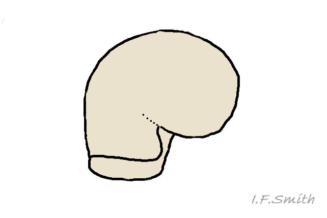
01 Patella pellucida. Shell of veliger larva, Growth Stage 1 (GS1). Drawing based on Lebour (1937).
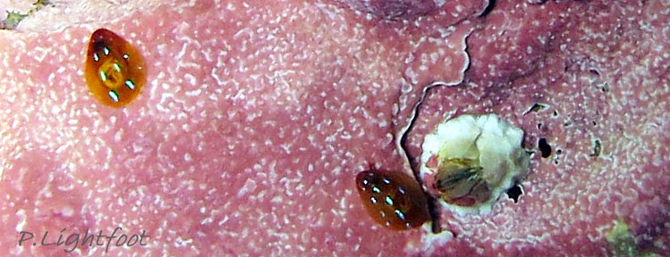
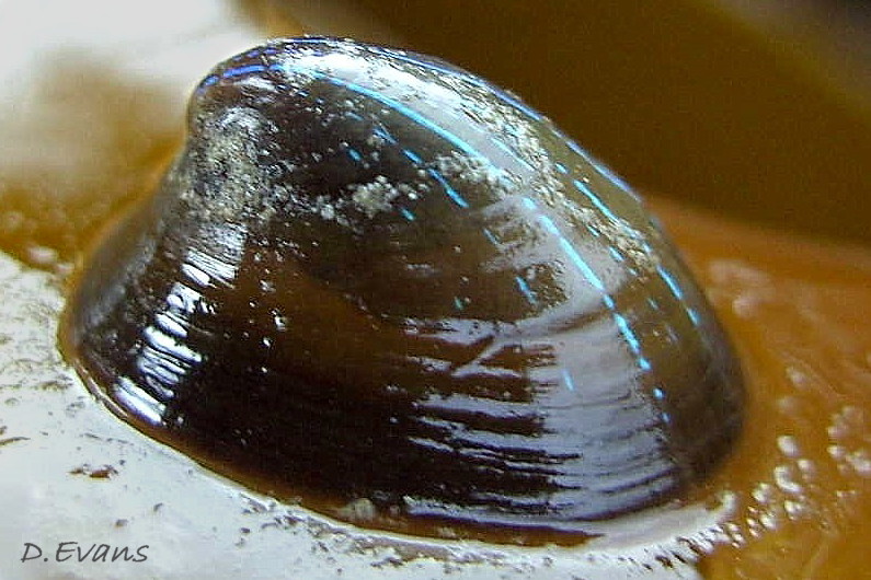
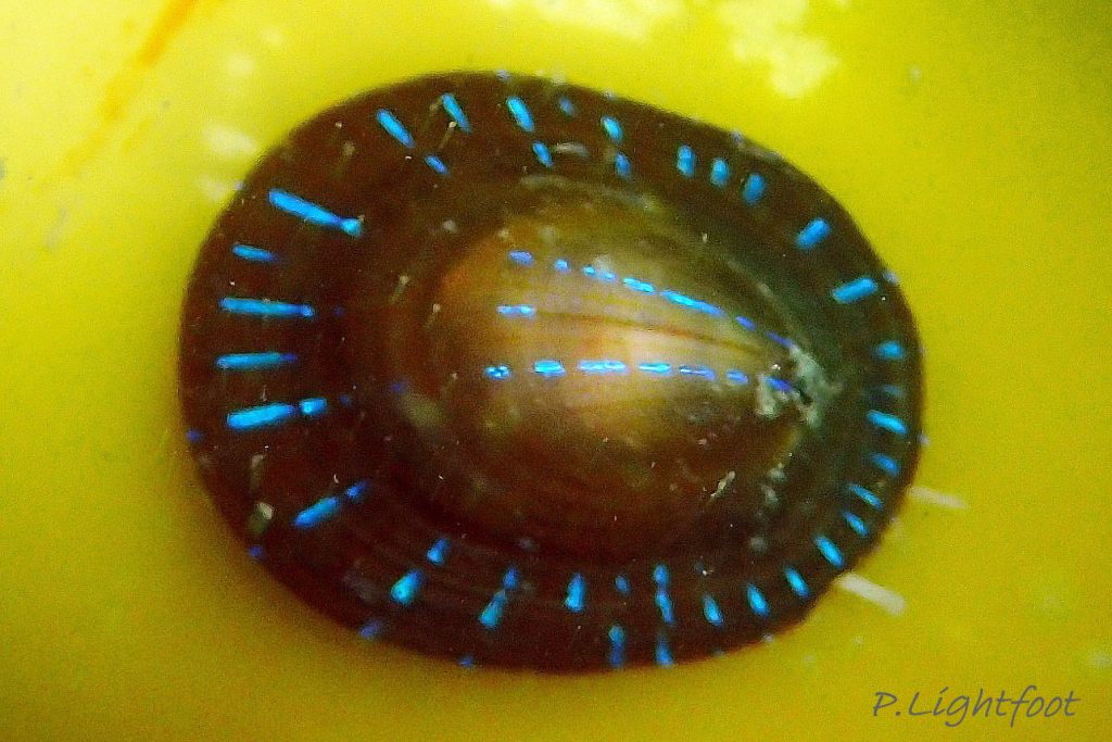
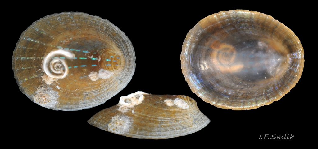
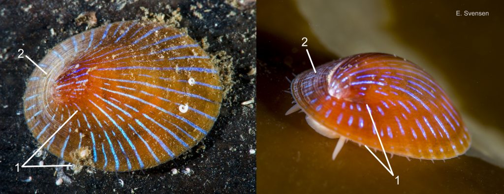
02 Patella pellucida. Growth Stage 2 (GS2). July 2012. Littoral. North Yorkshire, England. © P. Lightfoot.
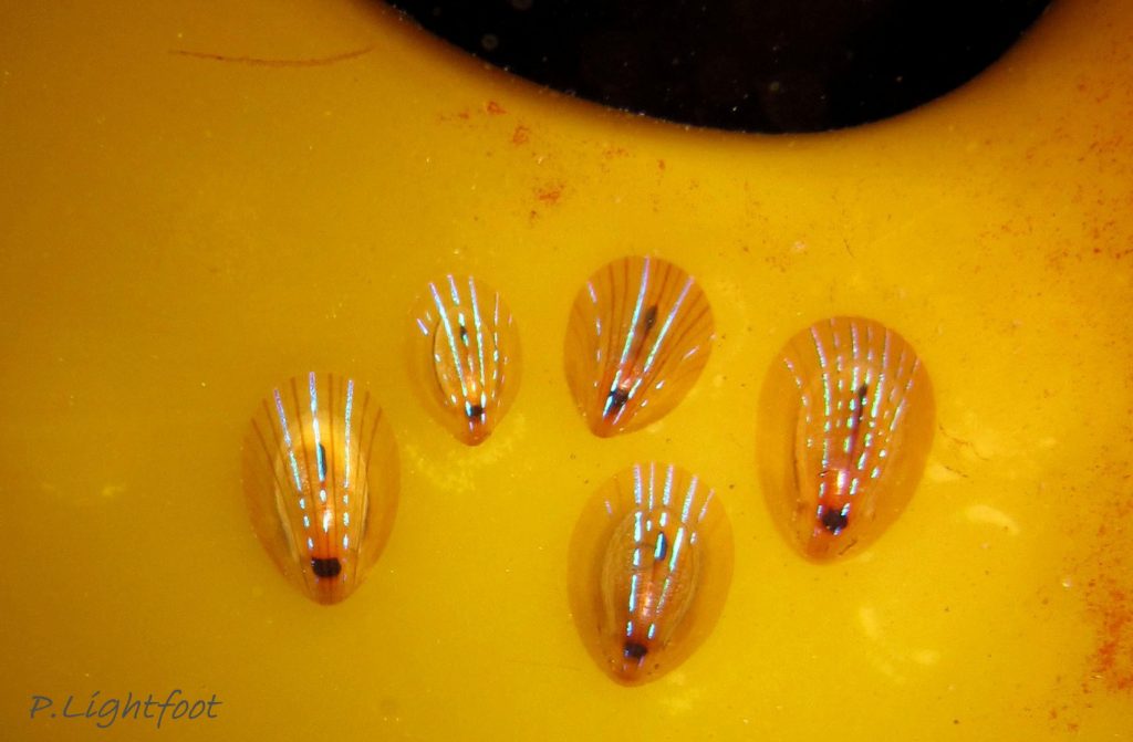
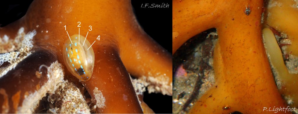
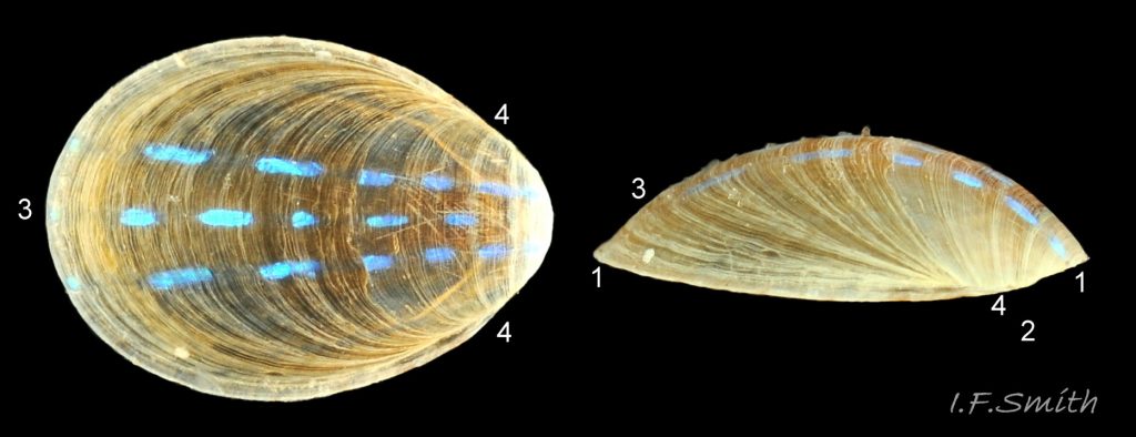
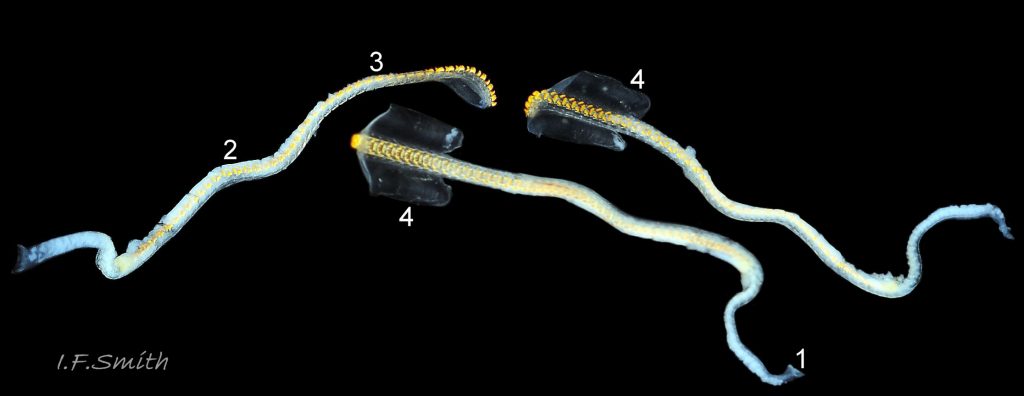
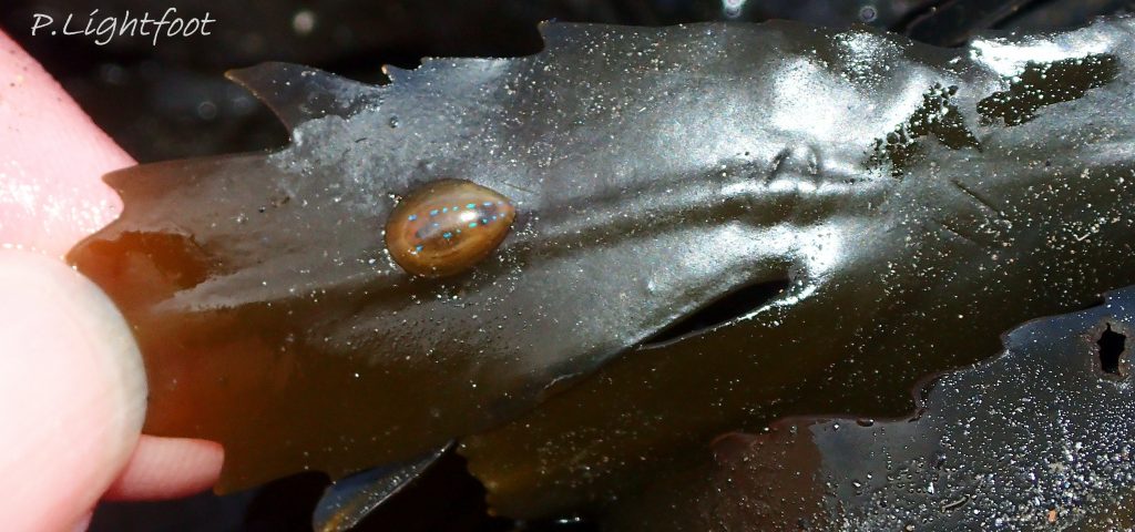
03 Patella pellucida. Growth Stage 3 (GS3). 2009. Sublittoral, Farne Islands, Northumberland, England. © P.Lightfoot.
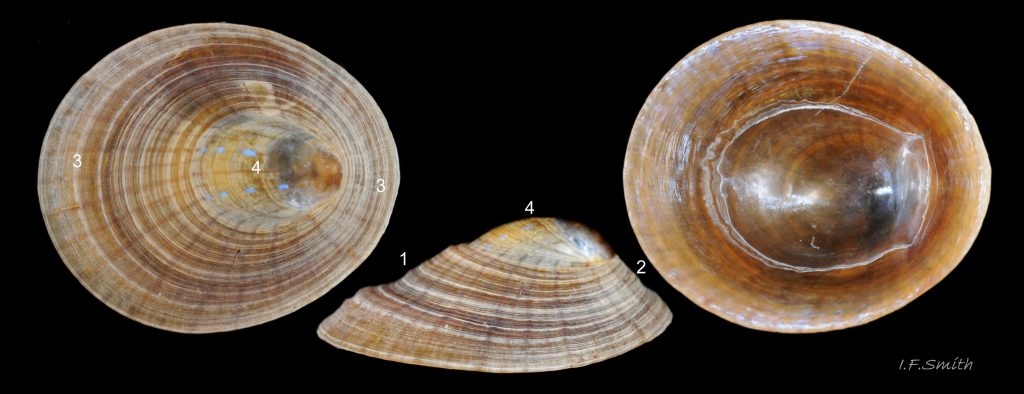
04 Patella pellucida. Length 4.6mm. Growth Stage 3 (GS3). September 2016. Yorkshire, England. © P.Lightfoot.
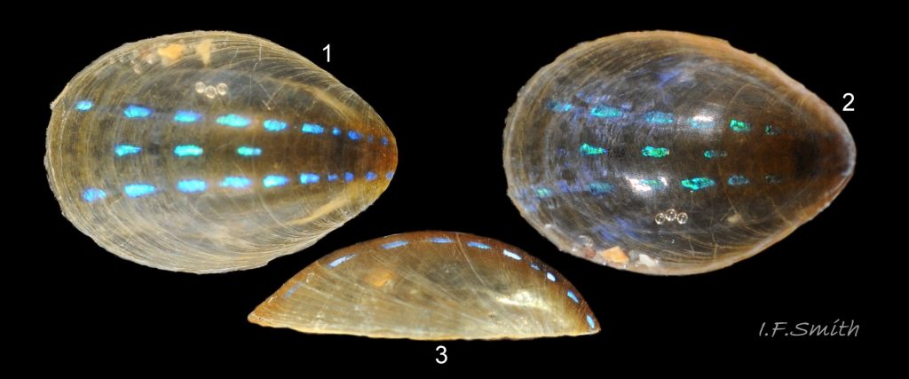
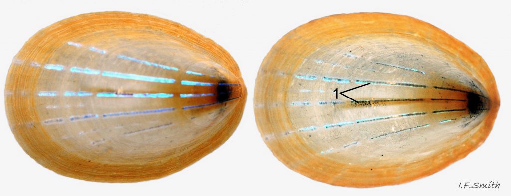
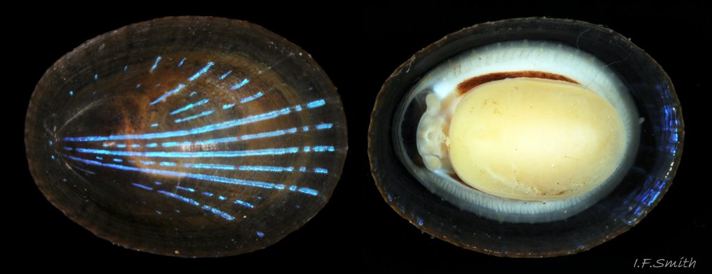
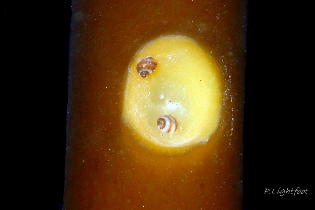
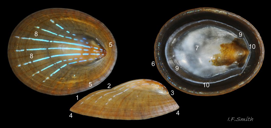
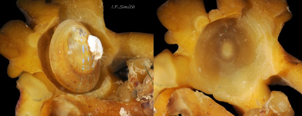
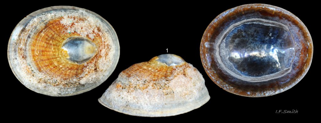
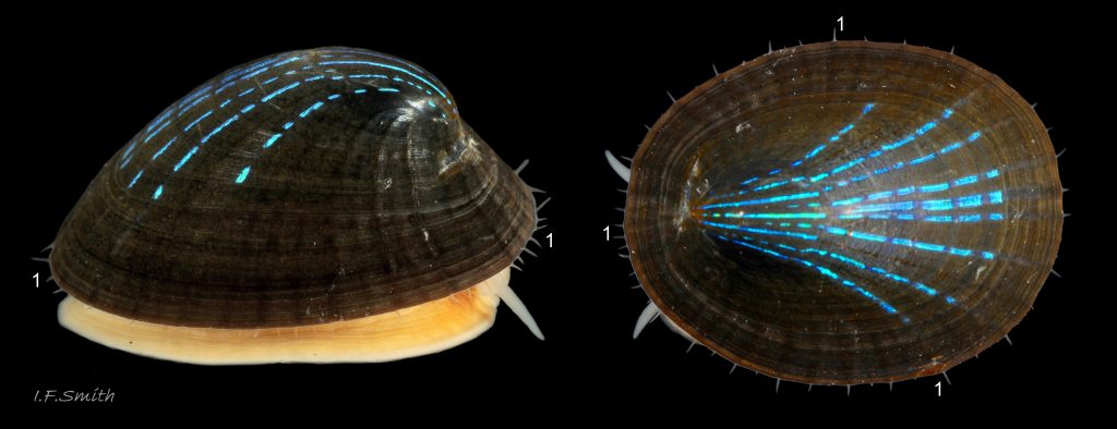
05 Patella pellucida. Growth Stage 3 (GS3). Left: March 2017. W. Anglesey, Wales. Right: at smaller scale, September 2017. Tyne & Wear, England.
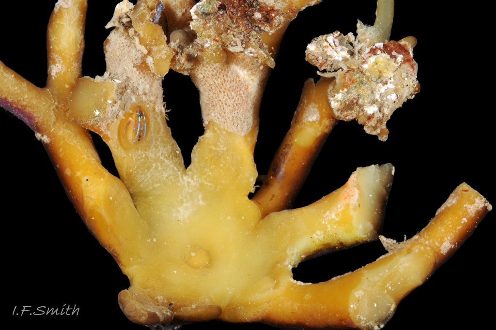
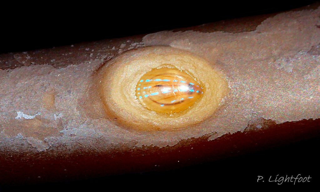
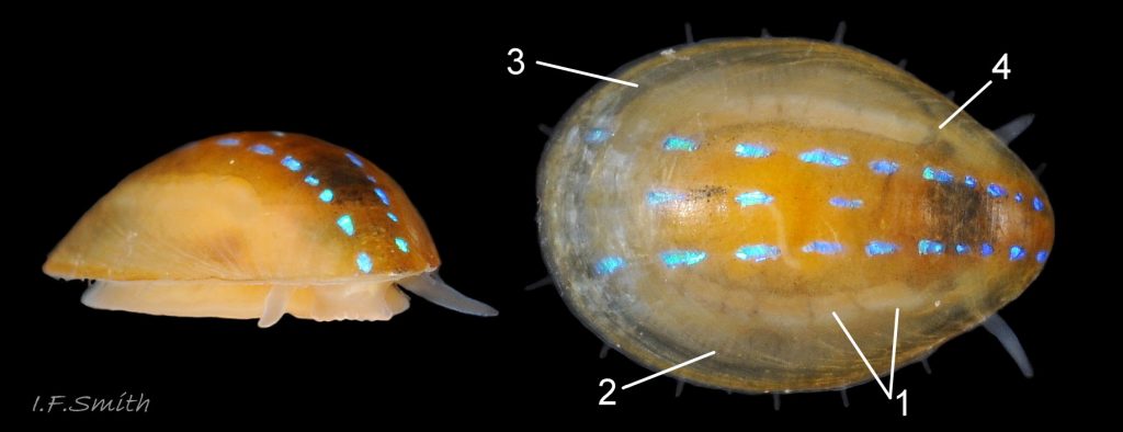
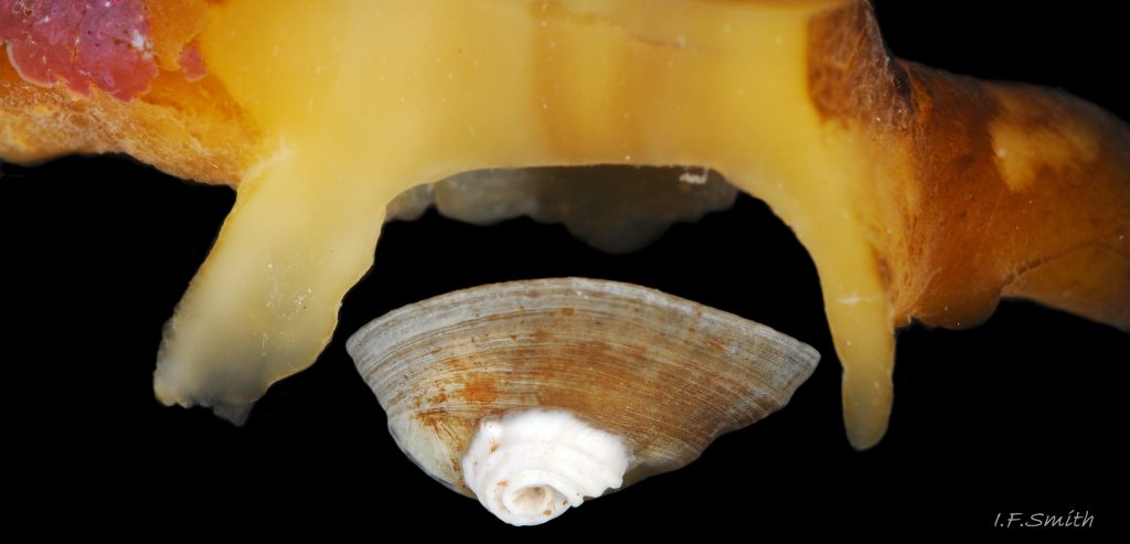
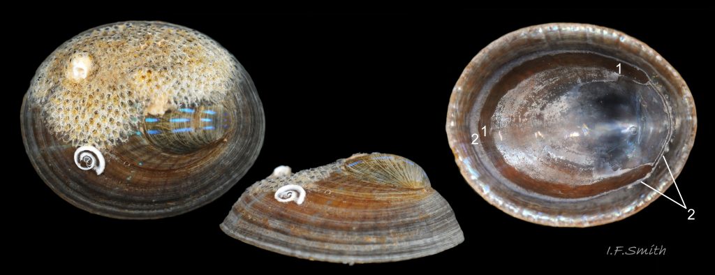
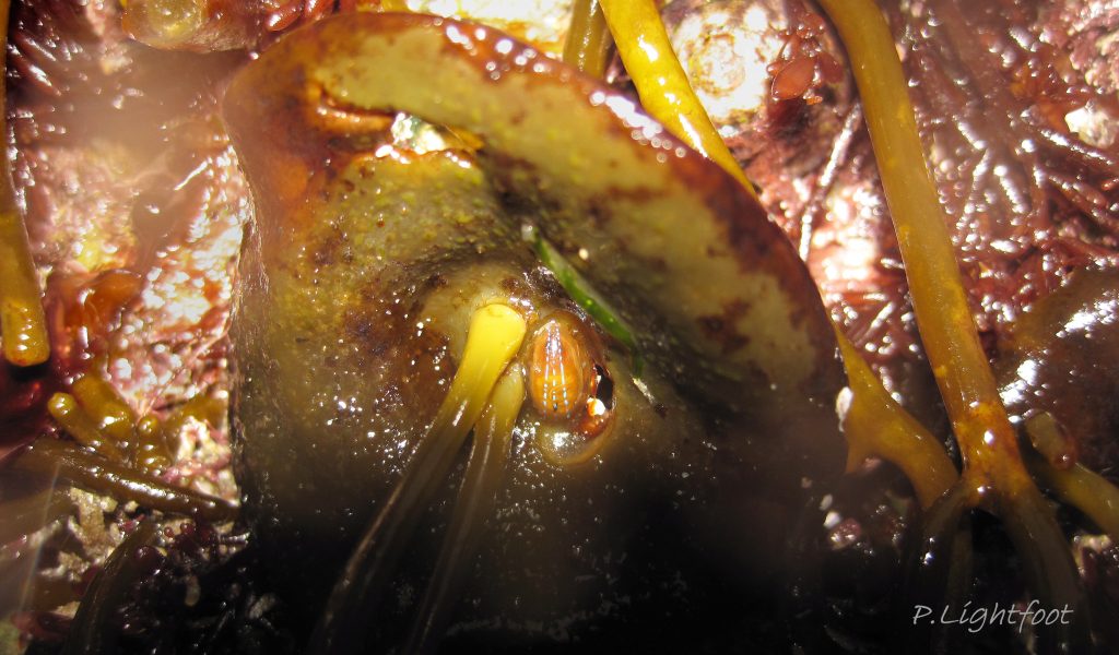
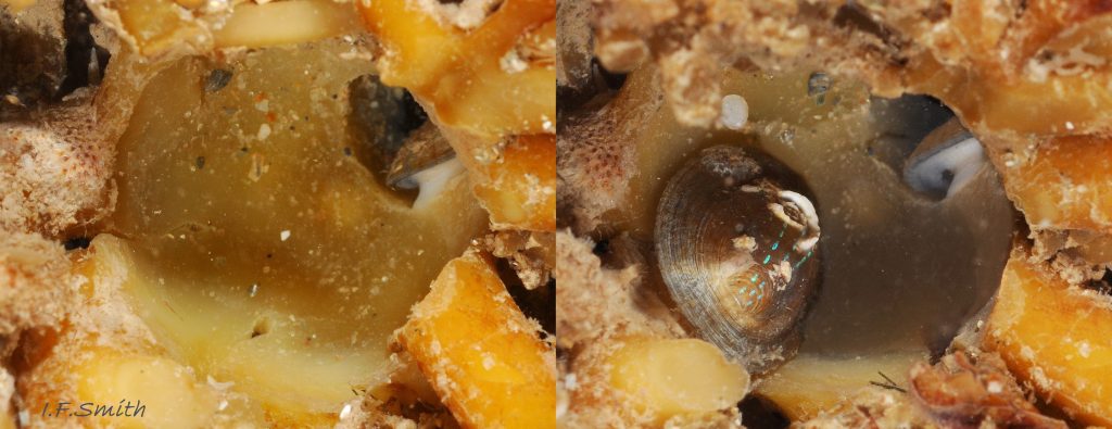
06 Patella pellucida. Length 4.7mm. Growth Stage 3h (GS3h). Littoral. March 2017. West Anglesey, Wales.
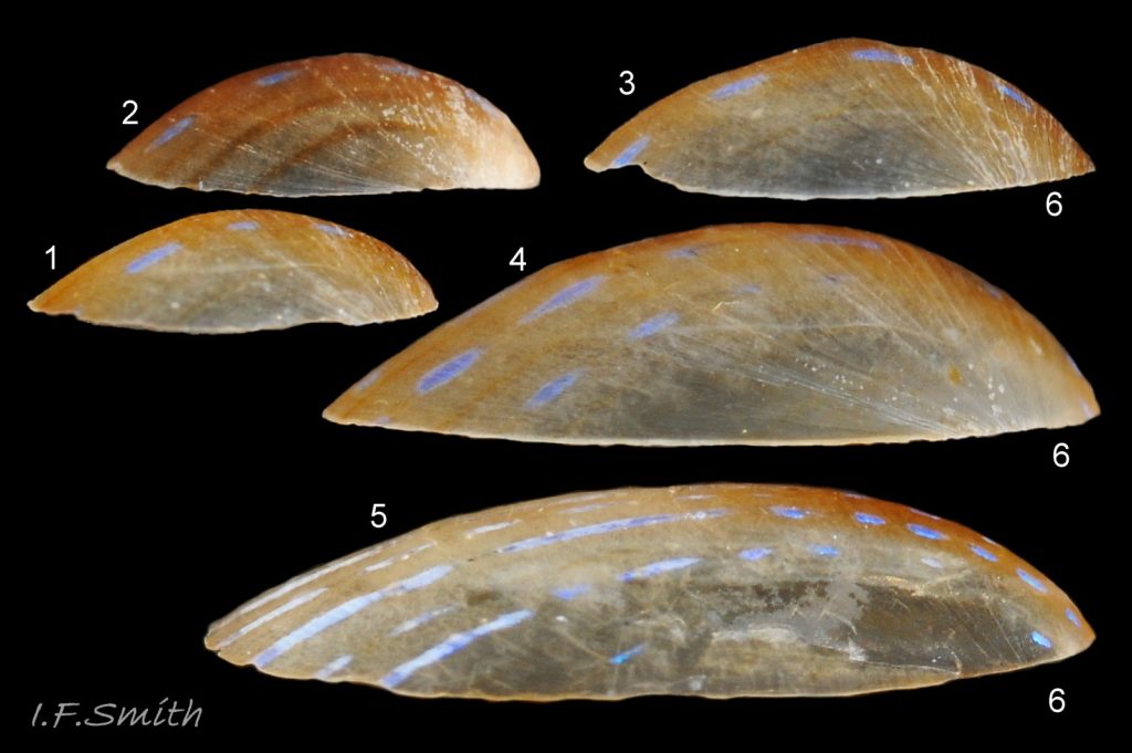
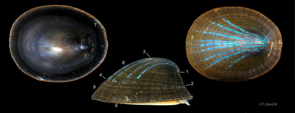
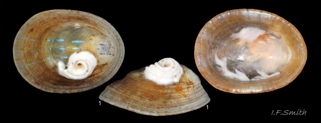
07 Patella pellucida. Growth Stage 3 (GS3). Littoral. Aberdeen, Scotland, 1971, except 5 from Llŷn, N. Wales, April 1969.
08 Patella pellucida. Juveniles, Growth Stage 3 (GS3). Littoral. Length 6.7mm, Llŷn, N. Wales, April 1969. Length 3.2mm. Aberdeen, Scotland, 1971.
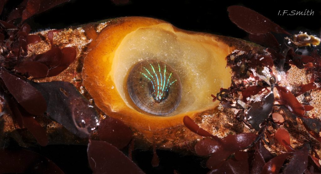
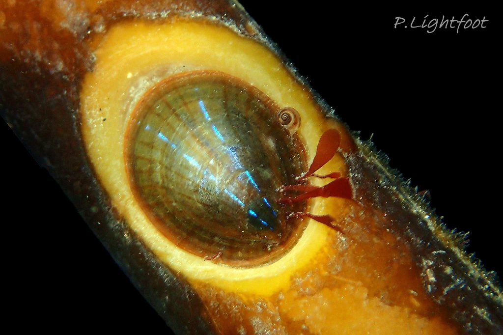
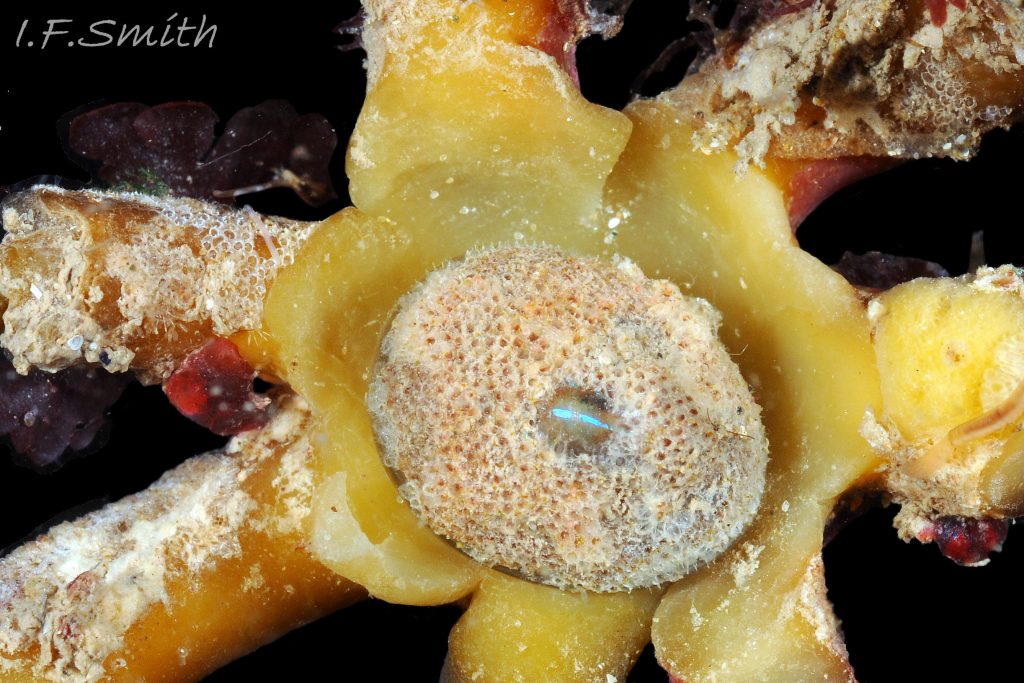
09 Patella pellucida. Growth Stage 3 holdfast dweller (GS3h) on Laminaria. March 2017. Littoral. West Anglesey, Wales.
10 Patella pellucida. Growth Stage 3f (GS3). March 2017. Littoral. West Anglesey, Wales.
11 Patella pellucida. 4,6mm long. Juvenile, Growth Stage 3 (GS3). From exterior of Laminaria holdfast. March 2017. Littoral. West Anglesey, Wales.
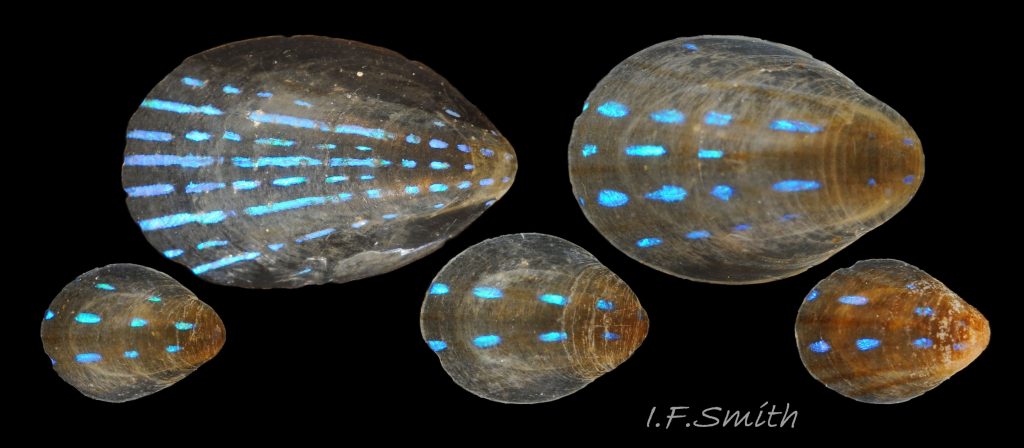
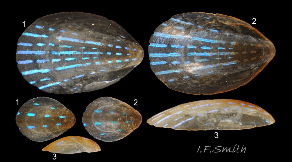
12 Patella pellucida. Juveniles, Growth Stage 3 (GS3). Littoral. Largest, length 6.7mm, from Llŷn, N. Wales, April 1969. Others, shortest length 3.2mm, from Aberdeen, Scotland, 1971.
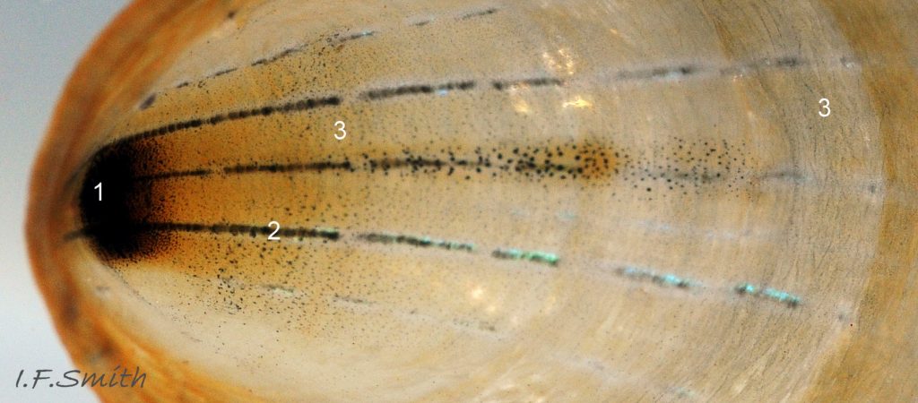
13 Patella pellucida. Length 6.7mm. Juvenile, Growth Stage 3 (GS3). April 1969. Llŷn, N. Wales.
14 Patella pellucida. Juveniles, Growth Stage 3 (GS3). Littoral. Largest, length 6.7mm, from Llŷn, N. Wales, April 1969. Others, shortest length 3.2mm, from Aberdeen, Scotland, 1971.
15 Patella pellucida. Length 10.2mm. Early adult, Growth Stage 4 frond dweller (GS4f), on Laminaria. March 2014. Littoral. West Anglesey, Wales.
16 Patella pellucida. Length 10.4mm. Growth Stage 4 frond dweller (GS4f), on Laminaria. March 2014. Littoral. West Anglesey, Wales.
17 Patella pellucida. Length 10.2mm. Early adult, Growth Stage 4 frond dweller (GS4f), on Laminaria. March 2014. Littoral. West Anglesey, Wales.
20 Patella pellucida. Length about 15mm. Growth Stage 5 frond dweller (GS5f). March 2010. Menai Strait, Wales. © D. Evans.
21 Patella pellucida. Length L19.3mm, H7.7mm. Growth Stage 5 frond dweller (GS5f). March 2014. Menai Strait, Wales.
22 Patella pellucida. Length 14mm, Growth Stage 5 frond dweller (GS5f). February 2011. Menai Strait, Wales.
23 Patella pellucida. Exceptional form of Growth Stage 5 frond dweller (GS5f). August 2015. Tyne and Wear, England. © P.Lightfoot.
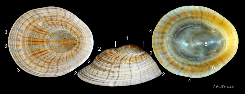
24 Patella pellucida. Length 11mm, Growth Stage 5 stipe dweller (GS5s). March 2011. Menai Strait, Wales.
25 Patella pellucida. July 2017. Tyne & Wear, England. © P.Lightfoot.
26 Patella pellucida. Length 11mm. Growth Stage 5 stipe dweller (GS5s). March 2011. Menai Strait, Wales.
27 Patella pellucida. Growth Stage 4 stipe dweller (GS4s). July 2017. Tyne & Wear, England. © P.Lightfoot.
28 Patella pellucida. Length 8.3mm. Growth Stage 4 holdfast dweller (GS4h). March 2017. West Anglesey, Wales.
29.1 Patella pellucida, growth stage 5h. Length 23.5mm. Embleton, Northumberland, England. March 2018. © P. Lightfoot.
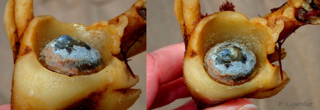
29.2 Patella pellucida, growth stage 5h. Length 23.5mm. Embleton, Northumberland, England. March 2018. © P. Lightfoot.
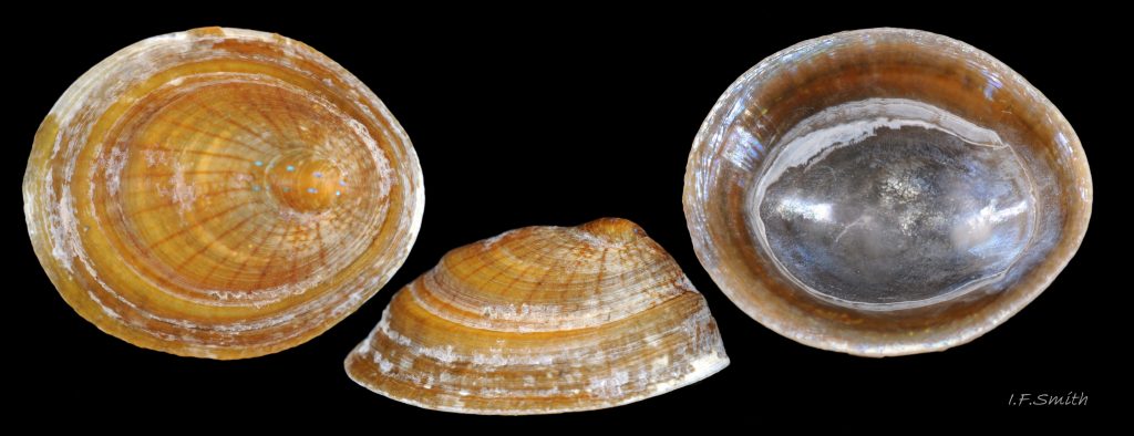
30 Patella pellucida. Growth Stages 4&5 holdfast dwellers (GS4h&5h). September 2014. North Yorkshire, England.
31 Patella pellucida. Length 13.3mm. Growth Stage 5 holdfast dweller (GS5h). September 2014. North Yorkshire, England.
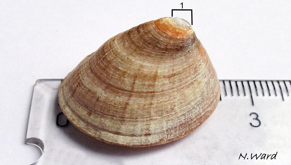
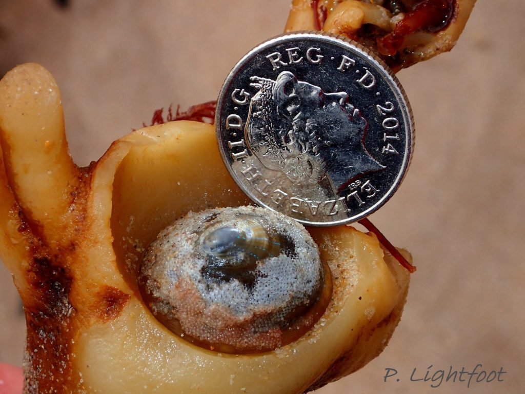
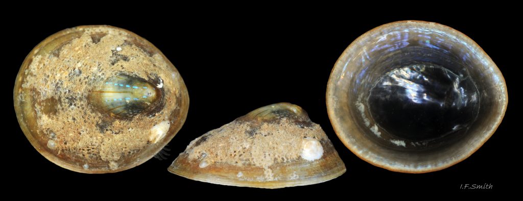
32.1 Patella pellucida. September 2017. Length 26mm. Growth Stage 5 holdfast dweller (GS5h). Northumberland, England. Leg. Katie Ward. © N. Ward.
33 Patella pellucida. Length 13.3mm. Growth Stage 5 holdfast dweller (GS5h). September 2014. North Yorkshire, England.
34 Patella pellucida. Length 12.7mm. Growth Stage 5 holdfast dweller (GS5h). September 2014. North Yorkshire, England.
35 Patella pellucida. Length 9.2mm. Growth Stages 4 holdfast dweller (GS4h). March 2017. West Anglesey, Wales.
36 Patella pellucida. Length 10.9mm. Growth Stage 5 holdfast dweller (GS5h). March 2017. West Anglesey, Wales.
37 Patella pellucida. Length 10.1mm. Growth Stage 5 holdfast dweller (GS5h). March 2017. West Anglesey, Wales.
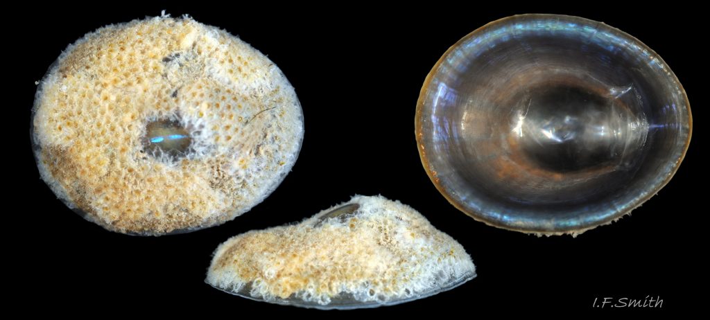
38 Patella pellucida. Length 15mm. Growth Stage 5 holdfast dweller (GS5h). September 2014. North Yorkshire, England.
39 Patella pellucida. Growth Stage 5 holdfast dweller (GS5h). September 2014. North Yorkshire, England.
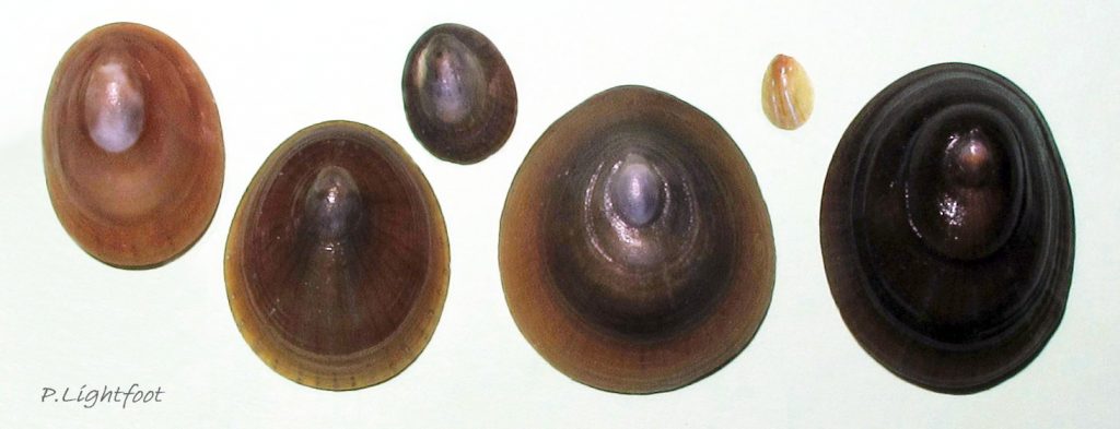
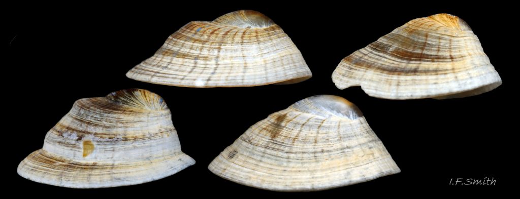
40 Patella pellucida. Range of specimens from juvenile (GS3) to adult (GS5). Vacated beachworn shells. Northumberland, England. © P.Lightfoot.
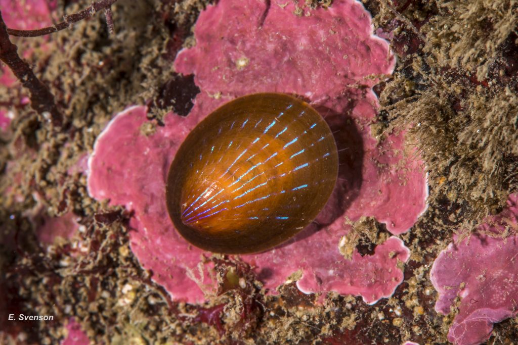
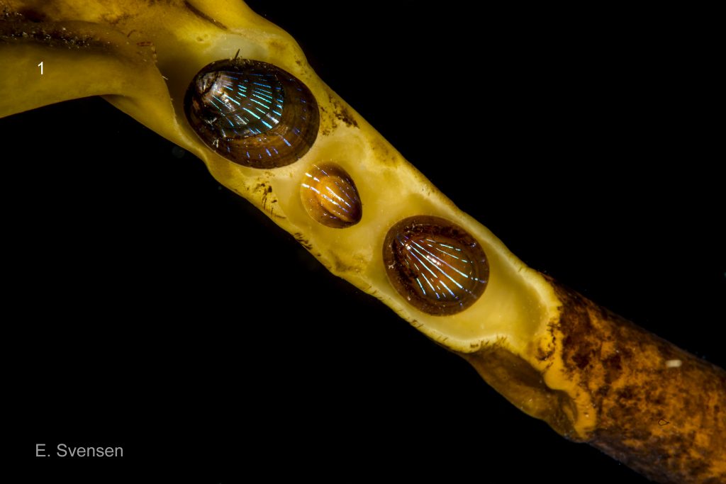
41 Patella pellucida. Early adult, Growth Stage 4 (GS4) . January 2015. Sublittoral. Egersund, S. Norway. © E. Svensen.
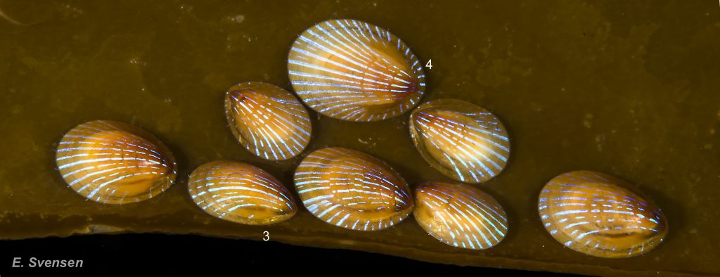
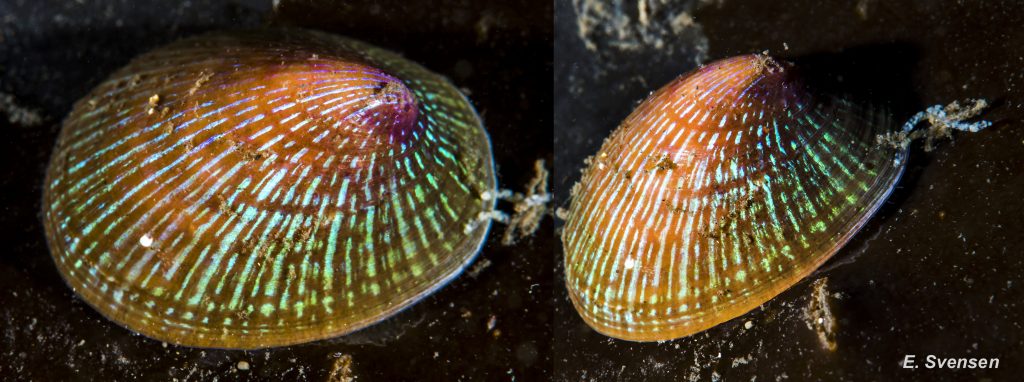
42 Patella pellucida. Juveniles and early adults, Growth Stages 3 & 4 (GS3 & 4). October 2013. Sublittoral. Egersund, S. Norway. © E. Svensen.
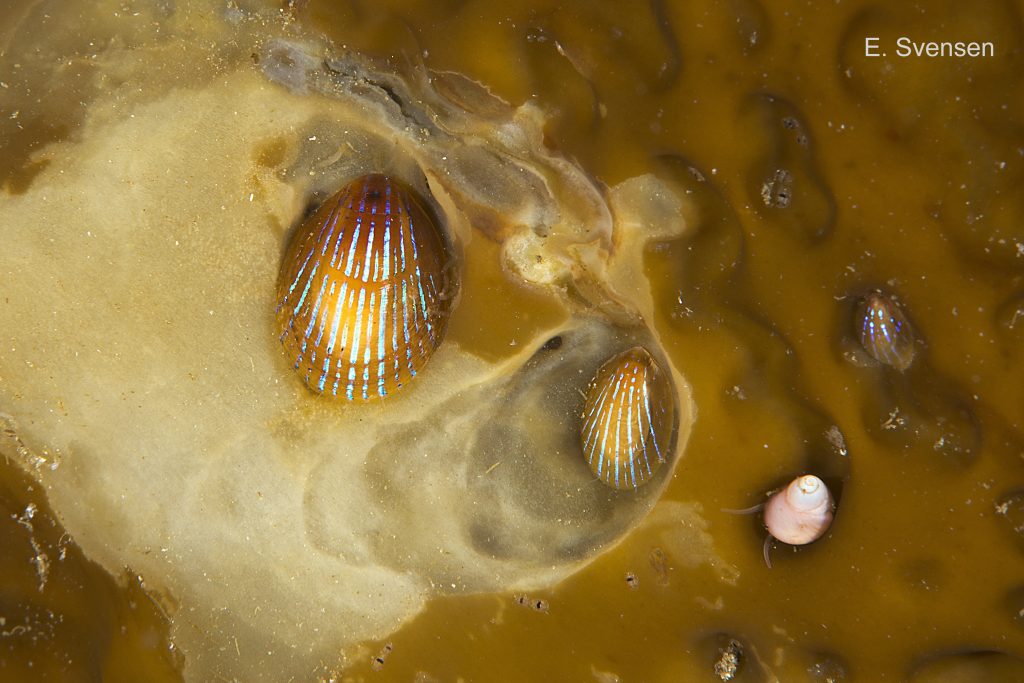
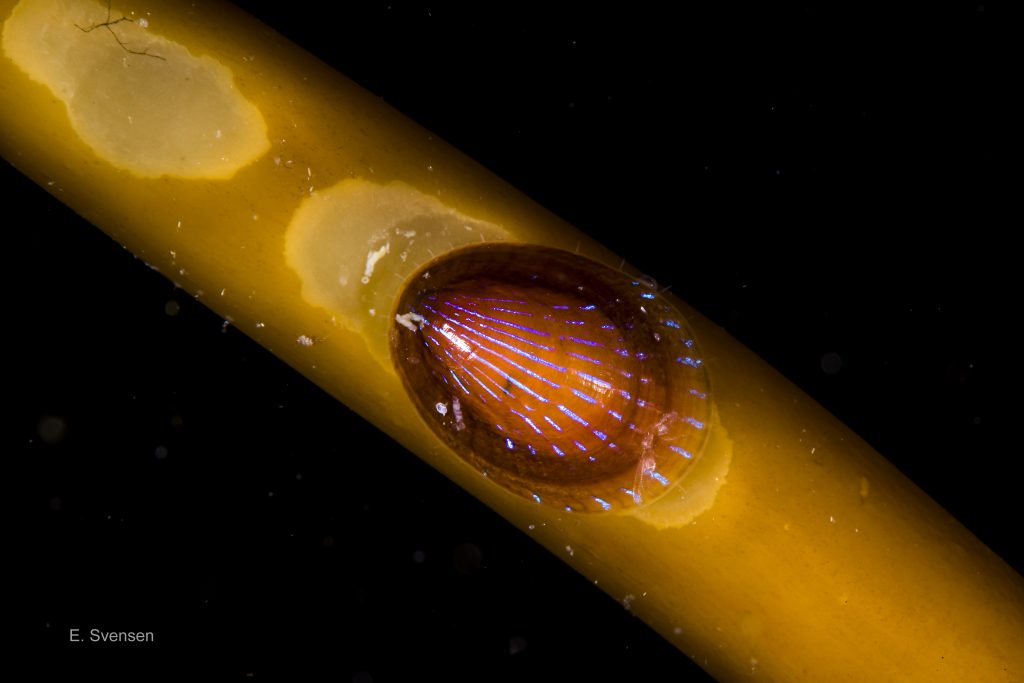
43 Patella pellucida. Juveniles, Growth Stage 3 (GS3) . October 2012. Sublittoral. Egersund, S. Norway. © E. Svensen.
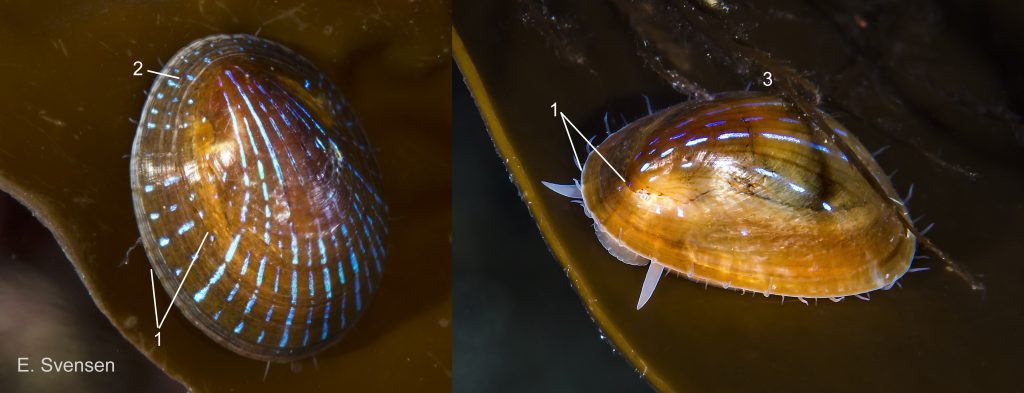
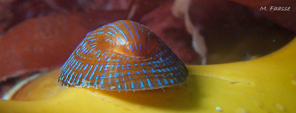
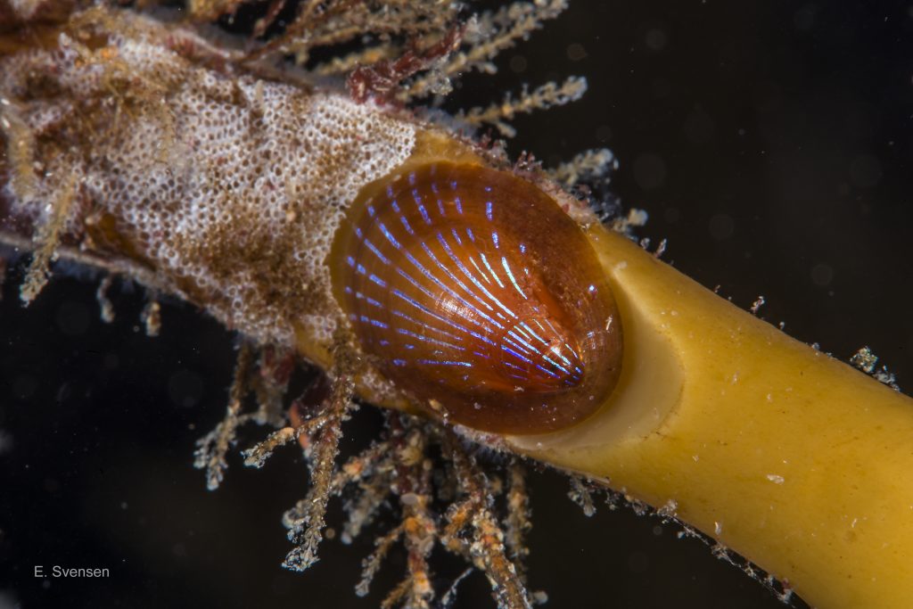
44 Patella pellucida. Early adults, Growth stage 4 (GS4), on Laminaria hyperborea frond. February 2013 (left) and July 2015. Sublittoral. Egersund, S. Norway. © E. Svensen.
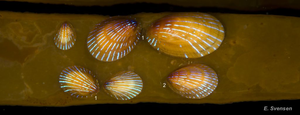
45 Patella pellucida. Growth Stages 3 (top left) & 4 (GS3 & 4) on Laminaria hyperborea frond. October 2013. Sublittoral. Egersund, S. Norway. © E. Svensen.
46 Patella pellucida. Growth stage 5 (possibly GS4). January 2014 (left) and March 2017. Sublittoral. Egersund, S. Norway. © E. Svensen.
47 Patella pellucida. Growth stage 5 (late adult) on Laminaria hyperborea frond. January 2016. Sublittoral. Egersund, S. Norway. © E. Svensen.
48 Patella pellucida. Growth stage 5 (late adult) on base of Laminaria hyperborea frond. August 2017. Sublittoral 5 to 10m. Not very exposed to wave action. Egersund, S. Norway. © M. Faasse.
49 Patella pellucida. Growth Stage 4s (early adult on stipe) . May 2014. Sublittoral. Egersund, S. Norway. © E. Svensen.
51 Patella pellucida. Growth Stages 3s & 4s (juvenile and early adults on stipe). April 2015. Sublittoral. Egersund, S. Norway. © E. Svensen.
52 Patella pellucida. Growth Stage 4s (early adult on stipe). May 2014. Sublittoral. Egersund, S. Norway. © E. Svensen.
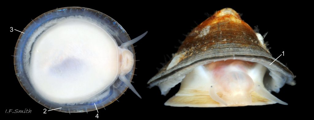
53 Patella pellucida. Shell length 11.9mm. Adult holdfast dweller, Growth Stage 5 (GS5h). September 2014. North Yorkshire, England.
54 Patella pellucida. Shell length 9.2mm. Adult holdfast dweller, Growth Stage 4 (GS4h). March 2017. Littoral. West Anglesey, Wales.
55 Patella pellucida. Shell length 4.6mm. Growth Stage 3 (GS3). March 2017. Littoral. West Anglesey, Wales.
56 Patella pellucida. Shell length 11.9mm. Growth Stage 5 holdfast dweller (GS5h) September 2014. North Yorkshire, England.
57 Patella pellucida. Shell length 10.2mm. Early adult, Growth Stage 4 frond dweller (GS4f). March 2014. Littoral. West Anglesey, Wales.
58 Patella pellucida. Shell length 19.3mm. Late adult, Growth Stage 5 frond dweller (GS5f). March 2014. Menai Strait, Wales.
59 Patella pellucida. Shell length 10.4mm. Early adult, Growth Stage 4 frond dweller (GS4f). March 2014. Littoral. West Anglesey, Wales.
60 Patella pellucida. Left & centre: Shell length 10.2mm. GS4h. March 2014. W. Anglesey, Wales. Right: Shell length 12.3mm.GS5h. September 2014. N. Yorkshire, England.
61 Patella pellucida. Shell length 11.9mm. Growth Stage 5 (GS5h). September 2014. North Yorkshire, England.
62 Patella pellucida. Left (not same scale as others): Shell length 8.3mm. GS4h. March 2017. W. Anglesey, Wales. Right: Shell length 10.2mm.GS4h. March 2014. W. Anglesey, Wales.
63 Patella pellucida. Shell length 11.9mm. Growth Stage 5 (GS5h). September 2014. North Yorkshire, England.
64 Patella pellucida. Shell length 19.3mm. Growth Stage 5 frond dweller (GS5f). March 2014. Menai Strait, Wales.
65 Patella pellucida. Shell length 9.2mm. Growth Stage 4 holdfast dweller (GS4h). March 2017. West Anglesey, Wales.
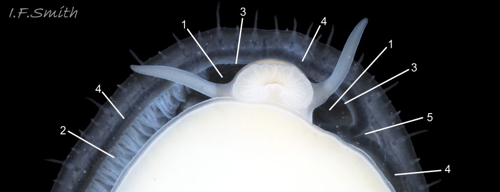
66 Patella pellucida. Left: L. 19.3mm. GS5f. Menai Strait, Wales. Right: L.4.6mm. GS3f. W. Anglesey, Wales.
67 Patella pellucida .Shell length 19.3mm. Growth Stage 5 frond dweller (GS5f). March 2014. Menai Strait, Wales.
68 Patella pellucida. Shell length 4.7mm. Growth Stages 3 (GS3). March 2017. Littoral. West Anglesey, Wales.

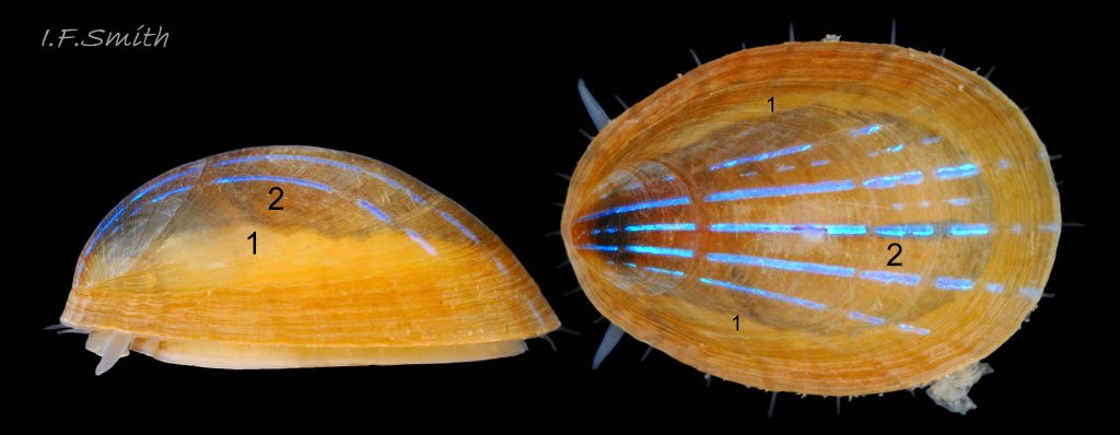
69 Patella pellucida. Growth Stages 5f (L.19.3mm) Menai Strait, 4f (L.10.2mm) and 3f (L.4.6mm) W. Anglesey; all frond dwellers.
70 Patella pellucida. Shell length 19.3mm. Growth Stage 5 frond dweller (GS5f). March 2014. Menai Strait, Wales.
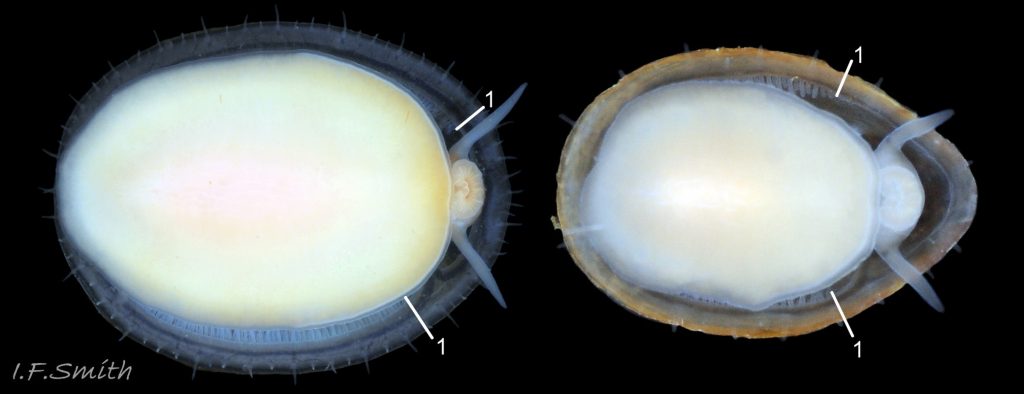
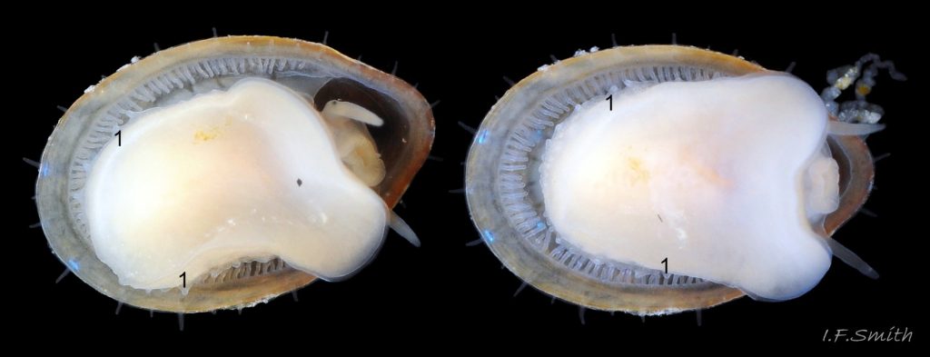
71 Patella pellucida. Shell length 4.5mm. Growth Stage 3 holdfast dweller (GS3h). March 2017. West Anglesey, Wales.
72 Patella pellucida. Shell length 9.2mm. Growth Stage 4 holdfast dweller (GS4h). March 2017. West Anglesey, Wales.
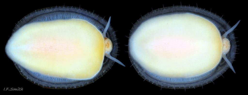
73 Patella pellucida. Shell length 9.2mm. Growth Stage 4 holdfast dweller (GS4h). September 2014. North Yorkshire, England.
74 Patella pellucida. Shell length 9.2mm. Growth Stage 4 holdfast dweller (GS4h). September 2014. North Yorkshire, England.
75 Patella pellucida. Shell length 19.3mm. Growth Stage 5 frond dweller (GS5f). March 2014. Menai Strait, Wales.
76 Patella pellucida. Shell length 9.2mm. Growth Stage 4 holdfast dweller (GS4h). March 2017. West Anglesey, Wales.
77 Patella pellucida. Shell length 9.2mm. Growth Stage 4 holdfast dweller (GS4h). March 2017. West Anglesey, Wales.
78 Patella pellucida. LEFT: L.10.2mm. Early adult, GS4f. March 2014. W. Anglesey. RIGHT: L. 11.9mm. Adult, GS5h. September 2014. N. Yorkshire.
79 Patella pellucida. Shell length 9.2mm. Growth Stage 4 holdfast dweller (GS4h). September 2014. North Yorkshire, England.
80 Patella pellucida. Shell length 9.2mm. Growth Stage 4 holdfast dweller (GS4h). March 2017. West Anglesey, Wales.
81 Patella pellucida. Shell length 9.2mm. Growth Stage 4 holdfast dweller (GS4h). March 2017. West Anglesey, Wales.

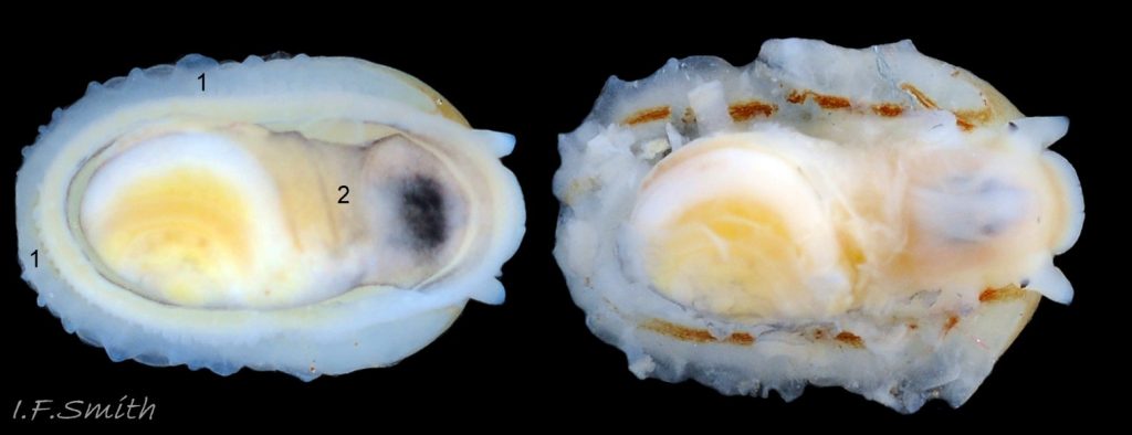
82 Patella pellucida. Shell length 4.5mm. Juvenile, Growth Stage 3 holdfast dweller (GS3h). March 2017. West Anglesey, Wales.
84 Patella pellucida. Shell length 9.2mm. Growth Stage 4 holdfast dweller (GS4h). March 2017. West Anglesey, Wales.
85 Patella pellucida. Shell length 9.2mm. Growth Stage 4 holdfast dweller (GS4h). March 2017. West Anglesey, Wales.
86 Patella pellucida. Shell length 9.2mm. Growth Stage 4 holdfast dweller (GS4h). March 2017. West Anglesey, Wales.
87 Patella pellucida. Shell length 9.2mm. Growth Stage 4 holdfast dweller (GS4h). March 2017. West Anglesey, Wales.
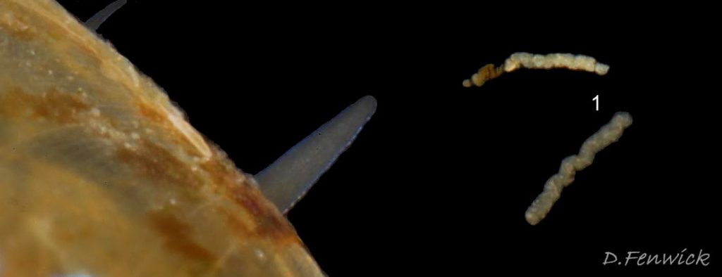
88 Patella pellucida. Faecal rods expelled from juvenile frond dweller (GS3f). October 2017. Cornwall, England. © D. Fenwick
89 Patella pellucida. Shell length 10.2mm. Growth Stage 4 holdfast dweller (GS4h). March 2017. West Anglesey, Wales.
8tella pellucida. Shell length 9.2mm. Growth Stage 4 holdfast dweller (GS4h). March 2017. West Anglesey, Wales.
90 Patella pellucida. Juvenile, Growth Stage 3. April 2012. Jersey, the Channel Islands. © P. Lightfoot.
91 Patella pellucida. Juvenile, Growth Stage 3. March 2017. West Anglesey, Wales. April 2016. North Yorkshire, England.
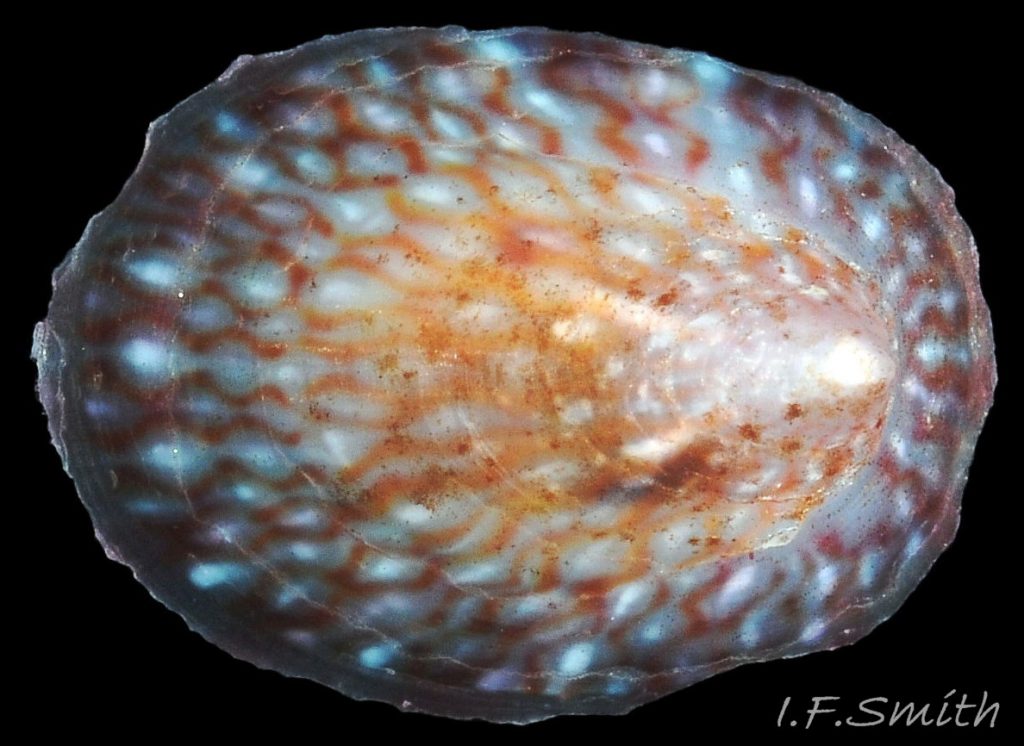
92 Patella pellucida. Length 10.1mm. Growth Stage 5 holdfast dweller (GS5h). March 2017. West Anglesey, Wales.
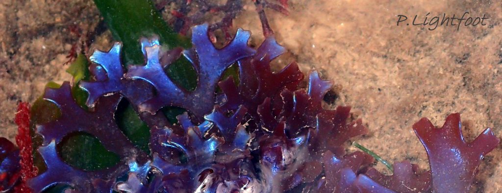
93 Patella pellucida. Iridescent Chondrus crispus.
Iridescent Chondrus crispus. Many specimens have much smaller amounts of iridescence. May 2017. North Yorkshire, England.Flashing on and off of the rays on P. pellucida living on the Laminaria fronds as they wave about and change the angle of light incidence, m…
94 Patella pellucida. Shell length 9.2mm. Growth Stage 4 holdfast dweller (GS4h). March 2017. West Anglesey, Wales.
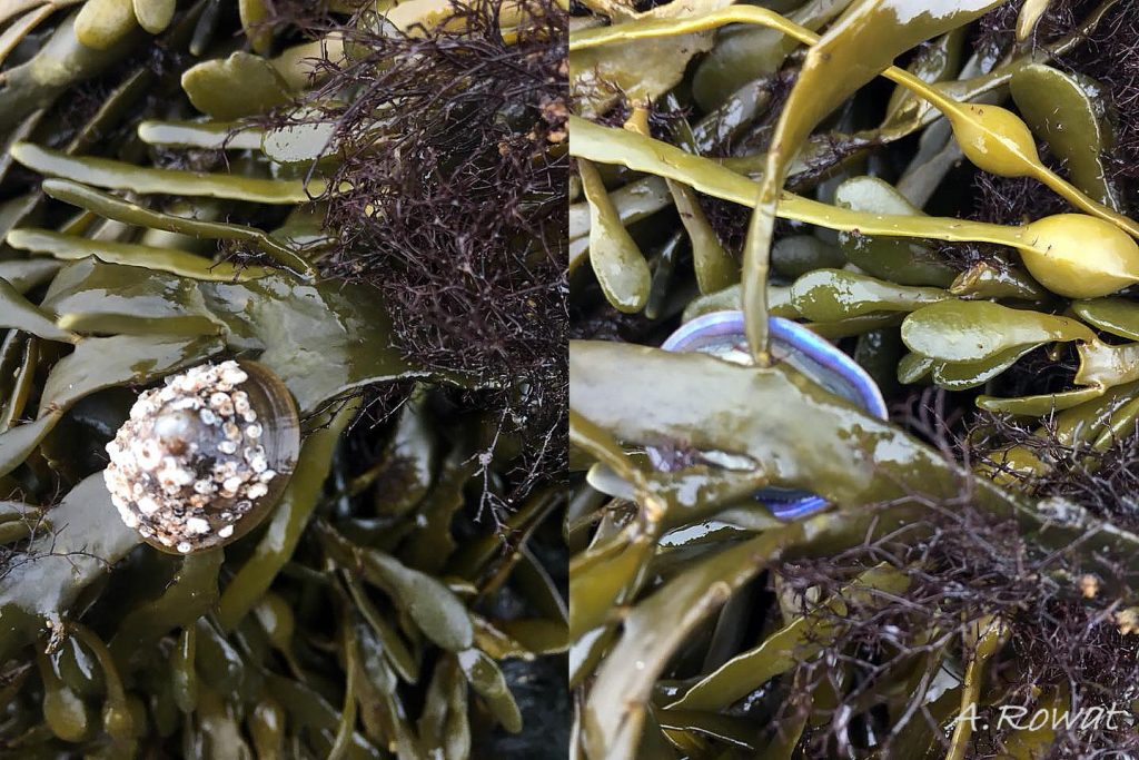
95 Patella pellucida. Growth Stage 5 holdfast dweller (GS5h) displaced by storm waves. October 2017. North-west Anglesey, Wales. © A. Rowat.
96 Patella pellucida album. COMPARISON image of Tectura virginea
Patella pellucida Linnaeus, 1758
Authors: Paula Lightfoot, Erling Svensen and Ian F. Smith (text) September 2017.
Introduction
In addition to when they metamorphose from veliger larva to juvenile, all individuals of P. pellucida undergo pronounced morphological change when they mature at about 7mm length. Then, the anterior of the shell ceases to be reduced and starts to grow, probably because of a change from resorption to secretion by the mantle edge. Juvenile growth lines, shaped like tilted horseshoes open at the cut away anterior, are succeeded by unbroken growth lines around the entire shell, and the profile starts to rise. Subsequent adult survival and shell morphology vary greatly with the local environment and microhabitats used. The following describes the resulting forms in detail for the majority of individuals in Britain, but exceptions occur.
Shell Descriptions (Britain)
Growth stage1: veliger larva.
Habitat: Plankton. Shell length: Up to 0.2mm. Shape: initially a smooth bowl shape, rapidly becoming slightly spiral with barely more than one whorl (Lebour, 1937). 01 Patella pellucida
Exterior surface: Smooth. Translucent.
Growth stage 2: newly settled juvenile.
Habitat: Usually on red, calcareous algae encrusting rock 02 Patella pellucida.
Shell length: Up to c. 2mm.
Shape: Very shallow saucer. The apex at the anterior intitially has a minute spiral protoconch (surviving larval shell) which is sealed off and lost soon after metamorphosis. The summit of the shell is further back. N.B. “apex” in this account refers to the protoconch location; the highest point of the shell above the substrate is referred to as the “summit”. Profile: Very low. Aperture outline: Pear-shape outline. Aperture rim: On a single plane conforming to the flat surface of encrusting algae. Exterior surface: Smooth, usually lacking epizoic growths. Translucence: Almost transparent, showing viscera. Colour: When live, the faintly horn-coloured shell is darkened by the visible viscera. Coloured rays: Two or three longitudinal rows of iridescent blue dashes .
Growth stage 3: juvenile.
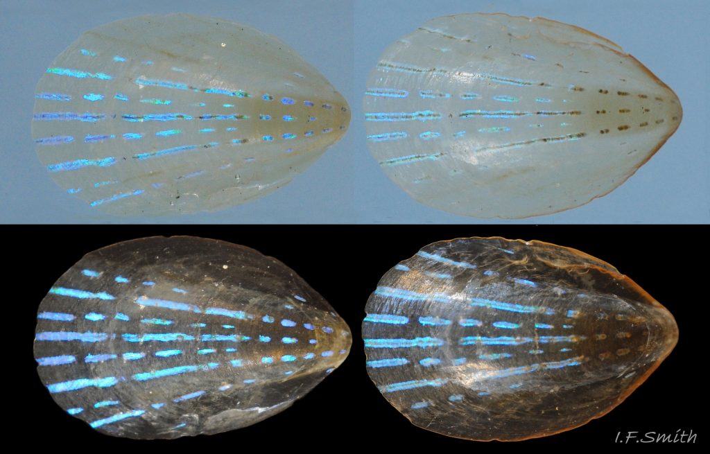
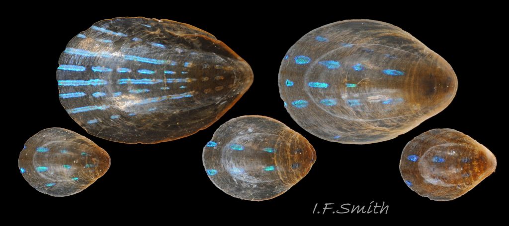
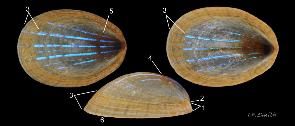
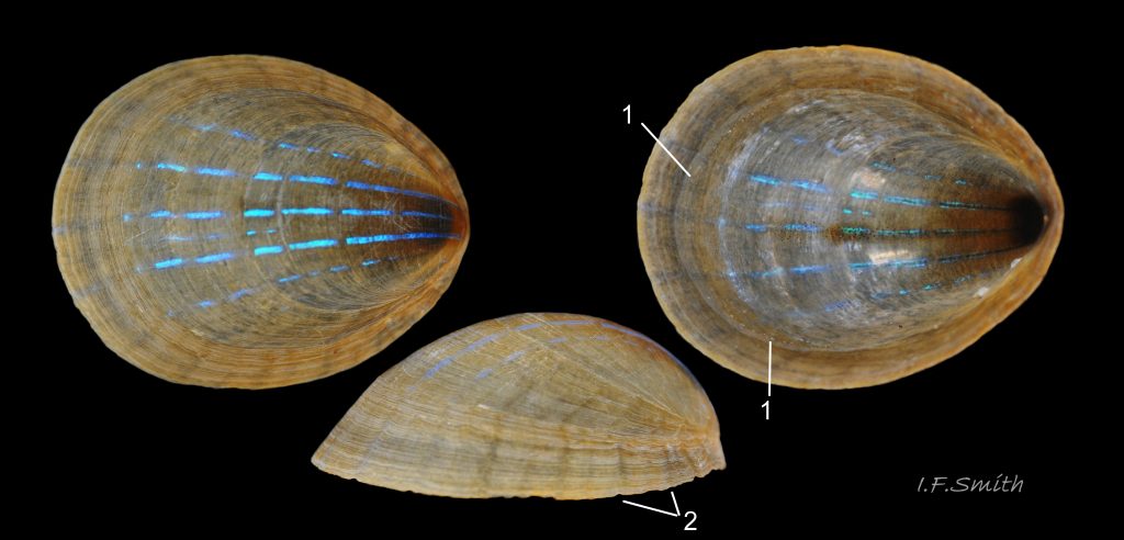
Habitat: Laminaria fronds 03 Patella pellucida stipes 04 Patella pellucida, and on 05 Patella pellucida and within 06 Patella pellucida holdfasts. Length: c. 2mm to c. 7mm. Height: Low, mean height 27% of length (Graham & Fretter,1947). Shape: Shallow saucer. Profile: Low, uninterrupted, streamlined 07 Patella pellucida. Aperture outline: Oval, narrowest at anterior. Aperture rim: On a single plane conforming to the flat surface of Laminaria frond 08 Patella pellucida. Those in holdfasts or depressions on stipes may have the rim curved up at the ends 09 Patella pellucida. Growth: Greatest at posterior, apex lost and anterior progressively removed, probably by resorption. Growth lines: horseshoe, open at anterior, in plan view. In side view, they slope downwards to where they end in the anterior quarter of shell. Most widely spaced at posterior 09 Patella pellucida . Thickness: very thin and fragile. Exterior surface: very smooth, no sculpture except fine growth lines 10 Patella pellucida, but less smooth with coarser growth lines if within a holdfast 09 Patella pellucida. Rarely, if ever, has any epibiotic growths. Translucence: almost transparent; body can usually be discerned through the shell 05 Patella pellucida. Colour: whitish, tinted horn to pale brown; partly due to the closely adhering, flimsy periostracum 08 Patella pellucida. Coloured rays: two or three rows of iridescent blue/green hyphens/lines on 2mm and 3mm long shells 11 Patella pellucida ; about eight rows when 7mm long 12 Patella pellucida Lines 0.1mm to 0.2mm wide. Occasionally, several red-brown rays among the blue ones 03 Patella pellucida. Interior: whitish, tinted horn to pale brown. Black, non-crystalline, colloidal particles underlie parts of the blue/green lines 13 Patella pellucida. Sometimes, the horseshoe-shaped pedal retractor muscle-scar is visible 14 Patella pellucida.
Growth stage 4f, early adult on Laminaria frond.
Habitat: Laminaria fronds. Shell length: c. 7mm to c. 10mm. Height: raised, about 35% of length when 10mm long 15 Patella pellucida. Shape: dome. Profile: smoothly rounded, except for break in slope at anterior where new adult growth joins juvenile growth 15 Patella pellucida. Anterior steeper than posterior. Aperture outline: usually a broad oval 15 Patella pellucida, or almost circular 16 Patella pellucida Aperture rim: on a single plane conforming to the flat surface of Laminaria. Growth: growth commences at anterior. Adult growth forms a distinct skirt around the juvenile shell 15 Patella pellucida. Growth greater at posterior so the skirt is wider there. Growth lines: in plan view, now continuous around the whole shell on adult skirt. In side view, they slope down on the skirt towards the anterior, but are not cut off. Thickness: adult growth thicker and, sometimes, slight radial ribs develop on it. Exterior surface: very smooth, no sculpture except fine growth lines. Rarely has any epibiotic growths. Translucence: the brown, adult skirt is almost opaque 17 Patella pellucida , but the pedal-retractor muscle and, within the muscle outline, the blackish mantle and fine spray of black particles on the shell interior are discernible through the juvenile growth in good light 18 Patella pellucida. Colour: usually a stronger brown than on juveniles 18 Patella pellucida. Coloured rays: blue/green rays persist on juvenile growth, may extend onto new adult growth, but usually feeble 15 Patella pellucidaor faint shadowy grey. Interior: juvenile area pale,translucent; adult skirt stronger brown. Increased number of black, colloidal particles under parts of the blue/green lines 19 Patella pellucida. Pedal retractor muscle-scar usually faint 16 Patella pellucida .
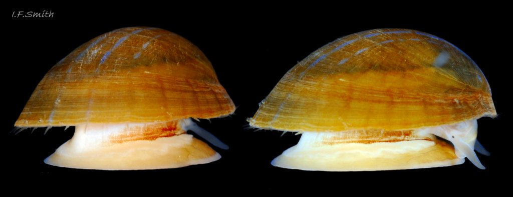
Growth stage 5f, late adult on Laminaria frond.
Habitat: on Laminaria frond. Shell length: c. 11mm to c. 19mm.
Height: up to 50%, or more, of length. Shape: conoid, steeper at anterior, often resembles a Phrygian cap 20 Patella pellucida. Profile: posterior a long smooth convex curve, anterior a short and steep concave curve with break of slope where juvenile growth meets adult growth, and, on two year old individuals, where first year adult growth meets second year growth. 21 Patella pellucida. Aperture outline: a broad oval 22 Patella pellucida sometimes approaching a circle 21 Patella pellucida. Aperture rim: on a single plane conforming to the flat surface of Laminaria frond. Growth: wide skirt of adult growth all around shell makes up the majority of it. Posterior growth still exceeds anterior growth, but less markedly. Growth lines: in plan view, continuous around the whole shell on adult skirt. Thickness: increases only slightly. Exterior surface: smooth, usually with no epibiotic growth, or a little 20 Patella pellucida. Translucence: less than on juveniles, but can be sufficient for viscera to be discerned 22 Patella pellucidaColour: usually dark brown on live shells 21 Patella pellucida. Coloured rays: blue lines persist on the juvenile growth and may continue on adult growth for part 21 Patella pellucida or all 22 Patella pellucida of the way to the posterior edge, but often continuing as faint shadowy grey lines 21 Patella pellucida. Rarely in Britain, bright blue lines continue on adult growth all around the shell 23 Patella pellucida. Interior: variable colour, often blackish, with some iridescence 21 Patella pellucida . Muscle scars vary in distinctness.
Growth stages 4s & 5s, adults on Laminaria stipe.
Habitat: Bowl-shape pit eaten into side of Laminaria stipe 24 Patella pellucida & 25 Patella pellucida. Shell length: c.7mm to 10mm (4s) and c.11mm to 19mm (5s). Height: Variable, c.30% of length. Shape: Conoid, less steep than on frond dwellers because adult skirt flares laterally. Skirt forms majority of shell at stage 5 26pp 26 Patella pellucida. Profile: Posterior is two smooth convex curves with a very distinct break of slope between them. Anterior is convex on juvenile growth and straight on adult skirt, with a break of slope where the two parts meet. Aperture outline: A broad oval, sometimes approaching a circle. Aperture rim: Strongly curved up from the horizontal at the ends. Growth: Wide skirt of adult growth all around shell makes up the majority of it by stage 5. Posterior growth exceeds anterior growth.
Growth lines: In plan view, continuous around the whole shell on adult skirt. Thickness: Increased on both juvenile and adult growth by mantle secretion over the whole of the interior. Exterior surface: Smooth, often with no epibiotic growth, but some shells have small amounts, including algae 27 Patella pellucida.
Translucence: Adult growth opaque; juvenile growth may retain some translucence. Colour: Usually dark brown on live shells. Coloured rays: Blue lines persist on the juvenile growth and may continue on adult growth for part or all of the way to the posterior edge. Red-brown radiating lines are frequent, often alternating with the blue lines 26 Patella pellucida . Interior: variable colour, often blackish or dark brown, with some iridescence. Muscle scars vary in distinctness; occasionally, there are a clearly marked, white mantle-attachment-scar, a distinct pedal-retractor-muscle scar and, within the muscle scar, a silky-white amphora mark 26 Patella pellucida..
Growth stages 4h& 5h, adults within Laminaria holdfast.
Habitat: Bowl shaped cavity eaten into base of stipe within Laminaria holdfast 28 Patella pellucida & 29 Patella pellucida. Shell length: c.7mm to 10mm (4h) and c.11mm to 19mm, exceptionally 30mm (5h) 29. 29.1 Patella pellucida & 29. 29.2 Patella pellucida.
Height: Mean height is 34% of length, varies17% to 57%. Shape: Wide variety of distorted conoids 30 Patella pellucida. Profile: very varied, but anterior steeper and shorter than posterior. Changes of gradient indicate change from juvenile to adult growth and, probably, annual pauses in growth. Aperture outline: a broad oval, sometimes approaching a circle 31 Patella pellucida. Aperture rim: Strongly curved up from the horizontal at the ends 28 Patella pellucida & 32 Patella pellucida. Growth: Wide skirt of adult growth all around shell makes up the majority of it by stage 5 33 Patella pellucida. Posterior growth exceeds anterior growth. Growth lines: In plan view, lines are continuous around the whole shell on the adult skirt, but form tilted horseshoes on juvenile growth 34 Patella pellucida . Thickness:Increased on both juvenile and adult growth by mantle secretion over the whole of the interior. Exterior surface: less smooth than on frond and stipe dwellers 35 Patella pellucida and is very often partly 36 Patella pellucida or completely 37 Patella pellucida coated with epizoic animals such as barnacles, bryozoa and tube worms. Smoother juvenile growth usually stays free of epizoa.
Translucence: Adult growth opaque 38 Patella pellucida. Colour: Brown or dark brown.
Coloured rays: Blue lines persist on the juvenile growth, unless eroded 31 Patella pellucida and may, occasionally, continue on adult growth for part or all of the way to the posterior edge 35 Patella pellucida but the blue is often less vibrant on the thicker adult growth. Red-brown radiating lines are frequent, often alternating with the blue lines 33 Patella pellucida or replacing them 31 Patella pellucida & 39 Patella pellucida. The lines are often concealed under epizoic growths. Interior: Varies, often partly iridescent 37 Patella pellucida. Often the peripheral zone is brown, and the area within the pedal-retractor-muscle scar is grey or blackish 39 Patella pellucida. The distinctness of the p-r-m scar, and of the mantle-attachment scar, varies from very clearly defined 36 Patella pellucida to indiscernible 35 Patella pellucida. Usually, from the interior, any exterior blue lines hidden but, occasionally, they are faintly visible 33 Patella pellucida.
Stranded shells
The layer of zig-zag lamellae of calcite that iridesces the blue rays is close to the surface and often abraded from beachworn shells, which consequently often lack rays 40 Patella pellucida. Occasionally the rays are eroded while live 31 Patella pellucid .
Shell Descriptions (Norway)
The following is based mainly on information and images from the Egersund area in southern Norway, and on Kain & Svendsen (1969). Images from other areas of Norway might require revision of this account. There are several differences in appearance and behaviour from those in Britain.
No images are available of newly settled juveniles under 2mm length (Growth Stage 2) on encrusting red calcareous algae (usual habitat in Britain), but an early adult (GS4) was photographed on calcareous algae 41 Patella pellucida. Juveniles (GS3) were observed on sublittoral fronds of Laminaria hyperborea 42 Patella pellucidaand L. saccharina 43 Patella pellucida. They had nearly-transparent, blue-rayed, low, smooth, streamlined shells, cut way at the anterior, similar to British examples. Early adults (GS4), as in Britain, cease anterior shell-loss and commence thicker growth all around the perimeter, and the anterior rises to form a dome 44 Patella pellucida But, there is a marked difference from Britain because the new adult growth usually has blue rays, as vivid as on the juvenile, now on the anterior as well as the posterior 45 Patella pellucida. Those that survive as later-adults (GS5f) on fronds continue to have blue rays all around the shell as it grows 46 Patella pellucida, and a few gain sufficient height to approximate to a Phrygian cap in profile 47 Patella pellucida & 48 Patella pellucida. A minority live on stipes 49 Patella pellucida almost always on the top 10cm, including the junction of stipe with frond flic.kr/p/XAamFR . Photographs viewed showed juveniles (GS3) and early adults (GS4) in pits that sometimes coalesce 51 Patella pellucida. The shell-forms on stipes (GS3s & 4s) were similar to those on fronds, but they differed by usually lacking any, or many, blue rays on the anterior 52 Patella pellucida and so resembled British specimens. Not a single distorted holdfast-dweller (GS5h), nor a vacant P. pellucida pit in a holdfast, was discovered in large samples from exposed and sheltered sites in Espegrend, S.W. Norway and Tromsö, N. Norway, and it appears that this form is absent from Norway (Kain & Svendsen,1969) .
Current taxonomy: World Register of Marine Species (WoRMS) www.marinespecies.org/aphia.php?p=taxdetails&id=147459
Synonyms: Anastes pellucida (Linnaeus, 1758); Helcion pellucidum (Linnaeus, 1758); Patella laevis Pennant, 1777; Patina pellucida (Linnaeus, 1758);
Vernacular names: Blue-rayed limpet, Peacock’s feather (English); Brenigen lasresog, Brenigen rasen las (Welsh); Blåskæl (Danish); Blauwgestreepte schaalhoren (Dutch); durchscheinende Häubchenschnecke (German); Blåsnigel (Norwegian); Laminaria-skålsnäcka (Swedish); Helcion transparent (French);
GLOSSARY below.
Key identification features of British P. pellucida Growth Stages
For quick reference; exceptions occur. Features should be used in combination; not singly. See full account for more detail and variations. Lengths approximate.
Veligers
GS1: planktonic, up to 0.2mm, spiral of just over one turn. 01 Patella pellucida
Juveniles
GS2: on calcareous encrusting algae, L. up to 2mm. Low profile, pear-shape. 2 or 3 vivid blue rays. 02 Patella pellucida
GS3: on Laminariafronds and stipes. 2mm to 7mm. Smooth, low profile, streamlined. Growth lines tilted horseshoe shape. Up to 8 vivid blue rays. 03 Patella pellucida
GS3h: in Laminariaholdfasts. As GS3 but surface, under magnification, less smooth as growth lines more prominent. 09 Patella pellucida.
Adults
GS4f: on Laminariafronds. L. 7mm to 10mm. Raised dome, smoothly rounded except slight break of slope at anterior. Steep peripheral skirt with growth lines encircling whole shell. Blue rays on summital juvenile growth may extend onto posterior of peripheral skirt, but often faint or absent. Horizontal rim. 15 Patella pellucida
GS5f: on Laminariafronds. L. 11mm to 19mm. High cone tending towards Phrygian cap. Steep peripheral skirt with growth lines encircling whole shell, comprises majority of shell. Blue rays on summital juvenile growth may extend onto posterior of peripheral skirt, but often faint or absent. Horizontal rim. 21 Patella pellucida
GS4s & GS5s: on Laminariastipes. No clear difference beween Growth Stages 4 & 5 except size; c7mm to 10mm (4s) and c.11mm to 19mm (5s). Peripheral skirt with growth lines encircling whole shell flares sidewards, not steep, somewhat like a broad brimmed hat. Skirt comprises majority of shell at GS5. Blue rays on summital juvenile growth may extend onto posterior of peripheral skirt, but often faint or absent. Rim curves up at ends. 26 Patella pellucida
GS4h & GS5h: in Laminariaholdfasts. No clear difference beween Growth Stages 4 & 5 except size; c7mm to 10mm (4s) and c.11mm to 19mm (5s). Very variably distorted conoid, usually with epizoic growths. Peripheral skirt with growth lines encircling whole shell.Skirt comprises majority of shell at GS5. Blue rays on summital juvenile growth may extend onto posterior of peripheral skirt, but often faint or absent, and often replaced partly or entirely by red-brown rays. Rim uneven, turned up at ends. 33 Patella pellucida
Variation from British P. pellucida by Norwegian specimens
Unlike Britain, there are apparently no holdfast dwellers with distorted shells at any growth stage in Norway.
GS1 & GS2: No information or images available.
GS3: Similar to British specimens, but often with more and brighter blue rays. 43 Patella pellucida
GS4f: Similar shell form to British specimens, but new adult growth on Norwegian frond dwellers usually has blue rays, as vivid as on the juvenile, on the anterior as well as the posterior. 42 Patella pellucida
GS4s: Those on stipes usually lacks blue rays on anterior of adult growth so they closely resemble British GS4. 52 Patella pellucida
GS5f: Frond dwellers occasionally approach Phrygian cap form, as in Britain, but differ by usually having bright blue rays on anterior of adult growth. 48 Patella pellucida
GS5s: No information or images available.
Similar species
No other British limpet has iridescent blue rays like those on Patella pellucida, but beachworn specimens that have lost the blue rays 40 Patella pellucida. may be confused with other limpet species. Sometimes specimens of Tectura virginea with blue markings are mistaken for P. pellucida 96 Patella pellucidabut they do not occur on brown algae.
Body description (Features visible on live animal.)
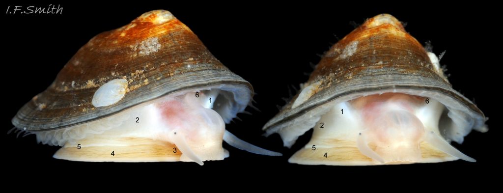
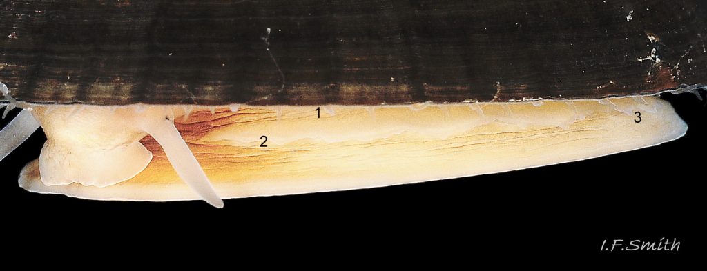
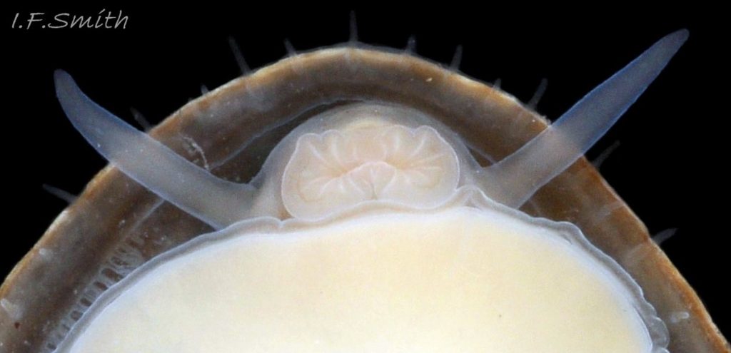
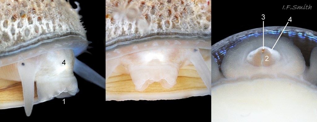
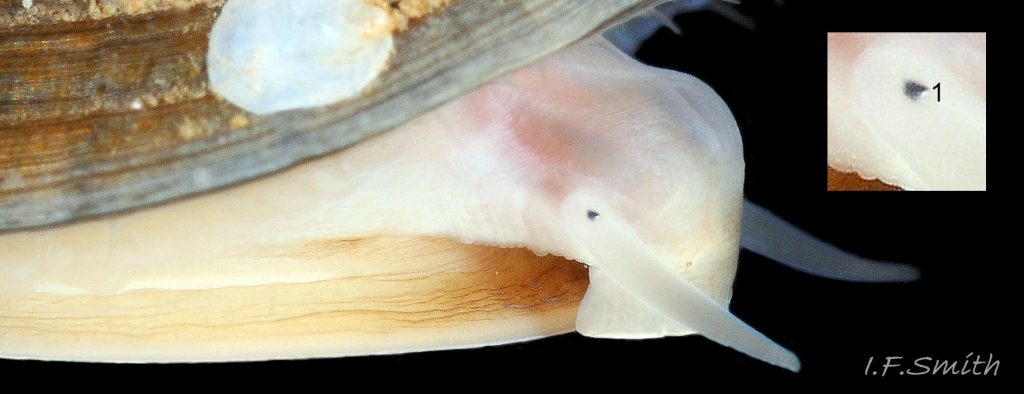
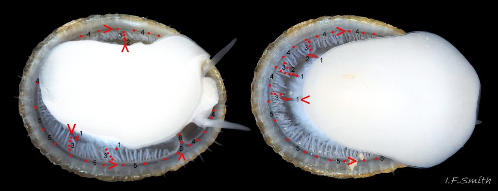
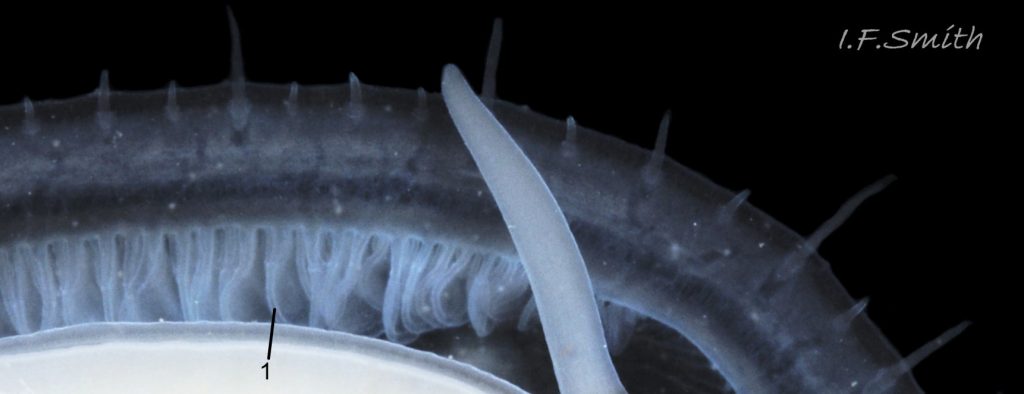
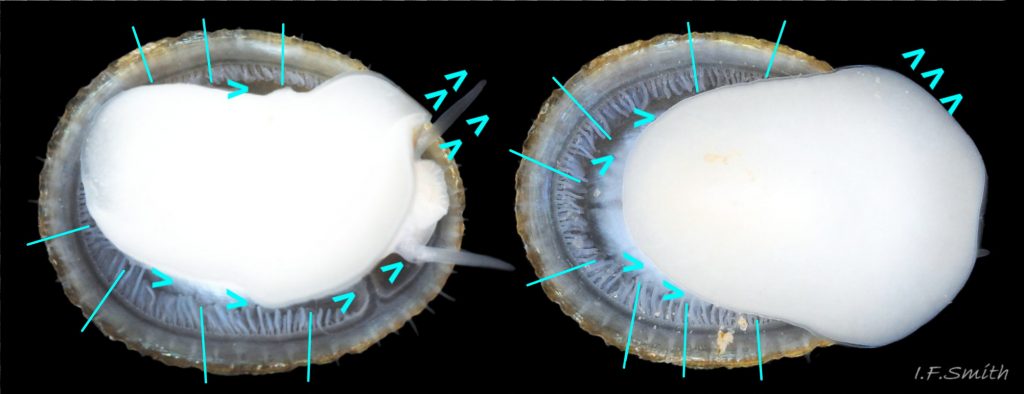
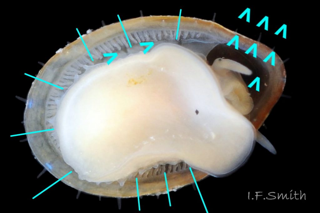
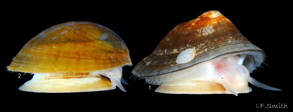
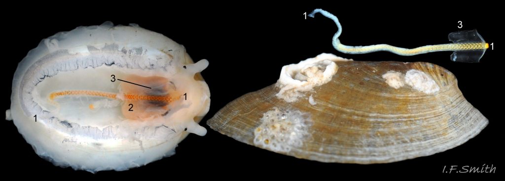
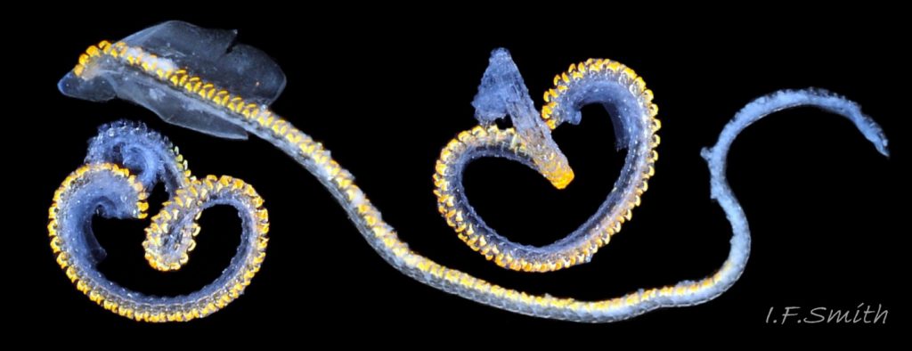
The main flesh colour is white or yellowish white. A horseshoe-shaped series of strong muscle bundles make up the pedal-retractor muscle 53 Patella pellucida. & 54 Patella pellucida . Juveniles have a distinct, longitudinal, dark chestnut band on the distal face of the pedal-retractor muscle that can often be seen through the translucent juvenile shell 03 Patella pellucida, 04 Patella pellucida & 05 Patella pellucida or by dissection 55 Patella pellucida. The chestnut band fades on adults inside holdfasts 56 Patella pellucida . Some of the adults on fronds retain a strongly pigmented band 57 Patella pellucida & 22 Patella pellucida, but others lose it 58 Patella pellucida. Although the band fades on holdfast dwellers, the anterior dorsal surface of the foot, below the head, usually becomes yellowish with brown creases. This colouration may extend faintly over more of the foot dorsally 53 Patella pellucida. The head has short stout snout occupied by a large mouth surrounded by thick, white, wrinkled outer lips 59 Patella pellucida which extend the snout when protruded 60 Patella pellucida. A pink, internal odontophore and buccal mass can be seen through the translucent, white head 61 Patella pellucida. The odontophore is separated from the outer lips by dull orange inner lips that open laterally and close to a vertical line 61 Patella pellucida. When they open, the radula with rust-coloured, iron-rich teeth is protruded through the arch of the opaque white jaw 60 Patella pellucida The tapered, translucent white cephalic tentacles each have a black eye with a tiny opening on the dorsum of the slightly swollen tentacle-base 61 Patella pellucida . The opening is at the apex of an internal cone of black pigment that, because visible through the translucent flesh, gives the false impression of a larger opening. Some or all of the black may be pigmented retinal cells. The eye can probably differentiate light from shade, and detect the direction of the light source, but cannot discern shapes. The mantle-skirt is translucent whitish, often showing the colour of underlying shell when viewed ventrally 63 Patella pellucida. The mantle cavity consists of a nuchal cavity over the head 64 Patella pellucida and a groove, containing pallial gills, around the periphery of foot but, unlike other British Patellidae, not around the anterior of the head 65 Patella pellucida. The gill-less anterior gap appears (small sample) to be proportionally of greatest extent on juveniles 66 Patella pellucid. A red (vestigial?) osphradium is located within the nuchal cavity next to each terminal bundle of the pedal-retractor muscle Pp53 53 Patella pellucida. The efferent pallial vessel, less whitish than the rest of the mantle skirt, runs around the entire animal at the base of the pallial gills 65 Patella pellucida and in front of the head to a point left of the head where its two ends unite in a trunk 64 Patella pellucida that passes through the roof of the nuchal cavity to the heart. The mantle skirt is fringed with translucent, white pallial tentacles that can protrude beyond the rim of the shell when the mantle is fully extended 67 Patella pellucida. Along the whole length of each side of the dorsal surface of the foot there is a lateral glandular streak 53 Patella pellucida with an overhanging upper lip. The lip has lobe-like retractile tentacles 68 Patella pellucida and is usually paler than the rest of the foot when the latter is tinted yellowish.
58 Patella pellucida. The sole is pure white on juveniles and early adults on fronds, often becoming yellowish, especially at the anterior, on frond dwelling, large adults 69 Patella pellucida. Adults in holdfasts have white soles even when the dorsal surface of the foot is yellowish with brown creases 65 Patella pellucida
The intensity of colour on coloured soles is diminished when the sole is expanded laterally 70 Patella pellucida . The anterior of the foot is bilaminate with the dorsal lamina extending beyond the sole 59 Patella pellucida. . The anterior-pedal mucous gland is between the laminae. As with other patellids, there is no penis as fertilization is external.
Internal functional anatomy
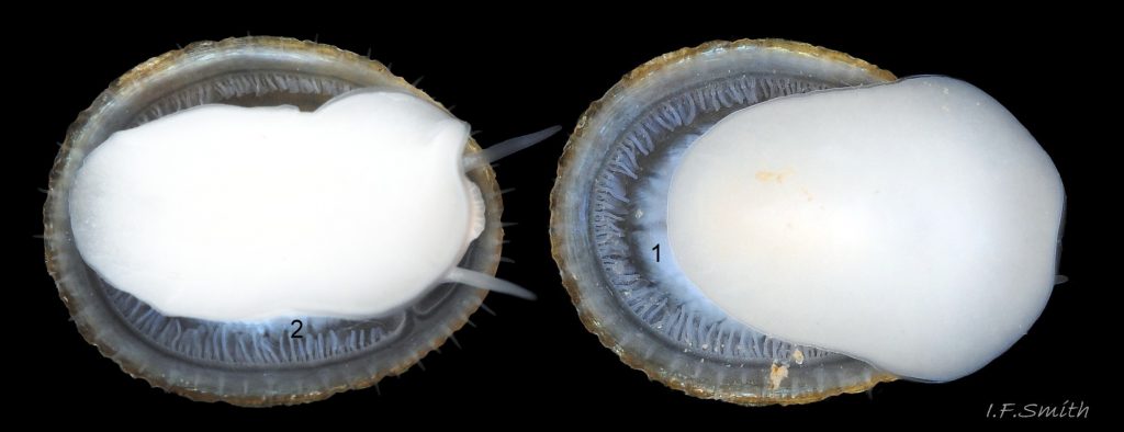
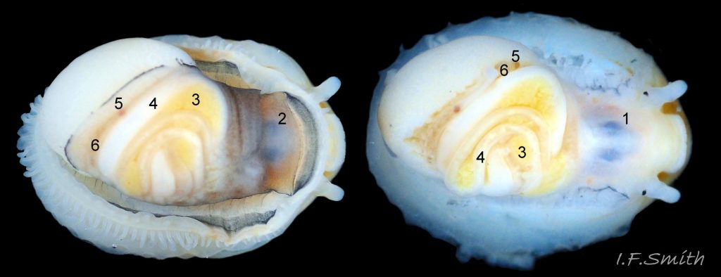
When the body is removed from the shell the entire mantle is visible. The peripheral mantle-skirt is white translucent and may reveal the white foot below 71 Patella pellucida. The rest of the mantle is semi-transparent with a fine spray of black pigment of varying intensity. Many features of the viscera can be discerned through it.
Blood circulation and respiration
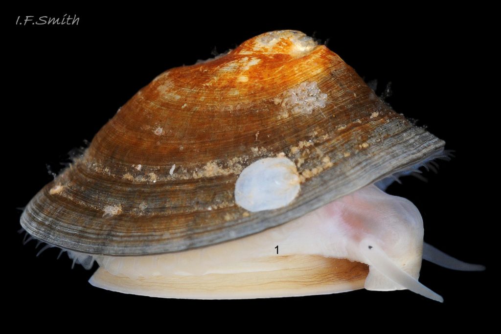
Image 72 Patella pellucida illustrates paragraph 1.
After it has oxygenated organs in the visceral mass, the colourless blood, depleted of oxygen, collects in visceral sinuses. It then passes in vessels through gaps in the pedal-retractor muscle into an afferent branchial vessel that runs through the mantle skirt around the edge of the distal face of the muscle . The afferent branchial vessel distributes blood to all the gill leaflets which re-oxygenate it as it passes through them. The oxygenated blood passes into a large efferent pallial vessel (a.k.a. efferent branchial vessel) and passes forwards on both right and left sides of the animal. The
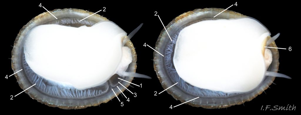
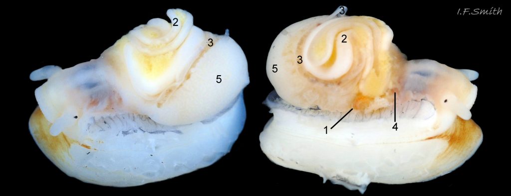
flow of blood in the efferent pallial vessel (e.p.v.) on the right continues round the front of the head to join the left of the e.p.v. in a trunk that enters the left side of the nuchal cavity to connect with the heart in the pericardial cavity behind the left of the nuchal cavity 55 Patella pellucida & 73 Patella pellucida . The heart consists of three parallel chambers; the auricle, ventricle and bulbous aorta. Blood is pumped out via the bulbous aorta to oxygenate organs in the head and visceral hump before returning to the visceral sinuses to recommence the cycle.
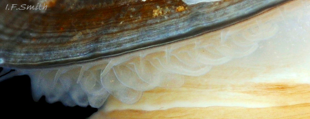
The gills consist of a large number of leaflets, alternating large and small, hanging from the roof of the pallial groove 74 Patella pellucida. The leaflets vary in shape with free ends that can be rounded, flat or pointed, but all narrow where they attach to the roof of the pallial groove and have a deep and densely ciliated groove around the rim 75 Patella pellucida. The respiratory inhalant water current is created by the motion of cilia on each leaflet of the gills drawing water in all around the shell, except at the gill-less anterior (X5000 scanning electron microscope images of cilia on a chiton at 18 Lepidochitona cinerea ). Sediment entering with the water is trapped by mucus from the lateral glandular streak 53 Patella pellucida and carried forwards and out by the exhalant current along the pallial groove. The oxygen-bearing water is drawn across the surface of the leaflets in the opposite direction to the blood flow within, so creating a counter current-system (Fretter and Graham, 1994). The dense cilia in the rim-groove are reported (Fretter and Graham, 1994) to propel debris particles caught on the leaflet surface to the free end where they drop off, but this would seem unlikely for those in holdfasts feeding upside down into the core of the stipe. After passing the leaflets, the current, now exhalant, is propelled by cilia forwards along the pallial groove to exit the gill-less anterior on the right of the animal 76 Patella pellucida & 77 Patella pellucida . It is unlikely that the red features referred to as the osphradia 53 Patella pellucida have the usually attributed function of water-quality testing as there are no associated ctenidia to forwarn, and they are located in the path of the exhalant water after it has flowed over the pallial gills and shortly before it exits the anterior of the shell. There are anatomical signs that they may be vestiges of a lost double osphradium-ctenidium complex like that still found in Diodora graeca 20 Diodora graeca (Spengel, 1881, in Fretter & Graham, 1962, p314). Graham & Fretter (1947) had a ‘suspicion’ that the leaflets on frond-dwellers are smaller than those on holdfast-dwellers, and that this can be correlated with the greater exposure to water movement of frond dwellers. This seems to be supported by some images78 Patella pellucida that show large leaflets projecting from the shell of holdfast dwellers, but no leaflets projecting from the shell of frond dwellers. But, measurement of live, expanded, gill area is difficult, and individuals can extend or retract the mantle and gills.
Alimentary and excretory features .
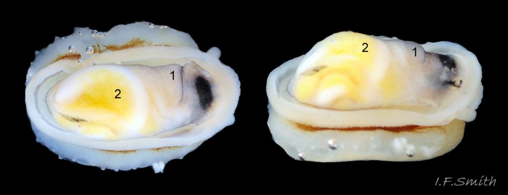
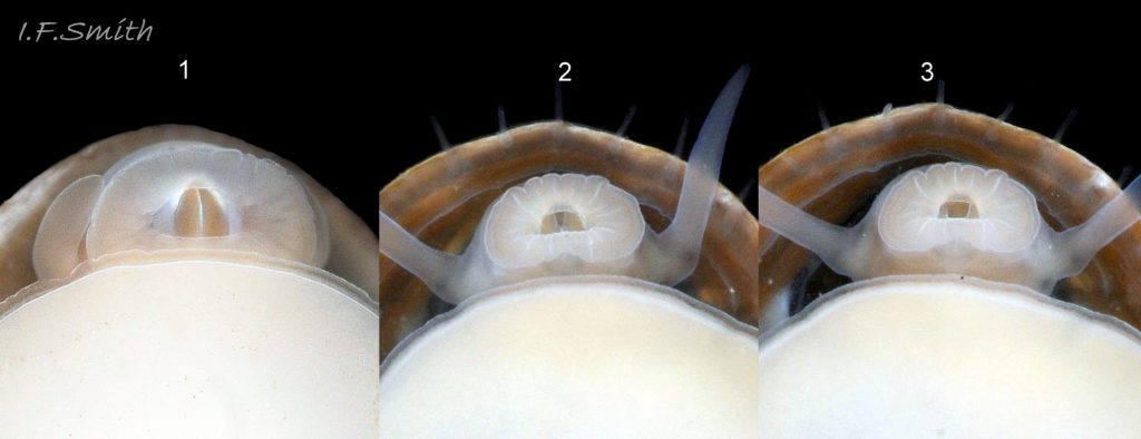
The inner lips of the mouth, described above, open into the buccal cavity. The anterior wall of the cavity is reinforced by a pliable, white, chitinous, antero-dorsal plate, called the “jaw” though it is not articulated and does not bite 60 Patella pellucida. It serves as an attachment for several muscles and has two lateral wings that meet dorsally at an angle and form an anterior shield for the inner lips when they are open. Within the buccal cavity there is a pink odontophore 61 Patella pellucida & 79 Patella pellucida consisting of a right and left bolster which is covered in thick cuticle. Much of a bolster is made of strong cartilage-like material. The odontophore occupies a large amount of the body, 25% of its length. There is a black mark within its anterior half that is visible through the dorsal surfaces of it 80 Patella pellucida the epithelium of the head and the overlying mantle 81 Patella pellucida, and, on many translucent shells, it underlies and adds to the intensity of a patch of black pigment on the anterior of the shell’s interior 82 Patella pellucida.
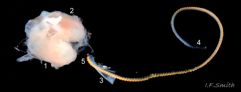
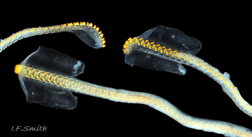
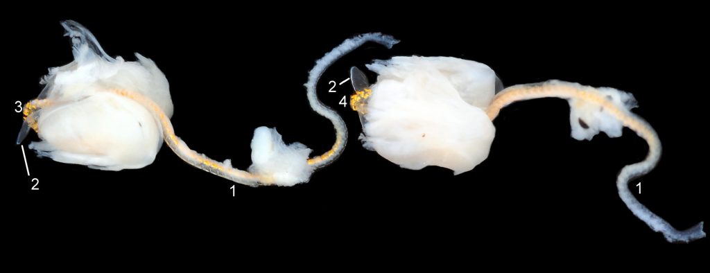
The radula is created in a semi-translucent sac 8tella pellucida. Its exposed anterior, widened into a hyaline shield 84 Patella pellucida, rests on the dorsum of the odontophore 85 Patella pellucida . The strong, iron-impregnated, unarticulated teeth are firmly fixed in a backwardly inclined position on the radula 86 Patella pellucida, but the anterior tip of the radula bends over the front of the odontophore so that the front rows of four teeth are inclined forwards like a chisel 85 Patella pellucida. To feed, the strong muscles of the large odontophore thrust it forwards against the front of the buccal cavity which, reinforced by the jaw, restrains the odontophore but allows the front teeth to project strongly from the narrow vertex of the gap between the wings of the jaw 60 Patella pellucida. When applied to Laminaria, the teeth effectively chisel into the tough frond or stipe, and the curve of the withdrawing teeth acts as a scoop to lift particles back to the oesophagus in the buccal cavity. The action is surrounded by the outer lips which prevent the escape of loosened food fragments 60 Patella pellucida. Like other limpets and some sea snails that graze rock surfaces, P. pellucida has a long, powerful radula, but it is shorter than most, being about 75% the length of the shell 37 Patella pellucida, requiring only a single fold to fit it inside the body 87 Patella pellucida. which is about the same length as it 80 Patella pellucida. Other British patellids have radulae between about 100% and 250% of shell length (Fretter & Graham, 1962). The cause of the correlation between length and rock grazing is uncertain. It may be that a lengthy process is needed for the teeth to acquire the required hardening mineralization, so less length/time would be needed to form a radula for grazing alga that is tough, but not as hard as rock. Tooth creation starts at the slightly bifid, white, inner end of the radular sac 8tella pellucida with secretion of colourless transparent cuticular material by odontoblast cells. As each new tooth commences, the previous one is pushed forwards along the sac. Cells along the roof of the sac make incremental additions, including the hardening salts of iron and silicon, to the teeth as they travel along the sac, with a progressive change from colourless through darkening shades of yellow/orange visible through the translucent sac walls. Creation is complete by the time the tooth reaches the buccal cavity, where the radula emerges from the sac onto the odontophore 85 Patella pellucida.
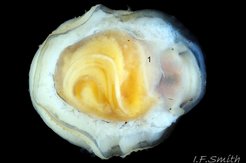
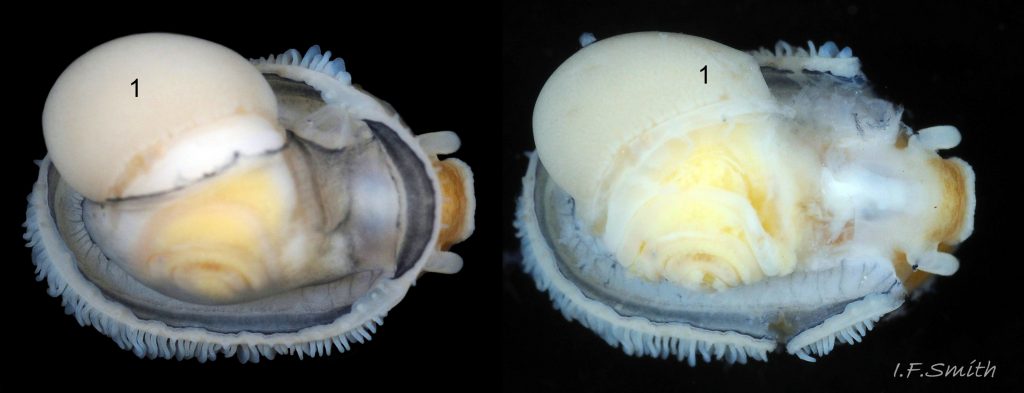
The yellow digestive gland 55 Patella pellucida & 81 Patella pellucida composed of a mass of tubules is usually the most obvious organ on the surface of the visceral mass when the shell is removed. Digestive cells in the tubules ingest particulate food to digest it intracellularly (Fretter & Graham, 1994, p. 219). The tubules extend into the blood filling the haemocoel, and their very thin covering of connective tissue allows the passage of nutrients into the blood. Food material passes along the long coiled intestine which is usually white like the consumed white inner parts of Laminaria 81 Patella pellucida & 87 Patella pellucida. Faecal boluses pass through the rectum 81 Patella pellucida and are compressed and bound with mucus to emerge as faecal rods 88 Patella pellucida from the anus at the rear right of the nuchal cavity 87 Patella pellucida and are conveyed to, and expelled from, the right anterior of the shell by cilia and the flow of exhalant water 77 Patella pellucida. Compression of faeces assists removal without fouling the gills. Excreta from the nephridipores by the anus are expelled by the same route. Particulate matter removed by cilia on the gill leaflets, and sediment caught in mucus from the lateral glandular streak, are conveyed along the pallial groove by cilia and exhalant water, to join the anterior expulsion.
Reproductive organs
The gonads are situated between the viscera and foot. During the breeding season, they may spread over much of the viscera 87 Patella pellucida. & 89 Patella pellucida, but on juveniles , and on adults out of breeding condition, they are usually hidden or nearly so 55 Patella pellucida & 82 Patella pellucida, on an animal removed from the shell. Female ovaries are granular, and male testes have numerous interconnected tubules. Fertilization is external, so the male has no penis. Gametes leave both sexes through the right nephridium (kidney) via its nephridipore at the rear right of the nuchal cavity, and carried from the shell by cilia and the forward flowing exhalant current.
Habits and ecology
Occurs on rocky shores, usually where Laminaria is present. Some wave action or current is preferred; it is often scarce or absent on extremely sheltered shores. It may occur in holdfasts on exposed shores where few, if any, can be found on fronds in some months. A current of 1.3 m/s (2.9 km/h) was found to be optimal for those on Saccorhiza fronds at Loch Hine, Eire, with a marked decline in numbers at lower and higher speeds (Ebling, 1948, in Kain & Svendsen, 1969), but it is difficult to be precise as other factors such as depth and competition for space by encrusting epibiota on the fronds can affect numbers. Depth limits are LWST to, presumably, the depth where Laminaria growth ceases, but presence/frequency within that zone varies. In the Isle of Man, the percentage of occupied holdfasts of large L. hyperborea was about 50% at 1m below LWST [where wave action is strongest], less than 10% at 11m and 0% at 20m (Kain & Svendsen, 1969), but abundance is affected by the interplay of other factors, including whether the local population utilizes holdfasts or, as in Norway, not.
Occurs and feeds primarily on Laminaria spp. and Saccorhiza and, when young, on diatoms etc caught on the surface of Laminaria 03 Patella pellucida. Newly settled juveniles, less than 4mm long, feed on small red algae, such as Rhodymenia palmata, and on calcareous red algae encrusting rocks 02 Patella pellucida Other plants sometimes eaten, mainly by juveniles, include Alaria, Himanthalia 90 Patella pellucida. and Fucus serratus 91 Patella pellucida(Wigham & Graham, 2017). It is uncertain whether these foodplants are adequate for development as adults are only occasionally found on them. They may be those that have failed to reach Laminaria or have been swept off it by storms 95 Patella pellucida. The hard iron-mineralized teeth of the radula 86 Patella pellucida, about 75% length of shell 80 Patella pellucida, enables larger specimens to eat, and excavate caves in, the base of a Laminaria stipe 92 Patella pellucida.
Defence: When on Laminaria fronds or stipes, the translucent juvenile shells show the colour of the underlying alga and the dark chestnut band on the muscle. This, and the brown shell-colour of adults, is cryptic when on brown algae, except for the iridescent blue/green rays 03 Patella pellucida. The colour of the rays is created by the light interference of zig-zag lamellae of calcite selectively reflecting blue/green in water. Saturation of the blue/green is intensified, and contrast with the visible white body within the shell is increased, by black, non-crystalline, colloidal particles underlying parts of the rays (Li et al., 2015) 17 Patella pellucida & 19 Patella pellucida. If the shell is rotated, different lines appear and disappear as the light’s angle of incidence on them changes (Li et al., 2015, “supplementary movie1” at www.nature.com/articles/ncomms7322#supplementary-information ). Flashing on and off of the rays as the Laminaria waves about and changes the angle of light incidence, may distract from the animal’s outline, or it may camouflage by resembling neighbouring iridescent weeds such as Chondrus crispus 93 Patella pellucida and en.wikipedia.org/wiki/Chondrus_crispus#/media/File:Chondr… .
Other species, such as Lacuna vincta and Steromphala cineraria take advantage of pits made by P. pellucida to feed on the exposed, nutritious core of Laminaria stipes 25 Patella pellucida.
Li et al. (2015) suggested that P. pellucida may have Batesian mimicry of nudibranchs with iridescence assumed to warn of repugnant secretions or stinging sequestered cnidocytes. But Batesian mimics are usually less numerous than their models as predators learn from the more numerous example and apply their learning to all. Iridescent nudibranchs are usually much less numerous than P. pellucida, so are unsuitable as models to mimic. Adults concealed in holdfasts have little need for camouflage or warning colouration, and are often coated with epizoic growths 92 Patella pellucida . The use of the rays for sexual attraction in a non-copulating species is unlikely, especially as the rays become much less obvious at maturity, and their primitive eyes, almost certainly, cannot distinguish the rays.
Breeds most months, with maximum in winter and spring; November to February in Plymouth. No copulation; female releases thousands of individual, planktonic ova with gelatinous coverings that swell on contact with water. Ova are fertilized by sperm shed into water by males. Eggs soon hatch into shell-less, rotating, planktonic trochophore larvae (stage passed within egg by most less “primitive” spp.)
Development: shell type numbers below refer to those in Part 1 – Shell Description.
After a few days, the trochophore larvae metamorphose into planktonic veligers with a weakly developed velum for swimming and food capture, and a slightly coiled shell (GS 1) 01 Patella pellucida.
After a short pelagic life, the veligers settle on the substrate and metamorphose into the juvenile limpet form (GS2) . Initially, they feed on small red weeds and can be found on rock surfaces 02 Patella pellucida By the time they attain shell length 4mm the radula is strong enough to rasp Laminaria as well as scrape up diatoms and other microscopic organisms that have settled on the fronds 03 Patella pellucida. To reach the Laminaria, the animals secrete mucus to form a ‘sail’ many times the size of the shell. The sails bring the animals to near neutral buoyancy and catch any water movement so they readily lift off from the substrate. As 1m² substrate carrying a Laminaria bed has about 8 to 12m² of Laminaria surface (Vahl, 1983), there is a good chance that the sails will strike a Laminaria plant and stick to it, as the mucus is a strong instant underwater adhesive. (While manipulating a supine specimen in water for photography, the 0.5mm wide tip of my forceps struck the invisible mucus being exuded. When I lifted the forceps, the animal followed with a piece of adhering Laminaria four times its weight, and was detatched only with difficulty). Any that fail to contact Laminaria, or a sufficient substitute, will die; the scarcity of the species in very sheltered conditions may be because of the lack of water movement sufficient for lift off. The great majority of the survivors (juveniles GS3) take up residence on the frond where their position is maintained by negative geotaxy (Vahl,1983) and their orientation may be prow-like anterior towards on coming wave action or current 03 Patella pellucida., but the evidence for the cause of orientation is not clear as the movement of waves over waving fronds is difficult to ascertain. A few of the settling juveniles land on the stipe or get into a holdfast where they initially feed on a finger of the holdfast 06 Patella pellucida . All, regardless of where they live, have similar juvenile shells, except that those in holdfasts may start to develop a rougher surface 09 Patella pellucida . The extremely low profile of a smooth, juvenile shell on a frond or stipe, orientated with narrow anterior facing the current, has minimal drag 03 Patella pellucida, and the broad foot with strongly adhesive mucus has a very firm grip, so wave backwash up to 20m/s (c.70km/h) can be withstood at this stage (Denny, 1984, in Fretter & Graham, 1994). The low, streamlined shell profile is maintained by loss of shell material at the anterior as the posterior grows. The loss may be caused by physical wear, but as the substrate is plant surface it seems unlikely. Resorption locally by the anterior of the mantle is a possibility.
Sexual maturity is reached at shell lengths of about 5 mm and upwards (Graham & Fretter, 1947). A change from shell loss to growth at the anterior commences shortly after maturation, at shell length 7 or 8mm. By 10mm length there is a considerable band of new shell around the whole shell with encircling growth lines, parallel to the substrate, (early adult GS4) that have an angular unconformity where they meet the earlier tilted growth lines 15 Patella pellucida. Blue lines are usually fewer, less distinct or absent on the new shell. The profile, viewed from the side, becomes a higher dome with a nick on the anterior profile where the convex early surface meets a nearly straight new surface 15 Patella pellucida.. Drag in water currents becomes much greater than on the low profiles of GS3 shells, and most on the exposed frond are swept off before they reach 11mm length to die or, possibly, reach other Laminaria in deeper, calmer water with the aid of a mucus sail.
In very sheltered waters with small amounts of wave action, some on fronds survive to lengths of 14mm or 15mm and form impressive, smooth, high, conoid shells resembling a Phrygian cap (GS5) 20 Patella pellucida. But this form is relatively scarce, though easily seen when present, as the sheltered conditions required for its survival are not conducive to large populations of the species. On both sheltered shores and more exposed one, a few individuals find less turbulent conditions on the stipe 24 Patella pellucida which has a much shorter arc of movement, diminishing to zero at its base, than the fronds that thrash back and forth in storms through a curve with about 2m diameter. The shell may resemble GS5, but if a wide pit is eaten into the stipe the newly deposited shell may flare out more widely so that the conoid has a lower profile with a break in slope at the junction of new and earlier shell 26 Patella pellucida.
By far the largest number of those who survive into (GS5) are those who found their way into holdfasts at an early stage and have grown secure from wave impact, unless the plant is torn away. On parts of some shores, including exposed ones, about 50% of Laminaria holdfasts may be inhabited. Occupation is usually restricted to a single adult (GS4 or 5h) per holdfast . Occasionally, when several holdasts are intermingled, one pit make break through into another 94 Patella pellucida. For those on fronds, lifespan is about a year or less. Those in holdfasts may live for two or more years, if the plant survives. Occasional stranded specimens, perhaps from holdfasts cast up from deep calm water, have exceptionally large shells with growth rings suggesting an age of four years 25. 32.1 Patella pellucida.
The absence from Norway of P. pellucida living in holdfasts, and of the associated distorted shell form lacking blue rays on most of the adult growth, suggests a lack of genetic interchange between Britain and Norway. It appears that the short planktonic larval stage is insufficient to survive the time needed for it to drift from Britain to Norway.
Distribution and status
Iceland and Murman Coast (N. Russia) to Straits of Gibraltar. Not resident further into Baltic than Øresund, south-west Sweden or continental coast of North Sea apart from on occasional strandings of washed in Laminaria. GBIF map www.gbif.org/species/5728509
Frequent around Britain and Ireland on hard substrate where Laminaria is present. Both P. pellucida and Laminaria are absent or rare in Liverpool Bay and Flamborough Head to Kent apart from occasional individuals washed in from other areas. Often overlooked at times when only those hidden in holdfasts are present. U.K. distribution map NBN species.nbnatlas.org/species/NHMSYS0021056396
Acknowledgements
I (I.F.S) should like to thank Debbie Evans, Dr Marco Faasse, David Fenwick, Allan Rowat and Neil Ward for information and use of their images, and Dr Ivan Nekhaev and Dr Julia Sigwart for advice with the text and interpretation of the images. I am extremely grateful to my co-authors Erling Svensen and Paula Lightfoot for their invaluable contributions of images, information and constructive criticism. Any errors or omissions are my responsibility.
Links and references
Forbes, E. & Hanley S. 1849-53. A history of the British mollusca and their shells. vol. 2 (1849), van Voorst, London. (As Acmaea virginea; Free PDF at archive.org/stream/historyofbritish02forb#page/428/mode/2up Use slide at base of page to select pp.429-433.)
Fretter, V. and Graham, A. 1962. British prosobranch molluscs. Ray Society, London.
Graham, A. 1988. Molluscs: prosobranch and pyramidellid gastropods: keys and notes for the identification of the species. Brill & Backhuys, for Linn. Soc. Lond. & Estuarine and Brackish-water Sciences Assoc. Synopses of the British Fauna (New Series) no.2. Edition 2 (662pages). Leiden. (Edition 1 of series, 1971, 112 pages, is no substitute.)
Graham, A. & Fretter, V. 1947. The life history of Patina pellucida (L.) J. mar. biol. Ass. U.K. 26: 590 to 601.
plymsea.ac.uk/1257/1/The_life_history_of_Patina_pellucida…
Jeffreys, J.G. 1862-69. British conchology. vol. 3 (1865). London, van Voorst. (As Tectura virginea) Free PDF at archive.org/stream/britishconcholog03jeff#page/248/mode/2up . Use slide at base of page to select pp.248- 250.
Kain, J.M. & Svendsen, P.1969. A note on the behaviour of Patina pellucida in Britain and Norway. Sarsia, 38:1, 25-30. dx.doi.org/10.1080/00364827.1969.10411147
Lebour, M.V., 1937. The eggs and larvae of the British Prosobranchs with special reference to those living in the plankton. Journal of the Marine Biological Association of the United Kingdom, 22: 105 to166.
plymsea.ac.uk/953/
Li, L. et al. 2015. A highly conspicuous mineralized composite photonic architechture in the translucent shell of the blue-rayed limpet. Nat. commun. 6:6322 doi:10,1038/ncomms7322(2015). Free pdf (open access) at www.nature.com/articles/ncomms7322
and supplementary images and video at www.nature.com/articles/ncomms7322#supplementary-information
Wigham, G.D. & Graham, A. 2017. Marine gastropods 1: Patellogastropoda and Vetigastropoda. Synopses of the British Fauna (New Series) no.60. (172pages). Field Studies Council, Telford, England.
Yonge, C.M. and Thompson, T.E. 1976. Living marine molluscs. Collins, London.
Current taxonomy: World Register of Marine Species (WoRMS) www.marinespecies.org/aphia.php?p=taxdetails&id=147459
GLOSSARY
afferent (adj. of vessel) = carrying blood etc. towards an organ.
aperture = mouth of gastropod shell; outlet for head and foot.
apex (definition for this account) = position of the larval protoconch (see summit).
branchial (adj.) = of or relating to gills (branchiae).
cephalic = (adj.) of or on the head.
cnidocytes = explosive stinging cells of hydroids, jellyfish, sea anemones etc. en.wikipedia.org/wiki/Cnidocyte
ctenidium = comb-like molluscan gill; usually an axis with a row of filaments either side.
efferent (adj. of vessel) = carrying blood etc. away from an organ.
ELWS = extreme low water spring tide (usually near March and September equinoxes).
epizoic (of a plant or animal) = growing or living non-parasitically on the exterior of a living animal.
GS1, GS2 etc. = Growth Stage 1 (planktonic veliger larva), Growth Stage 2 (newly settled juvenile) etc.
GS5f = Growth Stage 5 (late adult) living on Laminaria frond.
GS5h = Growth Stage 5 (late adult) living in Laminaria holdfast.
GS5s= Growth Stage 5 (late adult) living on Laminaria stipe.
haemocoel = system of interconnected spaces (sinuses) containing blood within body of a mollusc.
holdfast = rootlike in appearance, but not in function, tendrils attaching seaweed to the substrate. (a.k.a. hapteron).
mantle = sheet of tissue that secretes the shell and forms a cavity for the gill in most marine molluscs.
MLWS = mean low water spring tide level (mean level reached by lowest low tides for a few days every fortnight; Laminaria or Coralline zone on rocky coasts).
osphradium (pl. osphradia) = organ for testing water quality (chemical and/or for particles) usually near approach of inhalant current to ctenidium or pallial gills. Structure varied; including comblike, papillate or ribbing.
pallial (adj.) = of, relating to, or produced by the mantle (pallium).
periostracum = thin horny layer of chitinous material often coating shells.
phylogenetic (of development) = of change due to genetic make up.
resorb = absorb again that which was previously produced.
resorption = the process of absorbing again that which was previously produced.
stipe = stem of some brown seaweeds that supports the fronds and may contain a core of cells that transports sugars and nutrients within the alga.
summit (definition for this account) = highest point of the shell above the substrate (see apex).
trochophore = spherical or pear-shaped larva that moves with aid of girdle of cilia that beat to cause rotation. Stage preceding veliger, passed within gastropod egg in most spp. but free in plankton for patellid limpets, most Trochidae and Tricolia pullus, and, with no veliger, chitons.
veliger = shelled larva of marine gastropod or bivalve mollusc which feeds and swims by beating cilia of a velum (small on P. pellucida).