Internal anatomy
When extracted from shell, organs in place but indistinctly seen through translucent mantle on anterior half of body. When mantle cut along pallial inhalent channel and opened out to the right of the animal like a page of a book, organs attached to its inner face are displaced but more clearly visible.
Key to items on images 35aNl 35a Nucella lapillus. Male removed from shell, dorsal view. Mouth of Mersey Estuary, England. July 2014. & 35bNl 35b Nucella lapillus. Male removed from shell, tilted to expose right side. Mouth of Mersey Estuary, England. July 2014. (extracted male), 36 Nucella lapillus. Male removed from shell; mantle cut on left and turned out to animal’s right. Mouth of Mersey Estuary, England. July 2014. (opened male) 37 Nucella lapillus. Female removed from shell. Mouth of Mersey Estuary, England. July 2014. (extracted female) 38 Nucella lapillus. Female removed from shell, and mantle cut on left and turned out to animal’s right. Mouth of Mersey Estuary, England. July 2014. (opened female). If key is read in order, the functions and inter-relationship of the organs may be understood.
Mantle
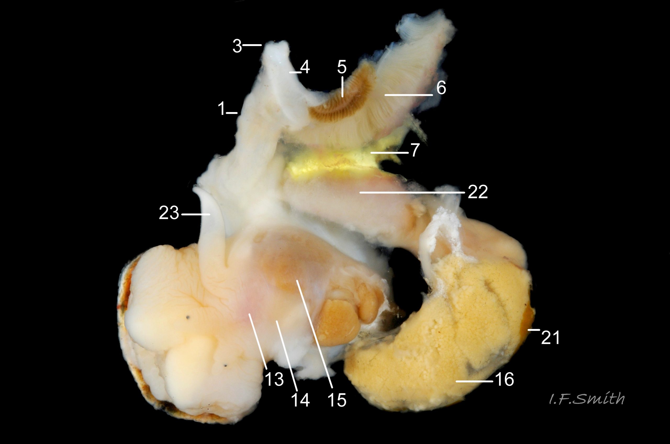
1: mantle edge – substantial, opaque, yellowish, slightly flounced, anterior border of mantle skirt. Only part that produces exterior layers of shell. Sometimes flounces exaggerated and make protruding shell-growth that forms imbricate sculpture. (Images 35a&b, 36, 37)
2: mantle skirt – forms roof of mantle cavity and, when uncut, partially obscures organs within. (Images 35a&b, 37)
Respiratory features
3: siphon – white, folded, extension of mantle that rests in shell’s siphonal canal and draws in inhalent current of water when animal extended. (Images 35a, 36, 37)
4: pallial inhalent channel – distinct, white fold in mantle forming a channel from siphon to osphradium. (Images 35a, 36, 37)
5: osphradium – dark, bipectinate chemo-receptor at inner end of inhalent channel that tests water approaching ctenidium for scent of food, prey, predators, mates and/or water quality. So highly developed on Nucella that Jeffreys (1867) thought it was second ctenidium. (Images 35a, 36, 37, 38)
6: ctenidium – substantial, buff-white, gill with many fine leaflets that receive oxygen from inhalent water, and oxygenate blood passed through them. (Images 35a&b, 36, 37, 38)
7: hypobranchial gland – puckered gland that produces mucous to trap particles from inhalent water before reaches ctenidium, and to transport particles out of mantle cavity. Other functions might be attraction of mates by scent, and acrid smell/taste to discourage predators. Initially cream-white, changing to yellow, green, purple etc when exposed to light and air. (Images 35a&b, 36,37)
Vascular features
8: kidney – de-oxygenated blood has urea etc. removed and excreted by kidney before passing to efferent renal vein [9]. (Images 35a&b)
9: efferent renal vein – carries blood away from kidney [8]. (Image 35b)
10: Hypobrachial vessels – carry blood from efferent renal vein [9] through hypobranchial gland [7]) to afferent vessel of ctenidium [11]. (Image 35b)
11: afferent vessel of ctenidium – receives blood from hypobranchial vessels [10] and passes it into leaflets of adjacent ctenidium [6] for oxygenation. (Image 35b)
12: rectal gland – long, linear gland; dark brown to purple-black in adults. Function uncertain, perhaps produces substances that supplement the excretory activity of the kidney. (Images 35a&b, 37, 38). (Images 35a&b, 37, 38)
Alimentary features
13: buccal mass – pink as rich in myoglobin; contains odontophore and anterior of radula used in rasping prey. (Image 36)
14: acinous salivary gland – compound gland of many small rounded sacs that secrete enzymes for external pre-digestion/ liquefaction of prey. (Image 36)
15: gland of Leiblein – secretes enzymes for internal digestion of ingested liquefied prey. (Image 36)
16: digestive gland – mass of branching tubules and ducts occupying majority of visceral mass (spiral ‘tail’ of body); most visible feature, except in breeding season when much may be covered by ovary or testis. Receives solution of digested food from stomach into tubules where taken up by absorbing digestive cells and passed into blood bathing the tubules. (Images 35a&b, 36, 37, 38)
Reproductive features
17: ovary- gland that produces eggs. (Image 37)
18: albumen gland – translucent whitish gland that secretes albumen for nutrition of developing eggs. (Images 37, 38)
19: sperm ingesting gland – dark brown blind tubules in female where sperm excess to requirements is engulfed and digested by cells; gland is used to discover sex of specimens as penis can occur on females affected by imposex. (Image 37, 38)
20: capsule gland – secretes fibrous wall of capsule containing ova, but does not give it its final shape. (Images 37, 38)
21: testis – gland that produces spermatozoa. (Images 35a&b, 36)
22: prostate – pink gland that secretes fluid which, with spermatozoa and seminal vesicle fluid, forms the semen. (Images 35b, 36)
23: penis – on males, and on females affected by imposex caused by tributyltin pollution. (Image 36)
36 Nucella lapillus. Male removed from shell; mantle cut on left and turned out to animal’s right. Mouth of Mersey Estuary, England. July 2014.