Main text for set is below image P2 02 Patella shell. Interior of Patella vulgata shell. Length 20.2mm. Specimen chosen for clarity. Many P. vulgata are not as clearly marked or so coloured. .
Half-grown specimen. Interior layers not yet thick enough to conceal pigmented exterior layer.
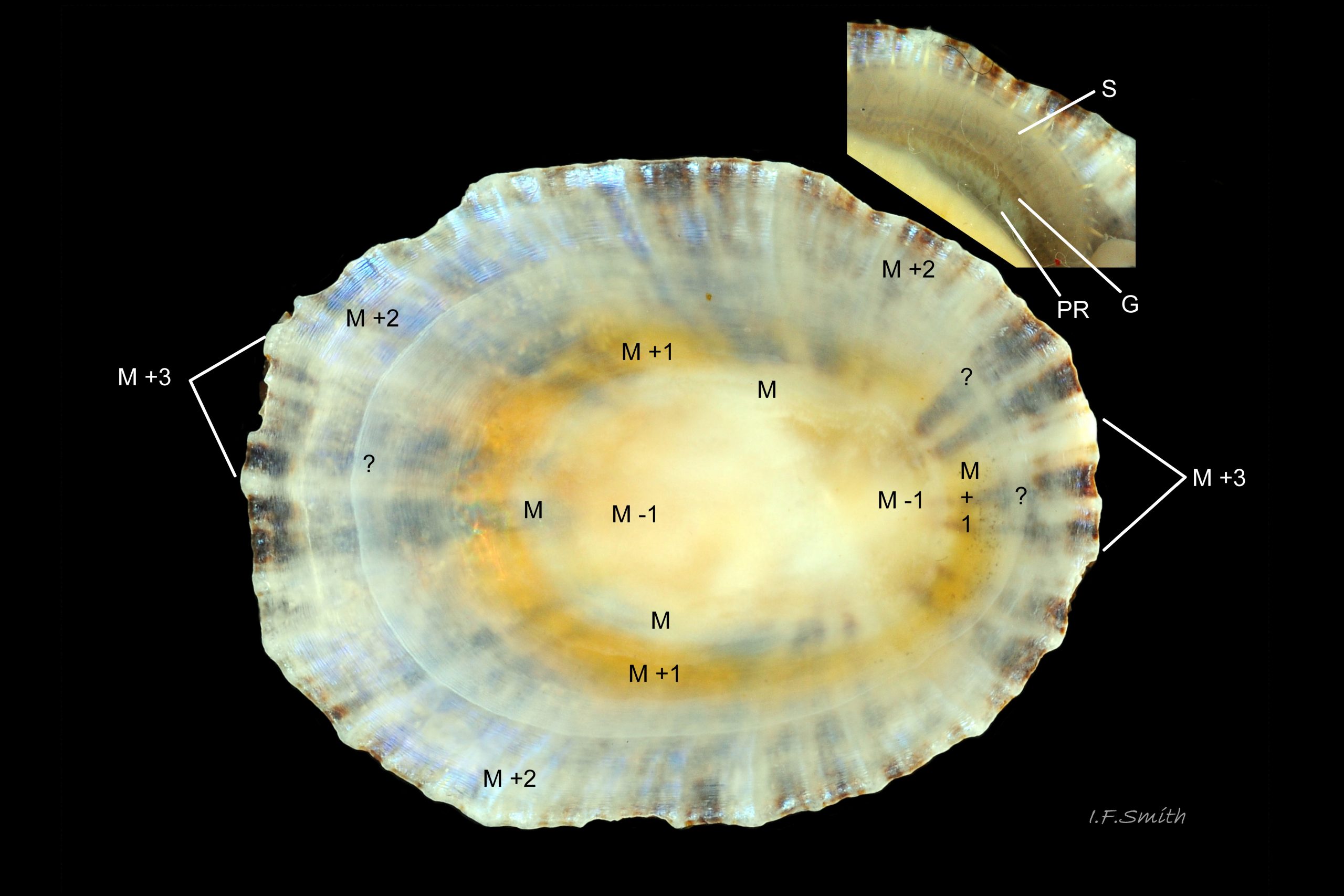
Growth of limpets slows/ceases at certain seasons when food is in short supply, and it is when at rest that scars and mantle marks become best defined. Consequently, during times of growth the body parts may not be located at strong marks that they caused previously. This seems to be the case with this specimen where the white pedal retractor muscle (PR), orange-brown gills (G) and free mantle skirt (S, not fully extended in image) are much closer to the rim of the shell than the shell-layers associated with them (M, M+1 and M+2). “?” marks an apparent part of M+2, but this would make M+2 extremely wide. It is possible that the thickened white line at “?” marks the start of deposition at the new position of the gills and will become the new M+1. This would match better the current distance from rim of the gills (G).
Shell structure of Patella species.
See 1P 01 Patella shell. Stylised transverse section through shell. Thickness of pigment-less interior layers exaggerated; colours selected to resemble those seen through the layers of specimen in image 2 of this set. for cross section diagram.
Pigment is incorporated into the substance of the shell during secretion only at the mantle edge creating the exterior layer (M +3). The rest of the mantle typically secretes five thin, overlapping, pigment-less layers that line the interior of the shell. The layers have varying crystalline structures with differing degrees of transparency and iridescence that affect the visibility through them of the colours in the exterior layer.
The layers are numbered from the scar made by the horseshoe-shaped shell-muscle (pedal retractor muscle). The scar is called the myostracum (abbreviation M). Working out from it, the layers are named M +1, M +2 and M +3. Working inwards, the layers are M -1 and M -2. Please note that this notation refers to relative position only; the number of layers and the crystal form of each layer varies from species to species. The crystal descriptions below are of Patella vulgata as an example, but the correlation of shell layers to soft parts applies to all of P. vulgata, P. depressa and P. ulyssiponensis.
M +3: exterior layer of shell produced by edge of mantle (E on 7P 07 Patella. Full grown (shell length 40 mm) Patella ulyssiponensis removed from shell to fully expose mantle. ). Only layer with pigment incorporated in its substance. Composed of large, irregular, foliated (flattened) crystals of calcite (mineral form of calcium carbonate) arranged radially (at right angles to rim of shell).
All the following layers are on the interior surface of the shell, thin and lack pigment when deposited.
M +2: formed by the free mantle skirt (S on 7P 07 Patella. Full grown (shell length 40 mm) Patella ulyssiponensis removed from shell to fully expose mantle. ). Composed of large, irregular foliated crystals of calcite arranged concentrically (parallel to rim of shell). Concentric arrangement can be seen in small concentric ripples that iridesce blue or green (close-up on 3P 03 Patella shell. Interior of Patella depressa shell. Length 24.9 mm. Inset is close-up of concentric iridescent ripples in M +2. ). Colours of pigmented exterior layer clearly seen through this single layer, especially in P. depressa and young specimens of P. vulgata and P. ulyssiponensis. This layer underlies all the others that have grown over it; it may be exposed on the external surface of the shell if M +3 is eroded 4P 04 Patella shell. Exterior of Patella depressa shell. Length 24.9 mm. .
M +1: formed by the mantle-roof of pallial groove where gills (G on 7P 07 Patella. Full grown (shell length 40 mm) Patella ulyssiponensis removed from shell to fully expose mantle. ) are suspended. Composed of small, lamellar crystals of aragonite (another mineral form of calcium carbonate) packed like leaves of a book, arranged concentrically.
M: myostracum; scar made by the horseshoe-shaped shell-muscle (a.k.a. pedal retractor muscle) (PR on 7P 07 Patella. Full grown (shell length 40 mm) Patella ulyssiponensis removed from shell to fully expose mantle. ). Extremely thin deposit of complex prismatic material. May show colours of exterior layer (less clearly than in M +2), and may include a line of different colour if located over a previous resting position of M +1, as in 3P 03 Patella shell. Interior of Patella depressa shell. Length 24.9 mm. Inset is close-up of concentric iridescent ripples in M +2. .
M -1: formed by mantle covering visceral hump and nuchal cavity (V on 7P 07 Patella. Full grown (shell length 40 mm) Patella ulyssiponensis removed from shell to fully expose mantle. ). Composed of small, lamellar crystals of aragonite, packed like leaves of a book, arranged radially. Porcelaneous, usually not transparent. May be partly or entirely stained with colour from digestive gland located immediately below mantle, so colour may vary with diet. For example, limpet in 2P 02 Patella shell. Interior of Patella vulgata shell. Length 20.2mm. Specimen chosen for clarity. Many P. vulgata are not as clearly marked or so coloured. was found feeding on red encrusting algae; has reddish M -1, perhaps because of its diet.
M -2: (not clearly visible or labelled on this image) layer may be well developed to absent on different individuals of same species. Formed near vertex of shell by part of mantle covering visceral hump. Composed of foliated crystals of calcite, arranged radially or irregularly. Often a reflective sheen or iridescence.
Links and references
Cohen, A.L. & Branch, G.M. 1992. Environmentally controlled variation in the structure and mineralogy of Patella granularis shells from the coast of southern Africa: implications for palaeotemperature assessments. Palaeogeography, palaeoclimatology, palaeoecology, 91: 49-57. www.whoi.edu/fileserver.do?id=163844&pt=2&p=36767
MacClintock, C. 1967. Shell structure of patelloid and bellerophontid gastropods (Mollusca). Peabody Museum of Natural History, Yale University. Bulletin 22. pdf at
www.google.co.uk/?gws_rd=ssl#q=MacClintock%2C+C.+1967.+Sh….
215 pages, may take a few minutes to download. Contents on page v.(= p.6 of pdf). To find pages on pdf add 1 to Roman numerals, and add 11 to modern numerals.